


![PREVALENCE OF VITAMIN D DEFICIENCY IN PATIENTS WITH ACUTE MYOCARDIAL
INFARCTION.
AmericanJournalofCardiology2011Jun1;107(11):1636-8.
Deficiencyin25-hydroxyvitaminD(25[OH]D)isatreatableconditionthathas
been associated with coronary artery disease and many of its risk factors. A
practicaltimetoassessfor25(OH)Ddeficiency,andtoinitiatetreatment,isat
the time of an acute myocardial infarction. The prevalence of 25(OH)D
deficiency and the characteristics associated with it in patients with acute
myocardial infarction are unknown. In this study, 25(OH)D was assessed in
239 subjects enrolled in a 20-hospital prospective myocardial infarction
registry. Patients enrolled from June 1 to December 31, 2008, had serum
samples sent to a centralized laboratory for analysis using the DiaSorin
25(OH)D assay. Normal 25(OH)D levels are ≥ 30 ng/ml, and patients with
levels <30 and >20 ng/ml were classified as insufficient and those with levels
≤ 20 ng/ml as deficient. Vitamin D levels and other baseline characteristics
were analyzed with the linear or Mantel-Haenszel trend test. Of the 239
enrolled patients, 179 (75%) were 25(OH)D deficient and 50 (21%) were
insufficient, for a total of 96% of patients with abnormally low 25(OH)D
levels. No significant heterogeneity was observed among age or gender
subgroups, but 25(OH)D deficiency was more commonly seen in non-
Caucasian patients and those with lower social support, no insurance,
diabetes, and lower activity levels. Higher parathyroid hormone levels (45.3
vs 32.7 pg/ml, p = 0.029) and body mass indexes (31.2 vs 29.0 kg/m(2), p =
0.025) were also observed in 25(OH)D-deficient subjects. In conclusion,
vitamin D deficiency is present in almost all patients with acute myocardial
infarctioninamulticenterUnitedStatescohort.
VitaminDdeficiencyispresentinalmost(96%)ofpatientswithacuteMI](https://image.slidesharecdn.com/vitamindmonograph-191205163935/85/Vitamin-d-monograph-28-320.jpg)
![VITAMIN D, PARATHYROID HORMONE, AND SUDDEN CARDIAC DEATH: RESULTS FROM
THE CARDIOVASCULAR HEALTH STUDY.
Hypertension.2011Dec;58(6):1021-8.
Recent studies have demonstrated greater risks of cardiovascular events and
mortality among persons who have lower 25-hydroxyvitamin D (25-OHD)
and higher parathyroid hormone (PTH) levels. We sought to evaluate the
association between markers of mineral metabolism and sudden cardiac
death (SCD) among the 2312 participants from the Cardiovascular Health
Study who were free of clinical cardiovascular disease at baseline. We
estimated associations of baseline 25-OHD and PTH concentrations
individually and in combination with SCD using Cox proportional hazards
models after adjustment for demographics, cardiovascular risk factors, and
kidney function. During a median follow-up of 14 years, there were 73
adjudicated SCD events. The annual incidence of SCD was greater among
subjects who had lower 25-OHD concentrations, 2 events per 1000 for 25-
OHD ≥20 ng/mL and 4 events per 1000 for 25-OHD <20 ng/mL. Similarly, SCD
incidence was greater among subjects who had higher PTH concentrations, 2
eventsper1000forPTH<65pg/mLand4eventsper1000forPTH≥65pg/mL.
Multivariate adjustment attenuated associations of 25-OHD and PTH with
SCD. Finally, 267 participants (11.7% of the cohort) had high PTH and low 25-
OHD concentrations. This combination was associated with a >2-fold risk of
SCD after adjustment (hazard ratio: 2.19 [95% CI: 1.17-4.10]; P=0.017)
compared with participants with normal levels of PTH and 25-OHD. The
combination of lower 25-OHD and higher PTH concentrations appears to be
associated independently with SCD risk among older adults without
cardiovasculardisease.
Lower 25-OHD and higher PTH concentrations appears to increases sudden
cardiacarrestriskby>2fold.](https://image.slidesharecdn.com/vitamindmonograph-191205163935/85/Vitamin-d-monograph-29-320.jpg)


![VITAMIN D STATUS AND OUTCOMES IN HEART FAILURE PATIENTS.
EuropeanJournalofHeartFailure2011Jun;13(6):619-25.
AIMS: Vitamin D status has been implicated in the pathophysiology of heart
failure (HF). The aims of this study were to determine whether a low vitamin
D status is associated with prognosis in HF and whether activation of the
renin-angiotensin system (RAS) and inflammatory markers could explain this
potentialassociation.
METHODS AND RESULTS: We measured 25-hydroxy-vitamin D (25(OH)D),
plasma renin activity(PRA), interleukin-6(IL-6), C-reactive protein (CRP), and
the incidence of death or HF rehospitalization in 548 patients with HF.
Median age was 74 (64-80) years, left ventricular ejection fraction was 30%
(23-42), and mean follow-up was 18 months. Low 25(OH)D levels were
associated with female gender (P< 0.001), higher age (P= 0.002), and higher
N-terminal pro-brain natriuretic peptide (NT-proBNP) levels (P< 0.001).
Multivariable linear regression analysis showed that PRA (P= 0.048), and CRP
levels (P= 0.006) were independent predictors of 25(OH)D levels. During
follow-up, 155 patients died and 142 patients were rehospitalized. Kaplan-
Meier analysis showed that
After adjustment in a multivariable Cox
regression analysis, low 25(OH)D concentration remained independently
associated with an increased risk for the combined endpoint [hazard ratio
(HR) 1.09 per 10 nmol/L decrease; 95% confidence interval (CI) 1.00-1.16; P=
0.040] and all-cause mortality (HR 1.10 per 10 nmol/L decrease; 95% CI 1.00-
1.22;P=0.049).
CONCLUSION: A low 25(OH)D concentration is associated with a poor
prognosis in HF patients. Activation of the RAS and inflammation may confer
theadverseeffectsoflowvitaminDlevels.
lower 25(OH)D concentration was associated
with an increased risk for the combined endpoint (all-cause mortality and HF
rehospitalization; log rank test P= 0.045) and increased risk for all-cause
mortality (log rank test P= 0.014).](https://image.slidesharecdn.com/vitamindmonograph-191205163935/85/Vitamin-d-monograph-32-320.jpg)


![BLOOD 25-HYDROXYVITAMIN D CONCENTRATION AND HYPERTENSION:
A META-ANALYSIS.
JournalofHypertension2011Apr;29(4):636-45.
OBJECTIVES: Increasing evidence indicates that vitamin D may influence the
risk of hypertension, which is a major risk factor for cardiovascular disease.
We conducted a meta-analysis to quantitatively review and summarize the
results on the association between blood 25-hydroxyvitamin D
concentrationsandhypertension.
METHODS: Relevant studies were identified by a search of PubMed and
EMBASE databases until November 2010. We also reviewed the references
of retrieved articles. We included prospective and cross-sectional studies
with blood 25-hydroxyvitamin D concentrations as the exposure and
hypertension as the outcome. Studies had to report results as a relative risk
oranoddsratio.Weusedrandom-effectsmodel.
RESULTS: Of the 18 studies included in the meta-analysis, 4 were prospective
studies and 14 were cross-sectional studies. The pooled odds ratio of
hypertension was 0.73 [95% confidence interval (CI) 0.63-0.84] for the
highest versus the lowest category of blood 25-hydroxyvitamin D
concentration. In a dose-response meta-analysis, the odds ratio for a 40
nmol/l (16 ng/ml) (approximately 2 SDs) increment in blood 25-
hydroxyvitaminDconcentrationwas0.84(95%CI0.78-0.90).
CONCLUSION: Findings from this meta-analysis indicate that blood 25-
hydroxyvitaminDconcentrationisinverselyassociatedwithhypertension.](https://image.slidesharecdn.com/vitamindmonograph-191205163935/85/Vitamin-d-monograph-36-320.jpg)
![RENIN-ANGIOTENSIN SYSTEM ACTIVITY IN VITAMIN D DEFICIENT, OBESE INDIVIDUALS
WITH HYPERTENSION: AN URBAN INDIAN STUDY.
Indian Journal of Endocrinology and Metabolism 2011 Oct;15 Suppl 4:S395-
401.
BACKGROUND: Elevated renin-angiotensin-aldosterone system (RAAS)
activity is an important mechanism in the development of hypertension.
Both obesity and 25-hydroxy vitamin D [25(OH)D] deficiency have been
associated with hypertension and augmented renin-angiotensin system
(RAS) activity. We tried to test the hypothesis that vitamin D deficiency and
obesity are associated with increased RAS activity in Indian patients with
hypertension.
MATERIALSANDMETHODS:Fiftynewlydetectedhypertensivepatientswere
screened. Patients with secondary hypertension, chronic kidney disease, or
coronaryarterydiseasewereexcluded.Patientsunderwentmeasurementof
vitamin D and plasma renin and plasma aldosterone concentrations. They
were divided into three groups according to their baseline body mass index
(BMI; normal <25 kg/m(2), overweight 25-29.9 kg/m(2) and obese ≥ 30
kg/m(2)) and 25(OH)D levels (deficient <20 ng/ml, insufficient 20-29 ng/ml
andoptimal≥30ng/ml).
RESULTS: A total of 50 (male:female - 32:18) patients were included, with a
mean age of 49.5 ± 7.8 years, mean BMI of 28.3 ± 3.4 kg/m(2) and a mean
25(OH)D concentration of 18.5 ± 6.4 ng/ml. Mean systolic blood pressure
(SBP) was 162.4 ± 20.2 mm Hg and mean diastolic blood pressure (DBP) was
100.2 ± 11.2 mm Hg.
Though all the three blood pressure
parameters (SBP, DBP and MAP) were higher among individuals with higher
BMIs, they were not achieving statistical significance. Increasing trends in
PRAandPACwerenoticedwithlower25(OH)DandhigherBMIlevels.
All the three blood pressure parameters [SBP, DBP and
mean arterial pressure (MAP)] were significantly higher among individuals
with lower 25(OH)D levels. The P values for trends in SBP, DBP and MAP were
0.009, 0.01 and 0.007, respectively.](https://image.slidesharecdn.com/vitamindmonograph-191205163935/85/Vitamin-d-monograph-37-320.jpg)

![SERUM 25-HYDROXYVITAMIN D LEVELS AND ALL-CAUSE AND CARDIOVASCULAR
DISEASE MORTALITY AMONG US ADULTS WITH HYPERTENSION: THE NHANES
LINKED MORTALITY STUDY.
JournalofHypertension2012Feb;30(2):284-9.
OBJECTIVES: Research suggests that serum concentrations of 25-
hydroxyvitamin D [25(OH)D] are inversely associated with hypertension
incidence. This study examined whether concentrations of 25(OH)D are
inverselyassociatedwithmortalityriskamongUSadultswithhypertension.
METHODS: We analyzed data from the 2001-2004 National Health and
Nutrition Examination Survey with mortality data obtained through 2006.
Hazard ratios with 95% confidence intervals (CIs) for all-cause and
cardiovascular disease (CVD) mortality were estimated using Cox
proportionalhazardmodels.
RESULTS:Of2609participantswithhypertension,191died(including68CVD
deaths) during an average of 3.7-year follow-up. Compared with participants
with 25(OH)D concentrations in the highest quartile (≥29ng/ml), the hazard
ratios for all-cause mortality were 1.93 (95% CI 1.06-3.49), 1.32 (95% CI 0.85-
2.04), and 1.36 (95% CI 0.84-2.22), respectively (P for trend <0.05), and the
hazard ratios for CVD mortality were 3.21 (95% CI 1.14-8.99), 2.42 (95% CI
0.85-6.90), and 2.33 (95% CI 0.88-6.12), respectively (P for trend <0.05), in
the first (<17ng/ml), second (17-<23ng/ml) and third (23-<29ng/ml)
quartiles of 25(OH)D after adjustment for potential confounding variables.
Additionally, concentrations of 25(OH)D as a continuous variable were
linearly and inversely associated with the risk of mortality from all causes
(P=0.012) and from CVD (P=0.010). These relationships were not affected
muchbyadjustmentforbaselinebloodpressureanduseofantihypertension
medications.
CONCLUSION: Concentrations of 25(OH)D were inversely associated with all-
cause and CVD mortality among adults with hypertension in the US.
Enhancing vitamin D intake may contribute to a lower risk for premature
death.](https://image.slidesharecdn.com/vitamindmonograph-191205163935/85/Vitamin-d-monograph-39-320.jpg)


![SERUM 25-HYDROXYVITAMIN D CONCENTRATION AND ARTERIAL STIFFNESS AMONG
TYPE 2 DIABETES.
DiabetesResClinPract.2012Jan;95(1):42-7
AIM: To evaluate the association between serum 25-hydroxyvitamin D
[25(OH)D]andarterialstiffnessinpatientswithtype2diabetes.
METHODS: Serum 25(OH)D was measured in a cross-sectional sample of 131
men and 174 women aged 30 years and over in Korea. Arterial stiffness was
assessed by pulse wave velocity (PWV) obtained with a VP-2000 pulse wave
unit. Fasting plasma glucose, insulin, lipid profile, HbA1c, calcium,
phosphorous,andHS-CRPweremeasured.
RESULTS: The prevalence of vitamin D deficiency was high (85.9%). Those
withlowervitaminDlevelshadincreasedPWV.Usingmultivariateregression
analysis, low 25(OH)D concentrations independently predicted PWV
(p<0.001) in people with type 2 diabetes after adjustment for other risk
factors such as age, smoking, hypertension, HS-CRP, diabetes duration,
hypertensionduration,HbA1c,andBMI.
CONCLUSIONS:
Vitamin D may influence the development of cardiovascular
disease. Clinical intervention studies are needed to clarify whether
treatment with vitamin D decreases the risk of cardiovascular disease in
patientswithtype2diabetes.
Vitamin D deficiency is common in type 2 diabetes, and a low
25(OH)D level is significantly associated with increased arterial stiffness in
these patients.](https://image.slidesharecdn.com/vitamindmonograph-191205163935/85/Vitamin-d-monograph-47-320.jpg)
![SERUM 25-HYDROXYVITAMIN D LEVELS AND PREDIABETES AMONG SUBJECTS FREE
OF DIABETES.
DiabetesCare.2011May;34(5):1114-9.
OBJECTIVE: Animal studies suggest that low serum 25-hydroxyvitamin D
(25[OH]D) may impair insulin synthesis and secretion and be involved in the
pathogenesis of diabetes. Results in studies in humans have not been
consistent, however. Prediabetes is a stage earlier in the
hyperglycemia/diabetes continuum where individuals are at increased risk
of developing diabetes and where prevention efforts have been shown to be
effective in delaying or preventing the onset of diabetes. However, previous
studies have not examined the association between low serum 25(OH)D
levelsandprediabetes.
RESEARCH DESIGN AND METHODS: We examined the 12,719 participants
(52.5% women) in the third National Health and Nutrition Examination
Survey aged >20 years who were free of diabetes. Serum 25(OH)D levels
were categorized into quartiles (≤ 17.7, 17.8-24.5, 24.6-32.4, >32.4 ng/mL).
Prediabeteswasdefinedasa2-hglucoseconcentrationof140-199mg/dL,or
a fasting glucose concentration of 110-125 mg/dL, or an A1C value of 5.7-
6.4%.
RESULTS: Lower serum 25(OH)D levels were associated with prediabetes
after adjusting for age, sex, race/ethnicity, season, geographic region,
smoking, alcohol intake, BMI, outdoor physical activity, milk consumption,
dietary vitamin D, blood pressure, serum cholesterol, C-reactive protein, and
glomerular filtration rate. Compared with quartile 4 of 25(OH)D (referent),
the odds ratio of prediabetes associated with quartile 1 was 1.47 (95% CI
1.16-1.85; P = 0.001 for trend). Subgroup analyses examining the relation
between 25(OH)D and prediabetes by sex, BMI, and hypertension categories
alsoshowedaconsistentpositiveassociation.
CONCLUSIONS: Lower serum 25(OH)D levels are associated with prediabetes
inarepresentativesampleofU.S.adults.](https://image.slidesharecdn.com/vitamindmonograph-191205163935/85/Vitamin-d-monograph-48-320.jpg)
![SERUM 25-HYDROXYVITAMIN D, CALCIUM INTAKE, AND RISK OF TYPE 2 DIABETES
AFTER 5 YEARS: RESULTS FROM A NATIONAL, POPULATION-BASED PROSPECTIVE STUDY
(THE AUSTRALIAN DIABETES, OBESITY AND LIFESTYLE STUDY).
DiabetesCare.2011May;34(5):1133-8.
OBJECTIVE: To examine whether serum 25-hydroxyvitamin D (25OHD) and
dietarycalciumpredictincidenttype2diabetesandinsulinsensitivity.
RESEARCH DESIGN AND METHODS: A total of 6,537 of the 11,247 adults
evaluated in 1999-2000 in the Australian Diabetes, Obesity and Lifestyle
(AusDiab) study, returned for oral glucose tolerance test (OGTT) in 2004-
2005. We studied those without diabetes who had complete data at baseline
(n = 5,200; mean age 51 years; 55% were women; 92% were Europids).
Serum 25OHD and energy-adjusted calcium intake (food frequency
questionnaire) were assessed at baseline. Logistic regression was used to
evaluate associations between serum 25OHD and dietary calcium on 5-year
incidence of diabetes (diagnosed by OGTT) and insulin sensitivity
(homeostasis model assessment of insulin sensitivity [HOMA-S]), adjusted
formultiplepotentialconfounders,includingfastingplasmaglucose(FPG).
RESULTS: During the 5-year follow-up, 199 incident cases of diabetes were
diagnosed. Those who developed diabetes had lower serum 25OHD (mean
58vs.65nmol/L;P<0.001)andcalciumintake(mean881vs.923mg/day;P=
0.03) compared with those who remained free of diabetes. Each 25 nmol/L
increment in serum 25OHD was associated with a 24% reduced risk of
diabetes (odds ratio 0.76 [95% CI 0.63-0.92]) after adjusting for age, waist
circumference, ethnicity, season, latitude, smoking, physical activity, family
history of diabetes, dietary magnesium, hypertension, serum triglycerides,
and FPG. Dietary calcium intake was not associated with reduced diabetes
risk. Only serum 25OHD was positively and independently associated with
HOMA-Sat5years.
CONCLUSIONS: Higher serum 25OHD levels, but not higher dietary calcium,
were associated with a significantly reduced risk of diabetes in Australian
adultmenandwomen.](https://image.slidesharecdn.com/vitamindmonograph-191205163935/85/Vitamin-d-monograph-49-320.jpg)
![PLASMA 25-HYDROXYVITAMIN D LEVELS ARE FAVORABLY ASSOCIATED WITH
B-CELL FUNCTION.
Pancreas.2012Jan17.
OBJECTIVE: The association of hypovitaminosis D with type 2 diabetes is well
recognized.AlthoughhypovitaminosisDisassociatedwithinsulinresistance,
thereismuchlessinformationaboutitsimpactonβ-cellfunctioninhumans.
METHODS: We enrolled 150 healthy, glucose-tolerant subjects for the
assessment of β-cell function (acute insulin response) and insulin sensitivity
index (ISI) using a hyperglycemic clamp. Adjusted β-cell function (ABCF) was
defined as the product of acute insulin response and ISI. The relations of
plasma 25-hydroxyvitamin D [25(OH)D] level with insulin sensitivity and
ABCFwereexamined.
RESULTS: Plasma 25(OH)D levels were positively associated with ABCF (P =
0.00004) and ISI (P < 0.00001). The associations remained significant after
adjustment for age, sex, body mass index, physical activity, ethnicity, and
seasonofstudy.
CONCLUSIONS:Plasma25(OH)Dlevelsarepositivelyassociationwithbothβ-
cell function and insulin sensitivity. Our observations suggest the roles of
vitaminDdeficiencyinthedualdefectoftype2diabetes.](https://image.slidesharecdn.com/vitamindmonograph-191205163935/85/Vitamin-d-monograph-50-320.jpg)
![VITAMIN D LEVELS AND ASYMPTOMATIC CORONARY ARTERY DISEASE IN TYPE 2 DIABETIC
PATIENTS WITH ELEVATED URINARY ALBUMIN EXCRETION RATE.
DiabetesCare.2012Jan;35(1):168-72..
OBJECTIVE: Coronary artery disease (CAD) is the major cause of morbidity
and mortality in type 2 diabetic patients. Severe vitamin D deficiency has
beenshowntopredictcardiovascularmortalityintype2diabeticpatients.
RESEARCH DESIGN AND METHODS: We investigated the association among
severevitaminDdeficiency,coronarycalciumscore(CCS),andasymptomatic
CAD in type 2 diabetic patients with elevated urinary albumin excretion rate
(UAER) >30 mg/24 h. This was a cross-sectional study including 200 type 2
diabetic patients without a history of CAD. Severe vitamin D deficiency was
definedasplasma25-hydroxyvitaminD(p-25[OH]D3)<12.5nmol/L.Patients
with plasma N-terminal pro-brain natriuretic peptide >45.2 ng/L or CCS ≥400
were stratified as being high risk for CAD (n= 133). High-risk patients were
examined by myocardial perfusion imaging (MPI; n = 109), computed
tomography angiography (n = 20), or coronary angiography (CAG; n = 86).
Patients' p-25(OH)D3 levels were determined by high-performance liquid
chromatography/tandemmassspectrometry.
RESULTS: The median (range) vitamin D level was 36.9 (3.8-118.6) nmol/L.
The prevalence of severe vitamin D deficiency was 9.5% (19/200). MPI or
CAG demonstrated significant CAD in 70 patients (35%). The prevalence of
CCS ≥400 was 34% (68/200). Severe vitamin D deficiencywas associated with
CCS ≥400 (odds ratio [OR] 4.3, 95% CI [1.5-12.1], P = 0.005). This association
persisted after adjusting for risk factors (4.6, 1.5-13.9, P = 0.007).
Furthermore,severevitaminDdeficiencywasassociatedwithasymptomatic
CAD(adjustedOR2.9,1.02-7.66,P=0.047).
CONCLUSIONS: In high-risk type 2 diabetic patients with elevated UAER, low
levelsofvitaminDareassociatedwithasymptomaticCAD.](https://image.slidesharecdn.com/vitamindmonograph-191205163935/85/Vitamin-d-monograph-51-320.jpg)


![Speaking at the 26th Annual Meeting of the North American Spine Society,
orthopaedic surgeons from Washington University School of Medicine in St.
Louis reported that more than half of 313 patients undergoing spinal fusion
surgery had inadequate levels of vitamin D; a quarter of them were severely
deficient[Stokeretal.2011].
One of the study authors, Jacob M. Buchowski, MD, said he became aware of
thevitaminDproblemwhenapatientinher40sexperiencedaslowrecovery
after spinal fusion surgery. While he was trying to determine why the
vertebrae did not fuse properly the woman mentioned that she had recently
been diagnosed with vitamin D deficiency. “It was like a 'light bulb' went off,”
he stated. Consequently, Buchowski and colleagues started routinely
screeningpatientspriortospinalfusionsurgeryforvitaminDdeficiency.
Low vitamin D levels are known to be common in elderly patients;
surprisingly, however, patients in this study most likely to have inadequate
levels were younger; on average, 55 years old. One quarter of the patients,
predominantly those who were older, had taken vitamin D supplements in
thepast.
The researchers found that the main risk factors for inadequate vitamin D
were smoking, obesity, disability prior to surgery, and never having taken
vitaminDormultivitaminsupplements.Althoughanearlierstudyhadshown
inadequate vitamin D levels in 43% of patients undergoing orthopedic
procedures,thisisthefirsttolooksolelyatpatientshavingspinesurgery.
Vitamin D is important for calcium absorption and patients with a deficiency
are at risk for osteomalacia that hinders new bone formation. As a follow-up
to this study, Buchowski and colleagues are planning an investigation of
whether there is a link between low vitamin D and poor outcomes following
spinal fusion. In the meantime, he recommends that patients should be
gettingsufficientvitaminDpriortoorthopedicsurgery.
VITAMIN D DEFICIENCIES HAMPER RECOVERY FROM SPINE SURGERY](https://image.slidesharecdn.com/vitamindmonograph-191205163935/85/Vitamin-d-monograph-56-320.jpg)
![This one from researchers at Johns Hopkins University, Baltimore — suggests
that vitamin D levels are significantly lower in patients with recurrent
inflammatory spinal cord disease, including transverse myelitis and
neuromyelitis optica [Mealy et al. 2011]. In transverse myelitis (TM) there is
involvement of the myelin sheath that protects nerve fibers; symptoms
include back pain and weakness in the legs. Neuromyelitis optica (NMO) is a
disease of the central nervous system that affects the optic nerves and spinal
cord.
Writing in an early online edition of the Archives of Neurology, Maureen A.
Mealy, RN, BSN, and colleagues report a retrospective analysis of vitamin D
levels among 77 patients, comparing monophasic versus recurrent
inflammatory diseases of the spinal cord (TM/NMO), and adjusting for
season,age,sex,andrace.TheyfoundthatvitaminDlevelsweresignificantly
deficient — 25(OH)D < 20 ng/mL — in patients who developed recurrent
spinal cord disease compared with those having nonrecurring monophasic
disease.
This is consistent with other recurrent autoimmune conditions and points to
a common link between low vitamin D levels and immunologic
dysregulation,” the researchers write. They suggest that further studies are
needed to assess the relationship between vitamin D and painful, recurrent
spinalcorddisease.
LOW VITAMIN D MAY INFLUENCE INFLAMMATORY SPINAL
CORD DISEASE](https://image.slidesharecdn.com/vitamindmonograph-191205163935/85/Vitamin-d-monograph-57-320.jpg)


![Effect of calcium plus vitamin D supplementation during pregnancy in
Brazilianadolescentmothers:arandomized,placebo-controlledtrial.
AmJClinNutr.2013Jul;98(1):82-91.
OBJECTIVE: We investigated the effect of calcium plus vitamin D
supplementation during pregnancy on bone mass during lactation in
Brazilianadolescentmotherswithlow-calciumdiets(∼600 mg/d).
DESIGN: Pregnant adolescents (14-19 y) randomly received daily calcium
(600 mg) plus vitamin D3 (200 IU) (n = 30) or a placebo (n = 26) from 26 wk of
pregnancy (baseline) until parturition. The bone mineral content (BMC),
bone area (BA), and bone mineral density (BMD) at the total body, lumbar
spine, and hip (total and femoral neck) were evaluated by using dual-energy
X-ray absorptiometry at 5 and 20 wk postpartum. Serum hormones and 25-
hydroxyvitamin D [25(OH)D] were measured. Group comparisons were
adjustedforsignificantcovariates.
RESULTS: The mean serum 25(OH)D concentration was 59 nmol/L at
baseline. In comparison with the placebo, 25(OH)D tended to be 14-15
nmol/L higher postpartum in the supplemented group (P = 0.08). Total body
and hip BMC and BMD decreased over time (P ≤ 0.005) in both groups with a
group × time interaction at the femoral neck (P < 0.04). Supplemented
mothers had higherlumbarspineBA(6.7%;P= 0.002)and lumbarspineBMC
(7.9%, P = 0.08) than did mothers who consumed the placebo at 5 wk
postpartum. At 20 wk postpartum, differences between groups were more
evident, with higher lumbar spine BMC (13.9%), lumbar spine BA (6.2%), and
lumbarspineBMD(10.6%)inthesupplementedgroup(P≤0.008).
CONCLUSIONS: Calcium plus vitamin D supplementation during pregnancy
of adolescents with low calcium intake results in higher lumbar spine bone
massandareducedrateoffemoralneckbonelossduringlactation.](https://image.slidesharecdn.com/vitamindmonograph-191205163935/85/Vitamin-d-monograph-79-320.jpg)
![Pharmacokinetics of high-dose weekly oral vitamin D3 supplementation
duringthethirdtrimesterofpregnancyinDhaka,Bangladesh.
Nutrients.2013Mar12;5(3):788-810.
A pharmacokinetic study was conducted to assess the biochemical dose-
response and tolerability of high-dose prenatal vitamin D3 supplementation
in Dhaka, Bangladesh (23°N). Pregnant women at 27-30 weeks gestation (n =
28) were randomized to 70,000 IU once + 35,000 IU/week vitamin D3 (group
PH: pregnant, higher dose) or 14,000 IU/week vitamin D3 (PL: pregnant,
lower dose) until delivery. A group of non-pregnant women (n = 16) was
similarly administered 70,000 IU once + 35,000 IU/week for 10 weeks (NH:
non-pregnant, higher-dose). Rise (∆) in serum 25-hydroxyvitamin D
concentration ([25(OH)D]) above baseline was the primary pharmacokinetic
outcome. Baseline mean [25(OH)D] were similar in PH and PL (35 nmol/L vs.
31 nmol/L, p = 0.34). A dose-response effect was observed: ∆[25(OH)D] at
modeled steady-state was 19 nmol/L (95% CI, 1 to 37) higher in PH vs. PL (p =
0.044). ∆[25(OH)D] at modeled steady-state was lower in PH versus NH but
the difference was not significant (-15 nmol/L, 95% CI -34 to 5; p = 0.13). In
PH, 100% attained [25(OH)D] ≥ 50 nmol/L and 90% attained [25(OH)D] ≥ 80
nmol/L; in PL, 89% attained [25(OH)D] ≥ 50 nmol/L but 56% attained
[25(OH)D] ≥ 80 nmol/L. Cord [25(OH)D] (n = 23) was slightly higher in PH
versusPL(117nmol/Lvs.98nmol/L;p=0.07).VitaminD3waswelltolerated;
there were no supplement-related serious adverse clinical events or
hypercalcemia. In summary,
Further research is required to establish the safety of high-
dose vitamin D3 in pregnancy and to determine if supplement-induced
[25(OH)D]elevationsleadtomaternal-infanthealthbenefits.
a regimen of an initial dose of 70,000 IU and
35,000 IU/week vitamin D3 in the third trimester of pregnancy was non-
hypercalcemic and attained [25(OH)D] ≥ 80 nmol/L in virtually all mothers
and newborns.](https://image.slidesharecdn.com/vitamindmonograph-191205163935/85/Vitamin-d-monograph-80-320.jpg)


![VITAMIN D IN CHILDREN
VITAMIN D IS a prohormone that is essential for normal absorption of
calcium from the gut, and deficiency of vitamin D is associated with rickets in
growing children. Vitamin D deficiency that presents as hypocalcemic
seizures or tetany is reported more frequently in infancy and adolescence
thaninchildhood.
VitaminDDeficiencyResultingFromPoorMaternalVitaminDStatus
In 1 US study, 12% of women 20 to 29 years old (peak childbearing years) had
serum 25(OH)-D levels below the accepted threshold of deficiency (37.5
nmol/L [15 ng/mL]), and in another study, vitamin D deficiency was reported
to be more common in black (42%) than white (4%) women.137 High rates of
vitaminDdeficiencyhavebeenreportedindarker-skinnedpregnantwomen,
particularlyinthewintermonthsand athigherlatitudes,140and lowvitamin
D levels during pregnancy have been associated with intrauterine growth
retardation,prematurelabor,andhypertension,allofwhichincreasetherisk
of low birth weight. Indeed black and Asian American mothers have higher
rates of low birth weight infants in the United States than do Americans of
European or Hispanic descent. Decreased vitamin D levels in the mother
resultindecreasedtransplacentaltransferofvitaminDandreducedstoresat
birth. Serum 25(OH)-D levels in infants correlate with maternal serum
25(OH)-D.
VitaminDDeficiencyResultingFromPrematurity
Prematurely born infants have a shorter duration in which to accumulate
vitamin D stores from transplacental transfer from the mother and also have
a higher requirement for vitamin D than term infants. Therefore, they are
more likely to be vitamin D deficient. They have been reported to be more
likely to have enamel defects in both primary and permanent teeth, because
vitaminDsufficiencyisnecessaryfornormalfetaltoothdevelopment](https://image.slidesharecdn.com/vitamindmonograph-191205163935/85/Vitamin-d-monograph-83-320.jpg)


![Correcting vitamin D insufficiency improves insulin sensitivity in obese
adolescents:arandomizedcontrolledtrial.
AmJClinNutr.2013Apr;97(4):774-81.
OBJECTIVE:Theobjectivewastodetermineinobeseadolescentstheefficacy
and safety of 4000 IU vitamin D3/d and whether subsequent increased
circulating concentrations of 25-hydroxyvitamin D [25(OH)D] are associated
with improved markers of insulin sensitivity and resistance and reduced
inflammation.
DESIGN: Obese adolescent patients [n = 35; mean ± SD age: 14.1 ± 2.8 y; BMI
(in kg/m(2)): 39.8 ± 6.1; 25(OH)D: 19.6 ± 7.1 ng/mL] were recruited from the
University of Missouri Adolescent Diabetes and Obesity Clinic and were
randomly assigned to receive either vitamin D3 (4000 IU/d) or placebo as
part of their standard care. Anthropometric measurements, inflammatory
markers (IL-6, TNF-α, C-reactive protein), adipokines (leptin, adiponectin),
fasting glucose, fasting insulin, and HOMA-IR values were measured at
baselineandat2follow-upvisits(3and6mo).
RESULTS: After 6 mo, there were no significant differences in BMI, serum
inflammatory markers, or plasma glucose concentrations between groups.
Participants supplemented with vitamin D3 had increases in serum 25(OH)D
concentrations (19.5 compared with 2.8 ng/mL for placebo; P < 0.001),
fasting insulin (-6.5 compared with +1.2 μU/mL for placebo; P = 0.026),
HOMA-IR (-1.363compared with +0.27for placebo;P = 0.033),and leptin-to-
adiponectin ratio (-1.41 compared with +0.10 for placebo; P = 0.045).
Inflammatorymarkersremainedunchanged.
CONCLUSION: The correction of poor vitamin D status through dietary
supplementation may be an effective addition to the standard treatment of
obesityanditsassociatedinsulinresistance.](https://image.slidesharecdn.com/vitamindmonograph-191205163935/85/Vitamin-d-monograph-88-320.jpg)
![VitaminDinsufficiencyandeffectofcholecalciferolinchildrenwithchronic
kidneydisease.
PediatrNephrol.2010Dec;25(12):2483-8.
Vitamin D insufficiency is common in patients with chronic kidney disease
(CKD) and may contribute to mineral bone disease. In a prospective
interventional study, we estimated the prevalence of vitamin D insufficiency
(serum 25-hydroxyvitamin D3 [25OHD] < 30 ng/ml), and examined the effect
of high-dose (600,000 IU) cholecalciferol supplementation after 6 weeks on
serum 25OHD and parathyroid hormone (PTH) levels in children with CKD
stages 2-4. Forty-two children (86% boys) with a mean age of 7.7±3.8 (range
2--5) years were studied. Thirty-seven children (82.1%) had vitamin D
insufficiency; 18 (42.8%) had 25OHD < 16 ng/ml. The median 25OHD
increased significantly from 16.7 (95% CI 11.3, 19.8) to 46.2 (34.5, 44.6)
ng/ml in patients with vitamin D insufficiency (P <0.001). The median PTH
decreased significantly from 51.3 (95% CI 46.7, 71.5) to 37.1 (29.0, 54.6)
pg/ml (P=0.003). Nineteen patients (47.5%) had >30% reduction in the PTH
after supplementation. Serum calcium, phosphorus, and estimated GFR did
not change significantly. We conclude that vitamin D insufficiency is highly
prevalent in children with CKD stages 2-4. High-dose cholecalciferol is safe
and effective in correcting vitamin D insufficiency and results in a significant
reductioninPTHlevelsinvitaminD-insufficientchildren.](https://image.slidesharecdn.com/vitamindmonograph-191205163935/85/Vitamin-d-monograph-89-320.jpg)


![Effect of two different doses of oral cholecalciferol supplementation on
serum 25-hydroxy-vitamin D levels in healthy Indian postmenopausal
women:Arandomizedcontrolledtrial.
AIM: To compare the effect of two different doses (500 and 1000 IU/day) of
oral vitamin D3 (cholecalciferol) on serum 25-hydroxy vitamin D [25(OH)D]
levelsinapparentlyhealthypostmenopausalIndianwomen.
MATERIALS AND METHODS: Serum 25(OH)D, calcium with albumin,
phosphorus, and alkaline phosphatase were measured in 92 apparently
healthy postmenopausal women. The subjects were randomly assigned to
one of the three groups and received supplementation for 3 months each.
Each group received 1000 mg calcium carbonate daily while groups B and C
received 500 and 1000 IU of cholecalciferol in addition, respectively. The
testswererepeatedafter3months.
RESULTS: At baseline, 83.7% subjects had vitamin D deficiency (≤20 ng/mL).
The difference in the percentage change in mean serum 25(OH)D levels from
baseline in group A (-30.5 ± 5.3%), group B (+8.9 ± 19.7%), and in group C
(+97.8 ± 53.3%) was statistically significant (P < 0.001) between the three
groups. Serum 25(OH)D level >20 ng/mL was achieved in 4.7% (1/21), 16%
(4/25), and 66.67% (12/18) subjects in groups A, B, and C, respectively. No
significant change was found in serum calcium, phosphorus, and alkaline
phosphataselevelsat3monthsineitherofthegroupsfrombaseline.
CONCLUSIONS: Standard dose of cholecalciferol available in "calcium
tablets" (250 IU per 500 mg calcium carbonate) is not adequate for achieving
optimum serum 25(OH)D levels in Indian postmenopausal women. Higher
dose of vitamin D supplementation with 1000 IU/day (500 IU per 500 mg
calcium carbonate) daily is superior to the standard dose therapy. For
achievement of optimum serum 25(OH)D levels (>30 ng/mL) in Indian
postmenopausal women, still higher doses of vitamin D are likely to be
required.
IndianJEndocrinolMetab.2013Sep;17(5):883-9.](https://image.slidesharecdn.com/vitamindmonograph-191205163935/85/Vitamin-d-monograph-107-320.jpg)


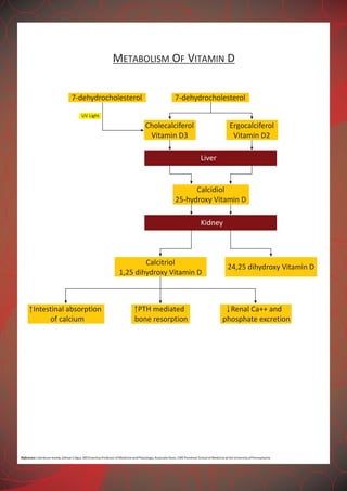
The document discusses the critical roles and history of vitamin D (cholecalciferol) in human health, including its synthesis, metabolism, and various physiological functions. It highlights the clinical significance of vitamin D in relation to conditions such as cardiovascular disease, diabetes, autoimmune diseases, and cancer prevention. Additionally, it addresses the prevalence of vitamin D deficiency in different populations and outlines the benefits of maintaining optimal vitamin D levels.


![VITAMIN D[ SUNSHINE VITAMIN] MEDICINAL CHEMISTRY BY P. RAVISANKAR, CHEMISTRY ...](https://cdn.slidesharecdn.com/ss_thumbnails/02fat-solublevitamins-130615192714-phpapp02-thumbnail.jpg?width=640&height=640&fit=bounds)


