This document discusses how lighter skin and weaker bones may have evolved as adaptations in human populations that migrated north out of Africa. It hypothesizes that natural selection favored these traits because they improved reproductive fitness in environments with less sunlight by minimizing risks of pelvic deformation during childbirth. Specifically, it suggests smaller, more rapidly mineralized bones reduced risks of cephalopelvic disproportion caused by vitamin D deficiency, allowing for greater reproductive success in northern latitudes despite higher osteoporosis risk later in life.
![REVIEW
Weaker bones and white skin as adaptions to improve
anthropological “fitness” for northern environments
R. Vieth1
Received: 13 May 2019 /Accepted: 11 September 2019 /Published online: 6 November 2019
#
Abstract
The vitamin D paradox relates to the lower risk of osteoporosis in people of sub-Saharan African ancestry (Blacks) compared
with people of European ancestry (Whites). The paradox implies that for bone health, Blacks require less vitamin D and calcium
than Whites do. Why should populations that migrated northward out of Africa have ended up needing more vitamin D than
tropical Blacks? Human skin color became lighter away from the tropics to permit greater skin penetration of the UVB light that
generates vitamin D. Lack of vitamin D impairs intestinal calcium absorption and limits the amount of calcium that can deposit
into the protein matrix of bone, causing rickets or osteomalacia. These can cause cephalopelvic disproportion and death in
childbirth. Whiter skin was more fit for reproduction in UV-light restricted environments, but natural selection was also driven
by the phenotype of bone per se. Bone formation starts with the deposition of bone-matrix proteins. Mineralization of the matrix
happens more slowly, and it stiffens bone. If vitamin D and/or calcium supplies are marginal, larger bones will not be as fully
mineralized as smaller bones. For the same amount of mineral, unmineralized or partially mineralized bone is more easily
deformed than fully mineralized bone. The evidence leads to the hypothesis that to minimize the soft bone that causes pelvic
deformation, a decrease in amount of bone, along with more rapid mineralization of osteoid improved reproductive fitness in
Whites. Adaptation of bone biology for reproductive fitness in response to the environmental stress of limited availability of
vitamin D and calcium came at the cost of greater risk of osteoporosis later in life.
Keywords Anthropology . Cephalopelvic disproportion . Cesarean section . Childbirth . Environment . Evolution . Natural
selection . Osteoporosis . Pelvis . Pregnancy . Ultraviolet light . Vitamin D
Introduction
Among the people living in temperate regions of the world,
those who are of Sub-Saharan-African ancestry (Blacks) tend
to possess bone of greater mineral density (if expressed as
g/cm2
) compared with those of European Ancestry (Whites)
[1–3]. Skin color is the obvious difference between Blacks
and Whites. Melanin determines skin color, and melanin
blocks the ultraviolet light that generates vitamin D in the skin.
To generate the same amount of vitamin D, Blacks require up
to six times more UVB light energy (acquired through either
duration or intensity of light) than do Whites [4, 5]. In tem-
perate regions, despite their lower serum 25-hydroxyvitamin
D (25(OH)D), Blacks generally have higher bone mineral
density (BMD) and higher parathyroid hormone (PTH) than
Whites [2]. This is generally referred to as the vitamin D
paradox, and in addition to more volumetric bone quantity
in Blacks [6], the paradox includes lower incidence of falls,
fractures, and osteopenia compared with Whites [7–9].
Since it was so common to find lower serum 25(OH)D in
Blacks, there has been a tendency to think this was normal for
them. Powe et al. attributed the vitamin D paradox to lower
serum levels of vitamin D–binding protein in Blacks com-
pared with Whites and concluded that despite lower total
25(OH)D, the free, bioavailable 25(OH)D was similar in
Blacks and Whites [10]. However, subsequent reports have
shown that the results of Powe et al. were probably an artifact
of the assay used for vitamin D–binding protein, which
underestimated the polymorphisms of vitamin D–binding pro-
tein that are more common in Blacks [11]. Subsequent reports
* R. Vieth
reinhold.vieth@utoronto.ca
1
Department of Laboratory Medicine and Pathobiology, and
Department of Nutritional Sciences, Faculty of Medicine, University
of Toronto, Medical Sciences Building, 5th Floor, Room 5253A 1
King’s College Circle, Toronto, Ontario M5S 1A8, Canada
Osteoporosis International (2020) 31:617–624
https://doi.org/10.1007/s00198-019-05167-4
The Author(s) 2019, corrected publication 2019](https://image.slidesharecdn.com/viethbonesvitd-200531200533/85/Vieth-bones-vit-d-1-320.jpg)
![showed that vitamin D–binding protein levels and the propor-
tion of serum 25(OH)D that is bioavailable are not different
between American Blacks and Whites. Without supplemental
vitamin D, American Blacks do indeed have both lower total
and free serum 25(OH)D than Whites [7, 12].
Some have suggested that Blacks are more likely to be
lactose intolerant, and that therefore, they have long adapted
to a lower calcium requirement. However, lactose tolerance
and high dairy intake correlate with agro-pastoral life of pop-
ulation subgroups, and it is not inherently specific to higher
latitude or European ancestry [13]. Tolerance for lactose in
milk does not distinguish Blacks from Whites; therefore, lac-
tose intolerance does not explain the vitamin D paradox either.
The teleological perspective, that it is normal for Blacks to
require less vitamin D, implies that the biology of human
populations living in sub-Saharan Africa somehow anticipat-
ed an eventual need to accommodate to lower 25(OH)D levels
compared with Whites living in the north. But tropical envi-
ronments have always provided consistent and substantial vi-
tamin D–generating ultraviolet light, for which the skin color
of Blacks is, and was appropriately suited [14, 15]. Highly
credible data on traditionally living Africans provide a reason-
able estimate of the vitamin D nutritional status of early
humans. The average 25(OH)D levels of early humans
exceeded 100 nmol/L (> 40 ng/mL) [16, 17]. Those values
are not unreasonable, given that they agree with published
25(OH)D concentrations in healthy non-human primates
[18]. Such ancestral levels of vitamin D nutrition are approx-
imately double the 25(OH)D levels reported for modern soci-
eties in North America and Europe today. For groups of peo-
ple in the sun-rich environment of the tropics, there was no
mechanistic reason as to why natural selection could have
favored a lower requirement for vitamin D and calcium than
those humans who migrated north into Europe.
A recent conference sponsored by the US Department of
Health was convened to review the pertinent information on
the paradox in the hope of developing insights that might
improve musculoskeletal health in all populations [8]. The
Institute of Medicine’s 2011 dietary guidelines for vitamin D
and calcium were quoted as a premise, “…emerging evidence
would suggest that there is perhaps a lower requirement for
calcium and vitamin D among African Americans relative to
ensuring bone health, at least compared with whites.” [19].
That statement begs the question: Why would people whose
ancestors migrated northward to Europe thousands of years
ago have ended up actually needing more calcium and vitamin
D than those who remained in the tropics? The appropriate
way to develop an answer to the question is to consider the
science through a progression that is forward through time,
from the perspective of evolutionary biology and
anthropology.
Previous attempts to understand the vitamin D paradox
have never addressed evolution or anthropology. I contend
that more-northerly-suitable human phenotypes must have
provided the advantage of making Whites more “fit” for a
relatively ultraviolet-and-vitamin D-deficient environment.
The hypothesis developed here is, that the health of the pelvis
during reproductive years is the key to understanding why the
vitamin D paradox exists.
From the perspective of evolution, it is not helpful to ask,
“Why are Black people different from Whites?” A more ap-
propriate approach to understanding differences between
Blacks and Whites is to start with the question, “What advan-
tage might there have been for the human populations that
migrated out of Africa towards temperate climates to select
for bones that—at least in the context of older adults—are of
poorer quality?”
Natural selection
It is genetic makeup that determines the phenotype, and it is
natural selection that eventually maximizes the fitness of phe-
notype to the environment. Like all primates, humans are a
species whose biology is best suited to inhabit the tropical
latitudes where our species originated [18]. The fitness of a
species for an environment is achieved through evolution. The
process of evolution involves two components: first, genetic
variation; second, natural selection. Genetic variation arises in
species because of the accumulation of random imperfections
that occur during the replication of genes. Those imperfec-
tions can be due to chemicals, radiation, or errors during the
copying of genes, such as rearrangement or deletion or inser-
tion of a single nucleotide or of nucleotide sequences. The
overall assembly of genes within a species is referred to as a
gene pool. Distinct differences in any specific gene from
among individuals are referred to as alleles. Alleles may or
may not alter the protein encoded by a gene. But as the num-
ber of alleles proliferates, the gene pool expands, to the point
where some alleles of certain genes may affect an aspect of the
phenotype of individuals, and potentially offer certain individ-
uals a specific survival advantage (fitness) over other individ-
uals who do not possess those alleles in their genome.
Natural selection is the process by which those individuals
of a species who possess genes that confer greater fitness for
their environment survive to the point of having offspring.
“Fitness”, in the context of natural selection, pertains to the
ability to produce more offspring that are viable to the extent
that they will likewise give birth to offspring of their own.
Natural selection increases the proportion of a population that
exhibits a genetic makeup more fit for an environment. Aside
from the indirect nurturing role of grandmothering [20], there
is no direct mechanism for natural selection beyond the years
of childbearing and parenting. Therefore, risk of osteoporosis
in older adults cannot have played a meaningful role in natural
selection among people migrating northward [21].
618 Osteoporos Int (2020) 31:617–624](https://image.slidesharecdn.com/viethbonesvitd-200531200533/85/Vieth-bones-vit-d-2-320.jpg)
![The original color of the skin of the human species is black,
type 6 skin, because Homo sapiens first appeared in Sub-
Saharan, tropical Africa and where that remains the predom-
inant skin type. As human populations migrated away from
the equator, both northward and southward, skin color light-
ened progressively with distance from the equator [22]. This
was not evolution in the full sense of the word, because a
diverse gene pool had already existed among those persons
migrating out of Africa tens of millennia ago. From that pool,
genes were selected that maximized fitness—the ability to
give birth and to grow healthy offspring. Random mutation
continued to affect the gene pools of all human sub-popula-
tions, both in sub-Saharan Africa and among those who mi-
grated toward the Arctic.
Away from the tropics, natural selection enriched the gene
pool of northward sub-populations with those traits most fit
for survival in temperate latitudes. The most widely accepted
explanation for how humans accommodated for the progres-
sively diminishing amounts of vitamin D–generating ultravi-
olet light is referred to as the vitamin D hypothesis, less dura-
tion and intensity of ultraviolet light resulted in less vitamin D
production in the skin and thereby lower levels of circulating
25(OH)D, the main index of vitamin D nutritional status. With
diminished vitamin D nutrition there was impaired absorption
of calcium from the diet, because lack of 25(OH)D as sub-
strate limits the ability to synthesize the vitamin D–derived
hormone, 1,25-dihydroxyvitamin D (1,25(OH)2D) that in-
creases efficiency of calcium absorption from food.
Together, the lack of vitamin D and lack of absorbed calcium
resulted in osteoid that was not completely mineralized in
infants and children [18, 23]. Moreover, 1,25(OH)2D im-
proves the skeletal microarchitecture of bone via a direct
mechanism, independent of its function to improve intestinal
absorption of calcium [24].
During adolescence, vitamin D intake has site-specific as-
sociations with bone mineral density, particularly at the pelvis
and spine [25]. It has been shown in a double-blind random-
ized controlled clinical trial, that in girls, supplementation
with 2000 IU/day of vitamin D increases not only bone min-
eral density but also improves the structural geometry of the
hip [26–28].
Pregnancy and the pelvis
Rickets can misshape a girl’s pelvis to a fatal degree [29], and
a healthy pelvis was the determining feature that drove natural
selection among human populations as they migrated into
temperate latitudes (Fig. 1). Without rickets to drive selection
for lighter skin color, the entire human population would al-
most certainly have remained deeply pigmented, with type 5
or 6 skin [14].
The growing pelvis is a far more complex bony structure
than are the long bones or the vertebrae. The pelvis comprises
seven centers of primary ossification that are mineralized by
age 9 years. However that stage is followed by a series of
secondary chondrification and ossification events that are
not completed until about 35 years of age [31]. Most of the
volume of the pelvic bone consists of trabecular bone that is
sandwiched between thin shells of cortical bone. The structur-
al, “sandwich behavior” of pelvic cortical bone means that this
cortical bone carries a stress load that is fifty-fold bigger than
pelvic trabecular bone inside it [32]. There are 21 different
muscles that attach to the pelvic bone, and those convey ad-
ditional support and strength for the pelvis [32]. Since vitamin
D deficiency causes proximal muscle myopathy [28], it is
reasonable to postulate that vitamin D deficiency–related mus-
cle weakness around the pelvis can add to the stress forces that
can deform pelvic bone, and together with unmineralized os-
teoid, vitamin D–related myopathy may contribute to risk of
cephalopelvic disproportion.
I have not been able to find any reports describing the
biomechanics of the rachitic or osteomalacic pelvis.
Published research about the pelvis has been directed at un-
derstanding pelvic fractures due to impacts or at understand-
ing the pelvis to optimize the quality of hip replacements [33].
What we do know relates to the structure of the normal pelvis.
The limited biomechanical data about the pelvis focus on the
forces related to standing and walking [32] or for use in de-
signing hip prostheses. No biomechanical analyses have been
directed at the forces of the sitting positions that probably
impose the kinds of forces that could diminish the width of
the pelvic opening. Future research to test the present pelvic
hypothesis for the vitamin D paradox could involve finite
element analysis of pelvic bone modeling, with adjustments
made for defects related to areas unmineralized osteoid of
rickets and osteomalacia.
If rickets develops in adolescents, the long bones may ap-
pear to be normal, but radiologic examination of the pelvis
will reveal excessive osteoid that is treatable if vitamin D is
available [34]. If rickets or osteomalacia continue through
pregnancy, then deformation of the pelvis becomes progres-
sively worse with each pregnancy and lactation, due to the
mineral demands of the growing fetus and infant [35].
Recent epidemiological data from the USA are consistent with
this, in that Black women (but not White women) develop a
progressively higher risk for cesarean delivery with each preg-
nancy [36] (Table 1).
Research into osteoporosis has focused on the vertebrae
and long bones. But ironically, the iliac crest is the classic site
for bone biopsy. Ethical reasons preclude comparative sam-
pling from other bone sites in humans, so it may not be rea-
sonable to assume that histology of the pelvis is representative
of all cortical and trabecular bones throughout the body. Most
of what we know about the relationship between vitamin D
Osteoporos Int (2020) 31:617–624 619](https://image.slidesharecdn.com/viethbonesvitd-200531200533/85/Vieth-bones-vit-d-3-320.jpg)
![nutritional status and unmineralized osteoid in a normal pop-
ulation comes from the work of Priemel et al. who obtained
blood samples and biopsied iliac crest from hundreds of vic-
tims of accidental deaths in Germany [50]. They showed a
steady decline in pelvic osteoid—improved mineralization—
as serum 25(OH)D levels increased toward 75 nmol/L.
Beyond 75 nmol/L, there was virtually no evidence of
unmineralized osteoid. These data by Priemel et al. were
highlighted by the Institutes of Medicine in their latest assess-
ment of dietary requirements for calcium and vitamin D [19].
But oddly, the evidence pertaining to the pelvis was ignored
when it came to the final recommendation for vitamin D [51,
52]. If the findings of Priemel et al. are true, then risk of
unmineralized osteoid extends well into the range of serum
25(OH)D recommended by the Institutes of Medicine, 50–
125 nmol/L, with risk particularly high if dietary calcium is
limited.
Through the natural course of pregnancy, serum 25(OH)D
levels are far higher in Black women who live a traditional
lifestyle consistent with that of early humans in Sub-Saharan
Africa, than are the 25(OH)D levels of White women who live
in Europe or North America. Luxwolda et al. reported serum
25(OH)D through pregnancy in five East African ethnic
groups: Maasai, Hadzabe, Same Sengerema, and Ukerewe.
The most striking observation was that despite no supplemen-
tal vitamin D, their serum 25(OH)D increased during the
Table 1 Risk of cesarean birth and features of bone and mineral metabolism for people of European ancestry (Whites) as compared with people of sub-
Saharan African ancestry (Blacks)
Cesarean birth risk
•White women in the USA are at lower risk of cesarean delivery, after correcting for sociodemographic confounders [36–40]
•White women have no increased risk of cesarean delivery with multiple pregnancies,
while risk of cesarean delivery increases with multiple pregnancies in Black women [36]
•White women have a wider pelvis in relation to stature height [41]
Osteoporosis risk
•White women have lower bone mineral density (BMD) based on cross-sectional area (g/cm2
) [1–3]
•White men have lower BMD (g/cm2
) for the whole body, tibia, hip, and femoral neck [42]
•White children have less tibial cortical bone density (g/cm3
) strength despite having higher levels of bone-promoting factors of physical activity,
dietary calcium intake, and serum 25(OH)D concentrations [43]
•Before puberty, vertebral trabecular number, thickness, and true BMD (volumetric, g/cm3
) do not differ by race or gender. Racial differences emerge
during puberty when male and female Blacks increase BMD from 250 to 260 mg/cm3 in Tanner stage 1 to 330–340 40 mg/cm3.
. In contrast, male
and female Whites increase BMD by half that amount, from 250 to 260 mg/cm3 in Tanner stage 1 to 290–300 mg/cm3
[6, 44].
•White women have longer hip axis length [45]
•White men and women have less favorable bone microarchitecture.
By young adulthood, their bone exhibits diminished plate-like morphology and less trabecular axial alignment [46] .
•White men have smaller bones with thinner cortices and less bending strength than Black men. [42]
•White men and women have weaker trabeculae and, in males, less bone quantity, and poorer bone quality. [47]
•White women have faster mineral apposition rate in iliac biopsy [48]
•Whites have higher levels of markers of bone turnover (osteocalcin, CTx,OHPro, BAP) [49]
•Whites have lower PTH [2, 49]
•Whites have greater skeletal response to PTH [49]
•Whites have lower levels of 1,25(OH)2D [49]
•Whites have a faster rate of bone loss [49]
Fig. 1 Drawing of the pelvis of a
healthy adult female (left)
compared with a photograph of
the pelvis of a woman with
untreated rickets and
osteomalacia (right) published by
Maxwell et al. [30] (with
permission). Both views are top-
down. Natural child-birth was not
possible with the narrow,
misshapen pelvis at the right that
illustrates an extreme example of
cephalopelvic disproportion
620 Osteoporos Int (2020) 31:617–624](https://image.slidesharecdn.com/viethbonesvitd-200531200533/85/Vieth-bones-vit-d-4-320.jpg)
![second and third trimesters of pregnancy, with concentrations
averaging 150 nmol/L [16]. Among African women living in
their tropical environment, without a vitamin D supplement,
the sharp increase in serum 25(OH)D attains double the serum
levels typically seen among White women in North America
and Europe, where 25(OH)D levels actually trend downward
during pregnancy [53, 54]. If women in sub-Saharan Africa
had extremely high vitamin D nutrition, as seen from our
present perspective, there must surely have been selection
pressure to adapt northwardly migrating populations to some-
how accommodate to lower amounts of ultraviolet light–
derived vitamin D.
Skin color is the obvious adaptation to accommodate to
diminishing ultraviolet light; however, there are many quali-
tative differences in muscuskeletal features between Black
and Caucasian adults, different bone densities, different mi-
crostructure, different rates of bone formation and different
rates of osteoporosis (Table 1). Another likely adaptation is
the comparatively wider pelvis of Arctic and European popu-
lations, compared with those of southern latitudes [41]. A
wider pelvis is a more robust phenotype, allowing for some
osteomalacic deformation of the pelvis while lowering the risk
of cephalopelvic disproportion. Although the preceding are
plausible features that may prevent cephalopelvic dispropor-
tion, they do not explain the cause of the differences in oste-
oporosis risk between Blacks and Whites.
Bone fibril strength
What survival advantage might there be to the diminished
quantity and quality of adult bone in northern populations?
The answer must surely have something to do with the “fit-
ness” in the sense of the phenotypes that maximize the number
of viable births. The quality of bone in the adult is secondary
to the characteristics of bone that minimize the risk of rickets
and osteomalacia. I contend that natural selection would have
increased prevalence of those phenotypes that prevented de-
formation of the pelvis of a growing girl or young woman
(Fig. 1).
Features of White bone evident from Table 1, such as less
bone quantity [1, 42, 43, 47, 48] and faster mineralization of
osteoid [48] serve as advantages that prevent pelvic deforma-
tion. This is because less bone growth through the initial step
of matrix formation requires less calcium to mineralize it fully,
making for more efficient use of the calcium that is available.
Proper mineralization is important for bone strength at both
the microfibril as well as at the nanofibrillar levels [55].
Recent research has characterized the biomechanical strength
of rachitic bone in the hypophosphatemic mouse model, com-
pared with the normal bone of wild-type mice [56].
Karunaratne et al. studied the functional link between altered
bone quality (reduced mineralization) and abnormal fibrillar-
level mechanics using real-time synchrotron X-ray nanome-
chanical imaging. They demonstrated a nanostructural mech-
anism in which incompletely mineralized fibrils of rachitic
mice are more extensible and less stiff, i.e., exhibiting greater
strain and bendability. “It is clear that the unmineralized or
partially mineralized fibrils will exhibit a much larger strain
than the fully mineralized fibril for the same force.” [56].
Similarly, Dardenne et al. demonstrated severely impaired
biomechanics of unmineralized long bone from rachitic and
osteomalacic mice, and showed that correction of the vitamin
D deficiency caused a rapid improvement in bone stiffness
toward normal [57]. Earlier work by Turner et al. showed that
the osteomalacia caused by excessive fluoride intake in rats
coincided with diminished bone strength as assessed by the
three-point–bending test [58]. In the macroscopic context,
what we know from animal studies is that incompletely min-
eralized bone is weaker, and much more flexible.
If these findings are taken in the context of the human
pelvis, we know that incompletely mineralized bone will
bend. If left untreated, this will result in severe pelvic defor-
mation, to the point that natural childbirth is impossible [30]
(Fig. 1). Pregnancy and breastfeeding impose strong demands
on the skeleton to supply calcium to the growing fetus and
infant. With poor vitamin D and calcium nutrition, osteoma-
lacia causes a progressive deformity of the pelvis with each
pregnancy, progressively increasing risk of death in childbirth
[30, 34–36].
Fig. 2 Illustrative images of biomechanical behavior of bone of Blacks
(top) compared to Whites (bottom). The horizontal white rectangles
represent long-bone samples, resting on solid blocks. The triangles
represent a stressing force, whose magnitude is represented by the size
of the triangle. a Thicker bone, but whose stiffness, or resistance to
bending is diminished during growth, because of an inability to acquire
sufficient calcium to the bone, leaving zones of unmineralized osteoid,
represented by the circles. b Sample of thinner bone, but where osteoid is
fully mineralized. With the smaller amount of total bone, less calcium is
needed for the more complete mineralization that makes bone resistant to
bending. Later in life, once all bone is suitably mineralized, c the thicker
bone resists an amount of stress that causes d failure and fracture of the
thinner bone
Osteoporos Int (2020) 31:617–624 621](https://image.slidesharecdn.com/viethbonesvitd-200531200533/85/Vieth-bones-vit-d-5-320.jpg)
![A key lesson, based on the laboratory-animal work, is that
in terms of bone stiffness, it is better to have less bone, but
bone which is fully mineralized, instead of more bone that
contains too many regions of incompletely mineralized os-
teoid (Fig. 2a, b). Many of the distinguishing features of
Whites are things that make for more efficient use of avail-
able calcium, because more rapid and more complete miner-
alization of osteoid improve stiffness of the pelvis,
preventing its deformation, cephalopelvic disproportion,
and death in childbirth.
The pelvis is the most important bone in the context of the
evolutionary fitness of the species. At the pelvis, less bone, but
bone which was fully mineralized would have been superior
to a situation with more bone, but where that bone contained
unmineralized osteoid. The benefit for the species of a pelvis
made up of stiffer bone, albeit with less total amount of bone,
was a higher probability that infants could be delivered vagi-
nally. However, the eventual consequence of less bone for the
older adult would be lower bone mineral density and weaker
bones (Fig. 2c, d). The mystery of the vitamin D paradox may
lie in the paradox that some of the adaptations that prevented
deformation of the pelvis in Whites also resulted in matured
skeletal bone that is of lesser quantity and quality. Adaptation
to less vitamin D involved a more rapid mineralization of bone
along with less bone accrual; these things optimized stiffness
of pelvic bone to accommodate for limited availability of cal-
cium absorbed from diet.
Bone density in the modern context
The perspective presented here predicts that compared with
Whites at identical margins of low calcium and low vitamin D
nutrition, Black children would be more likely to develop
rickets, while Black women during their childbearing years
would be more prone to osteomalacia. Given the same restrict-
ed calcium and vitamin D nutrition, Black and White women
can possess the same total amount of mineralized bone.
However, because of their larger total volume of bone,
Blacks at the margins of low calcium and low vitamin D
would be expected to have a lower mineral content per unit
volume of bone. Dual energy X-ray absorptiometry (DEXA)
which measures amount of mineral per cross-sectional area of
bone exposed to it (g/cm2
) cannot distinguish between osteo-
porosis and the osteomalacia that contributes to bone
bendability. Likewise, volumetric bone density based on com-
puted tomography (g/cm3
) has not been capable of
distinguishing impaired mineralization from porosity.
However, newer micro-CT technology is becoming capable
of estimating matrix mineral density [59]. It might be possible
to obtain enough micro-CT data to compare bone of young
Black and White women at equally marginal levels of
25(OH)D and calcium restriction. The margins for vitamin
D and calcium for such a comparison are likely to be serum
25(OH)D below 25 nmol/L, and a dietary calcium intake be-
low 500 mg/day [19, 60]. In this modern era, with readily
available sources of both vitamin D and calcium nutrition
for all who live in the north, the compromises of past millennia
are less evident in terms of rickets or pelvic disproportion.
However, although nutritional osteomalacia has been pre-
sumed to be rare in modern society [19, 60], this may be due
to the inability to detect it. The biopsy data from the German
population, as published by Priemel et al., imply that osteo-
malacia at the pelvis is disconcertingly common in people
with 25(OH)D below 75 nmol/L [50].
Conclusion
A sense as to why differences exist between the bones of
Blacks and Whites may help in developing better research
questions. The arguments presented here are an attempt to
explain why the osteoporosis-related vitamin D paradox ex-
ists. I have tried to approach the problem from the perspective
of anthropology and natural selection. Since anthropology is
an empirical science, it is not readily amenable to the experi-
mental and clinical methodologies generally expected in med-
icine. Research to test the present pelvic hypothesis to account
for the vitamin D paradox could involve bioengineering anal-
ysis of pelvic bone, with adjustments for effects of rickets and
osteomalacia. Emerging micro-CT technology could be used
to compare matrix mineral density between young Black and
White women matched for serum 25(OH)D and dietary calci-
um intake. Lastly the search for genetic insights could focus
on variants selected to construct less bone matrix or to make
the pelvis mineralize more quickly.
Based on the evidence presented here, the hypothesis is that
Whites are more prone to osteoporosis later in life, because of
the compromise during growth early in life, to accommodate
for diminished amounts of calcium absorbed via the gut. The
female pelvis is by far the most important part of the skeleton
in terms of “fitness” for producing offspring. In terms of nat-
ural selection, the suitability of the pelvis for a vaginal birth
took priority over the health of all other bones—to the point of
increasing the eventual risk of osteoporosis. Just as skin color
adapted to become whiter than that of the parent, Black pop-
ulation in Africa, bone and mineral metabolism also adapted
with less total amount of bone, and faster mineralization of
osteoid. In the face of less sunshine-derived vitamin D, these
metabolic adaptations made more efficient use of the dimin-
ished calcium absorbed from the diet, by making pelvic bone
stiffer and less susceptible to deformation, thereby making
Whites are more “fit” for non-tropical environments. But the
cost of that fitness to reproduce was weaker bone later in life
and greater risk of osteoporosis.
622 Osteoporos Int (2020) 31:617–624](https://image.slidesharecdn.com/viethbonesvitd-200531200533/85/Vieth-bones-vit-d-6-320.jpg)
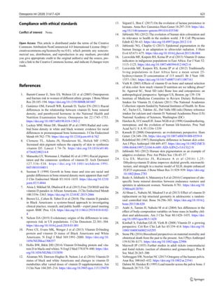
![33. Dalstra M (1993) Biomechanical aspects of the pelvic bone and
design criteria for acetabular prostheses. [Sl: sn]
34. Hunter GJ, Schneidau A, Hunter JV, Chapman M (1984) Rickets in
adolescence. Clin Radiol 35:419–421. https://doi.org/10.1016/
S0009-9260(84)80207-X
35. Chaim W, Alroi A, Leiberman JR, Cohen A (1981) Severe
contracted pelvis appearing after normal deliveries. Acta Obstet
Gynecol Scand 60:131–134
36. Min CJ, Ehrenthal DB, Strobino DM (2015) Investigating racial
differences in risk factors for primary cesarean delivery. Am J
Obstet Gynecol 212:814.e1–814.e14. https://doi.org/10.1016/j.
ajog.2015.01.029
37. Declercq E, Menacker F, Macdorman M (2006) Maternal risk pro-
files and the primary cesarean rate in the United States, 1991-2002.
Am J Public Health 96:867–872. https://doi.org/10.2105/AJPH.
2004.052381
38. Scott-Wright AO, Flanagan TM, Wrona RM (1999) Predictors of
cesarean section delivery among college-educated black and white
women, Davidson County, Tennessee, 1990-1994. J Natl Med
Assoc 91:273–277
39. Huesch M, Doctor JN (2015) Factors associated with increased
cesarean risk among African American women: evidence from
California, 2010. Am J Public Health 105:956–962. https://doi.
org/10.2105/AJPH.2014.302381
40. Penfield CA, Lahiff M, Pies C, Caughey AB (2017) Adolescent
pregnancies in the United States: how obstetric and sociodemo-
graphic factors influence risk of cesarean delivery. Am J Perinatol
34:123–129. https://doi.org/10.1055/s-0036-1584580
41. Wells JCK, DeSilva JM, Stock JT (2012) The obstetric dilemma: an
ancient game of Russian roulette, or a variable dilemma sensitive to
ecology? Am J Phys Anthropol 149(Suppl 55):40–71. https://doi.
org/10.1002/ajpa.22160
42. Zengin A, Pye SR, Cook MJ et al (2016) Ethnic differences in bone
geometry between White, Black and South Asian men in the UK.
Bone 91:180–185. https://doi.org/10.1016/j.bone.2016.07.018
43. Warden SJ, Hill KM, Ferira AJ et al (2013) Racial differences in
cortical bone and their relationship to biochemical variables in
black and white children in the early stages of puberty.
Osteoporos Int 24:1869–1879. https://doi.org/10.1007/s00198-
012-2174-8
44. Gilsanz V, Skaggs DL, Kovanlikaya A et al (1998) Differential
effect of race on the axial and appendicular skeletons of children.
J Clin Endocrinol Metab 83:1420–1427. https://doi.org/10.1210/
jcem.83.5.4765
45. Cummings SR, Cauley JA, Palermo L et al (1994) Racial differ-
ences in hip axis lengths might explain racial differences in rates of
hip fracture. Osteoporos Int 4:226–229. https://doi.org/10.1007/
BF01623243
46. Popp KL, Xu C, Yuan A et al (2019) Trabecular microstructure is
influenced by race and sex in Black and White young adults.
Osteoporos Int 30:201–209. https://doi.org/10.1007/s00198-018-
4729-9
47. Schnitzler CM, Pettifor JM, Mesquita JM et al (1990) Histomor-
phometry of iliac crest bone in 346 normal Black and White South
African adults. Bone Miner 10:183–199
48. Parisien M, Cosman F, Morgan D et al (1997) Histomorphometric
assessment of bone mass, structure, and remodeling: a comparison
between healthy Black and White premenopausal women. J Bone
Miner Res 12:948–957. https://doi.org/10.1359/jbmr.1997.12.6.
948
49. Aloia JF (2008) African Americans, 25-hydroxyvitamin D, and
osteoporosis: a paradox. Am J Clin Nutr 88:545S–550S. https://
doi.org/10.1093/ajcn/88.2.545S
50. Priemel M, von Domarus C, Klatte TO et al (2010) Bone mineral-
ization defects and vitamin D deficiency: histomorphometric anal-
ysis of iliac crest bone biopsies and circulating 25-hydroxyvitamin
D in 675 patients. J Bone Miner Res Off J Am Soc Bone Miner Res
25:305–312. https://doi.org/10.1359/jbmr.090728
51. Maxmen A (2011) Nutrition advice: the vitamin D-lemma. Nature
475:23–25. https://doi.org/10.1038/475023a
52. Vieth R, Holick MF (2018) Chapter 57B - the IOM—Endocrine
Society controversy on recommended vitamin D targets: in support
of the Endocrine Society position. In: Feldman D (ed) Vitamin D
(fourth edition). Academic press, pp 1091–1107
53. Bodnar LM, Platt RW, Simhan HN (2015) Early-pregnancy vitamin
D deficiency and risk of preterm birth subtypes. Obstet Gynecol
125:439–447. https://doi.org/10.1097/aog.0000000000000621
54. Wagner CL, Baggerly C, McDonnell S et al (2016) Post-hoc anal-
ysis of vitamin D status and reduced risk of preterm birth in two
vitamin D pregnancy cohorts compared with South Carolina March
of Dimes 2009-2011 rates. J Steroid Biochem Mol Biol 155:245–
251. https://doi.org/10.1016/j.jsbmb.2015.10.022
55. Li Y, Aparicio C (2013) Discerning the subfibrillar structure of
mineralized collagen fibrils: a model for the ultrastructure of bone.
PLoS One 8:e76782. https://doi.org/10.1371/journal.pone.0076782
56. Karunaratne A, Esapa CR, Hiller J et al (2012) Significant deterio-
ration in nanomechanical quality occurs through incomplete
extrafibrillar mineralization in rachitic bone: evidence from in-situ
synchrotron X-ray scattering and backscattered electron imaging. J
Bone Miner Res 27:876–890. https://doi.org/10.1002/jbmr.1495
57. Dardenne O, Prud’Homme J, Hacking SA et al (2003) Rescue of
the pseudo-Vitamin D deficiency rickets phenotype of CYP27B1-
deficient mice by treatment with 1,25-dihydroxyvitamin D3: bio-
chemical, histomorphometric, and biomechanical analyses. J Bone
Miner Res 18:637–643. https://doi.org/10.1359/jbmr.2003.18.4.
637
58. Turner CH, Owan I, Brizendine EJ et al (1996) High fluoride in-
takes cause osteomalacia and diminished bone strength in rats with
renal deficiency. Bone 19:595–601. https://doi.org/10.1016/S8756-
3282(96)00278-5
59. Chiang CY, Zebaze R, Wang X-F et al (2018) Cortical matrix min-
eral density measured noninvasively in pre- and postmenopausal
women and a woman with vitamin D-dependent rickets. J Bone
Miner Res 33:1312–1317. https://doi.org/10.1002/jbmr.3415
60. SACN (2016) Scientific advisory committee on nutrition vitamin D
and health. https://wwwgovuk/government/groups/scientific-
advisory-committee-on-nutrition. Accessed 20 Dec 2016
Publisher’s note Springer Nature remains neutral with regard to
jurisdictional claims in published maps and institutional affiliations.
624 Osteoporos Int (2020) 31:617–624](https://image.slidesharecdn.com/viethbonesvitd-200531200533/85/Vieth-bones-vit-d-8-320.jpg)


