Download to read offline


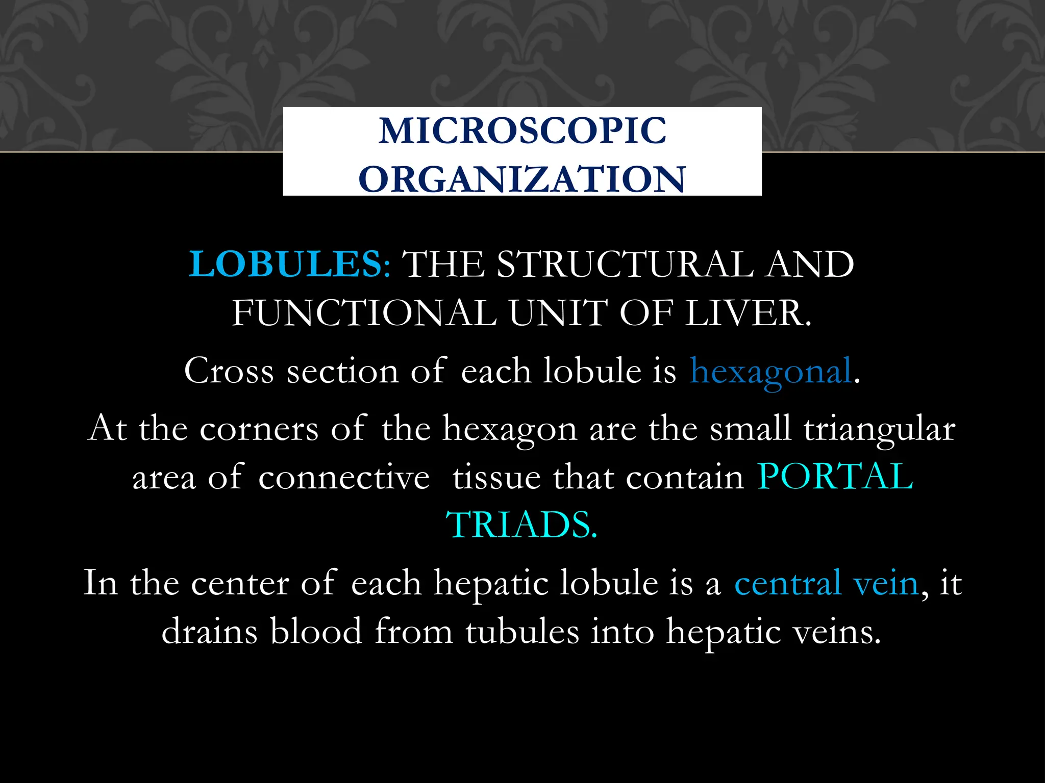


The document discusses the histology of the liver, describing it as a modified exocrine gland with a unique double blood supply and organized into lobes and lobules. It details the microscopic organization of hepatocytes, the structure of classical, portal lobules, and liver acinus, as well as the importance of different zones in relation to blood flow and degeneration patterns. Additionally, it mentions the presence of key cellular components like Kupffer cells and hepatic stellate cells, alongside various pathologies affecting liver architecture.


![Histology of the liver and gall bladder [compatibility mode]](https://cdn.slidesharecdn.com/ss_thumbnails/histologyoftheliverandgallbladdercompatibilitymode-131029194000-phpapp02-thumbnail.jpg?width=640&height=640&fit=bounds)


