SlideShare a Scribd company logo
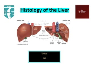
Histology of the Liver
Trishna Kisiju
Group
B3
under supervision of Dr : Reda Mohammed
The Liver
• Largest gland of the body.
• 1500 grams and 2.5% of total body weight.
• Location:
- Right hypochondrium
- Epigastric region
- Left hypochondrium
Vascular Supply of the Liver
• Receives dual vascular supply:
-Hepatic Portal Vein (75%)
- Hepatic Artery(25%)
• Both vessels enter the liver via Porta hepatis.
The Liver Stroma
• Consists of: - Connective Tissue Capsule
- Trabeculae
- Reticular fibers
1. Connective Tissue Capsule:
- Serous layer: Visceral Peritoneum
- Fibrous layer: Glisson’s capsule
• Trabeculae: The Glisson’s capsule extends into the
interior of liver as numerous branching trabeculae and
septa.
• Reticular fibers:
- Supporting connective tissue of the liver.
- Line the sinusoids, support the endothelial cells, and
form a denser network of reticular fibers in the wall of
the central vein.
- Also merge with the collagen fibers in the interlobular
septum, where they surround the portal vein and the
bile duct.
The Liver Parenchyma
• Organized as thousands of small hepatic lobules.
• Hepatic Lobules: Structural units of Liver.
Roughly hexagonal arrangement of irregular plates or
cords of hepatocytes radiating outward from a central
vein.
Concepts of Liver Lobules
• Classical Hepatic Lobule
• Portal Lobule
• Hepatic Acinus of Rappaport
Classical Hepatic Lobule
• Each lobule consists of a hexagonal mass of liver cells.
• Central axis occupied by:
Central vein.
• Is an independent venous unit.
• From the central vein, hepatocytes
radiate irregularly as plates known
as Hepatic lamina.
• Spaces between the hepatic
lamina are called Hepatic lacunae,
occupied by Hepatic sinusoids.
The Portal Area
• Peripherally, each lobule has 3 to 6 portal areas with more
fibrous connective tissue, each of which contains
interlobular structures that comprise the portal triad. They
include:
• A venule branch of the portal vein, with blood rich in
nutrients but low in O2.
• An arteriole branch of the hepatic artery that supplies O2.
• One or two small bile ductules of cuboidal epithelium,
branches of the bile conducting system.
Bile Canaliculi
• Formed by spaces present between plasma membranes of
adjacent liver cells.
• Form hexagonal networks around the liver cells.
• Borders around the canaliculi are sealed by tight junctions.
This forms the blood-bile barrier.
Bile Canaliculi
• The canaliculi pass to periphery of the hepatic lobules
where they form intralobular canal of Herring, that
finally drains into the interlobular duct of the portal
area.
• Bile canaliculi are intralaminar and centrifugal in
direction
Space of Disse / Perisinusoidal Space
• Potential space between the wall of sinusoids and
laminae of the liver cells.
• Filled with blood plasma and
chylomicrons that percolate
through the wall of sinusoids.
• Presence of Ito cells.
Ito cells
• Irregular outline with numerous lipid vesicles.
• Function of Ito cells:
- Secrete collagenous matrix
- Provide growth factor for regeneration of
damaged liver cells.
- Store Vitamin A in their lipid vesicles.
Space of Mall
• Potential space, between the glisson’s capsule of portal
area and the hepatic plates of the cells.
• Lymphatics of liver
begin here.
The Portal Lobule
• Territory of liver tissue centered around a portal triad.
• Drawn by joining the central veins of three adjacent
lobules.
• Nutritional lobule of the liver.
Hepatic acinus of Rappaport
• Diamond shaped area of liver parenchyma.
• Forms structural and metabolic functions of the liver.
• Numerous branches arise
at right angles from the
blood vessels of portal area,
these terminal vessels
form backbone of the liver
acinus.
The Hepatocytes
• Large cuboidal or polyhedral epithelial cells, with
large, round central nuclei and eosinophilic cytoplasm
rich in mitochondria.
THANK YOU!!
At the end of the presentation,
we hope that you like it and
gain satisfaction.
assigment Histology (B3)-Reduced-Reduced.pdf

More Related Content

Similar to assigment Histology (B3)-Reduced-Reduced.pdf

Anatomy of gall bladder
Anatomy of gall bladderAnatomy of gall bladder
Anatomy of gall bladder
prashanthsangu
 
Histology of liver by aravindh dpi
Histology of liver by aravindh dpiHistology of liver by aravindh dpi
Histology of liver by aravindh dpi
Aravindh Dpi
 

Similar to assigment Histology (B3)-Reduced-Reduced.pdf (20)

Renal Histology
Renal HistologyRenal Histology
Renal Histology
 
Liver anatomy and histology
Liver anatomy and histologyLiver anatomy and histology
Liver anatomy and histology
 
Washing machine of body
Washing machine of bodyWashing machine of body
Washing machine of body
 
Lecture urinary system histology
Lecture urinary system histologyLecture urinary system histology
Lecture urinary system histology
 
Histology of the liver.ppt this covers liver
Histology of the liver.ppt this covers liverHistology of the liver.ppt this covers liver
Histology of the liver.ppt this covers liver
 
Anatomy of kidneys
Anatomy of  kidneysAnatomy of  kidneys
Anatomy of kidneys
 
Anatomy of gall bladder
Anatomy of gall bladderAnatomy of gall bladder
Anatomy of gall bladder
 
Anatomy of gall bladder
Anatomy of gall bladderAnatomy of gall bladder
Anatomy of gall bladder
 
Ultrasonography of liver
Ultrasonography of liverUltrasonography of liver
Ultrasonography of liver
 
Histology of liver by aravindh dpi
Histology of liver by aravindh dpiHistology of liver by aravindh dpi
Histology of liver by aravindh dpi
 
Structure and function of Spleen
Structure and function of SpleenStructure and function of Spleen
Structure and function of Spleen
 
Excretory system
Excretory systemExcretory system
Excretory system
 
Hepatobilliary system
Hepatobilliary systemHepatobilliary system
Hepatobilliary system
 
Urinary system
Urinary systemUrinary system
Urinary system
 
liver and pancreas.pptx
liver and pancreas.pptxliver and pancreas.pptx
liver and pancreas.pptx
 
Section 3, chapter 17: liver and intestines
Section 3, chapter 17: liver and intestinesSection 3, chapter 17: liver and intestines
Section 3, chapter 17: liver and intestines
 
Urinary System.pptx
Urinary System.pptxUrinary System.pptx
Urinary System.pptx
 
The urinary system
The urinary systemThe urinary system
The urinary system
 
Urinary system
Urinary systemUrinary system
Urinary system
 
liver.pptx
liver.pptxliver.pptx
liver.pptx
 

Recently uploaded

一比一原版(CU毕业证)卡尔顿大学毕业证成绩单
一比一原版(CU毕业证)卡尔顿大学毕业证成绩单一比一原版(CU毕业证)卡尔顿大学毕业证成绩单
一比一原版(CU毕业证)卡尔顿大学毕业证成绩单
yhkoc
 
Opendatabay - Open Data Marketplace.pptx
Opendatabay - Open Data Marketplace.pptxOpendatabay - Open Data Marketplace.pptx
Opendatabay - Open Data Marketplace.pptx
Opendatabay
 
一比一原版(YU毕业证)约克大学毕业证成绩单
一比一原版(YU毕业证)约克大学毕业证成绩单一比一原版(YU毕业证)约克大学毕业证成绩单
一比一原版(YU毕业证)约克大学毕业证成绩单
enxupq
 
Investigate & Recover / StarCompliance.io / Crypto_Crimes
Investigate & Recover / StarCompliance.io / Crypto_CrimesInvestigate & Recover / StarCompliance.io / Crypto_Crimes
Investigate & Recover / StarCompliance.io / Crypto_Crimes
StarCompliance.io
 
Computer Presentation.pptx ecommerce advantage s
Computer Presentation.pptx ecommerce advantage sComputer Presentation.pptx ecommerce advantage s
Computer Presentation.pptx ecommerce advantage s
MAQIB18
 
一比一原版(IIT毕业证)伊利诺伊理工大学毕业证成绩单
一比一原版(IIT毕业证)伊利诺伊理工大学毕业证成绩单一比一原版(IIT毕业证)伊利诺伊理工大学毕业证成绩单
一比一原版(IIT毕业证)伊利诺伊理工大学毕业证成绩单
ewymefz
 
一比一原版(QU毕业证)皇后大学毕业证成绩单
一比一原版(QU毕业证)皇后大学毕业证成绩单一比一原版(QU毕业证)皇后大学毕业证成绩单
一比一原版(QU毕业证)皇后大学毕业证成绩单
enxupq
 
一比一原版(UPenn毕业证)宾夕法尼亚大学毕业证成绩单
一比一原版(UPenn毕业证)宾夕法尼亚大学毕业证成绩单一比一原版(UPenn毕业证)宾夕法尼亚大学毕业证成绩单
一比一原版(UPenn毕业证)宾夕法尼亚大学毕业证成绩单
ewymefz
 

Recently uploaded (20)

一比一原版(CU毕业证)卡尔顿大学毕业证成绩单
一比一原版(CU毕业证)卡尔顿大学毕业证成绩单一比一原版(CU毕业证)卡尔顿大学毕业证成绩单
一比一原版(CU毕业证)卡尔顿大学毕业证成绩单
 
Opendatabay - Open Data Marketplace.pptx
Opendatabay - Open Data Marketplace.pptxOpendatabay - Open Data Marketplace.pptx
Opendatabay - Open Data Marketplace.pptx
 
一比一原版(YU毕业证)约克大学毕业证成绩单
一比一原版(YU毕业证)约克大学毕业证成绩单一比一原版(YU毕业证)约克大学毕业证成绩单
一比一原版(YU毕业证)约克大学毕业证成绩单
 
Investigate & Recover / StarCompliance.io / Crypto_Crimes
Investigate & Recover / StarCompliance.io / Crypto_CrimesInvestigate & Recover / StarCompliance.io / Crypto_Crimes
Investigate & Recover / StarCompliance.io / Crypto_Crimes
 
Tabula.io Cheatsheet: automate your data workflows
Tabula.io Cheatsheet: automate your data workflowsTabula.io Cheatsheet: automate your data workflows
Tabula.io Cheatsheet: automate your data workflows
 
社内勉強会資料_LLM Agents                              .
社内勉強会資料_LLM Agents                              .社内勉強会資料_LLM Agents                              .
社内勉強会資料_LLM Agents                              .
 
Computer Presentation.pptx ecommerce advantage s
Computer Presentation.pptx ecommerce advantage sComputer Presentation.pptx ecommerce advantage s
Computer Presentation.pptx ecommerce advantage s
 
Innovative Methods in Media and Communication Research by Sebastian Kubitschk...
Innovative Methods in Media and Communication Research by Sebastian Kubitschk...Innovative Methods in Media and Communication Research by Sebastian Kubitschk...
Innovative Methods in Media and Communication Research by Sebastian Kubitschk...
 
Business update Q1 2024 Lar España Real Estate SOCIMI
Business update Q1 2024 Lar España Real Estate SOCIMIBusiness update Q1 2024 Lar España Real Estate SOCIMI
Business update Q1 2024 Lar España Real Estate SOCIMI
 
一比一原版(IIT毕业证)伊利诺伊理工大学毕业证成绩单
一比一原版(IIT毕业证)伊利诺伊理工大学毕业证成绩单一比一原版(IIT毕业证)伊利诺伊理工大学毕业证成绩单
一比一原版(IIT毕业证)伊利诺伊理工大学毕业证成绩单
 
一比一原版(QU毕业证)皇后大学毕业证成绩单
一比一原版(QU毕业证)皇后大学毕业证成绩单一比一原版(QU毕业证)皇后大学毕业证成绩单
一比一原版(QU毕业证)皇后大学毕业证成绩单
 
Q1’2024 Update: MYCI’s Leap Year Rebound
Q1’2024 Update: MYCI’s Leap Year ReboundQ1’2024 Update: MYCI’s Leap Year Rebound
Q1’2024 Update: MYCI’s Leap Year Rebound
 
How can I successfully sell my pi coins in Philippines?
How can I successfully sell my pi coins in Philippines?How can I successfully sell my pi coins in Philippines?
How can I successfully sell my pi coins in Philippines?
 
Slip-and-fall Injuries: Top Workers' Comp Claims
Slip-and-fall Injuries: Top Workers' Comp ClaimsSlip-and-fall Injuries: Top Workers' Comp Claims
Slip-and-fall Injuries: Top Workers' Comp Claims
 
一比一原版(UPenn毕业证)宾夕法尼亚大学毕业证成绩单
一比一原版(UPenn毕业证)宾夕法尼亚大学毕业证成绩单一比一原版(UPenn毕业证)宾夕法尼亚大学毕业证成绩单
一比一原版(UPenn毕业证)宾夕法尼亚大学毕业证成绩单
 
Using PDB Relocation to Move a Single PDB to Another Existing CDB
Using PDB Relocation to Move a Single PDB to Another Existing CDBUsing PDB Relocation to Move a Single PDB to Another Existing CDB
Using PDB Relocation to Move a Single PDB to Another Existing CDB
 
2024-05-14 - Tableau User Group - TC24 Hot Topics - Tableau Pulse and Einstei...
2024-05-14 - Tableau User Group - TC24 Hot Topics - Tableau Pulse and Einstei...2024-05-14 - Tableau User Group - TC24 Hot Topics - Tableau Pulse and Einstei...
2024-05-14 - Tableau User Group - TC24 Hot Topics - Tableau Pulse and Einstei...
 
Jpolillo Amazon PPC - Bid Optimization Sample
Jpolillo Amazon PPC - Bid Optimization SampleJpolillo Amazon PPC - Bid Optimization Sample
Jpolillo Amazon PPC - Bid Optimization Sample
 
Supply chain analytics to combat the effects of Ukraine-Russia-conflict
Supply chain analytics to combat the effects of Ukraine-Russia-conflictSupply chain analytics to combat the effects of Ukraine-Russia-conflict
Supply chain analytics to combat the effects of Ukraine-Russia-conflict
 
Pre-ProductionImproveddsfjgndflghtgg.pptx
Pre-ProductionImproveddsfjgndflghtgg.pptxPre-ProductionImproveddsfjgndflghtgg.pptx
Pre-ProductionImproveddsfjgndflghtgg.pptx
 

assigment Histology (B3)-Reduced-Reduced.pdf

  • 1. Histology of the Liver Trishna Kisiju Group B3 under supervision of Dr : Reda Mohammed
  • 2. The Liver • Largest gland of the body. • 1500 grams and 2.5% of total body weight. • Location: - Right hypochondrium - Epigastric region - Left hypochondrium
  • 3. Vascular Supply of the Liver • Receives dual vascular supply: -Hepatic Portal Vein (75%) - Hepatic Artery(25%) • Both vessels enter the liver via Porta hepatis.
  • 4. The Liver Stroma • Consists of: - Connective Tissue Capsule - Trabeculae - Reticular fibers 1. Connective Tissue Capsule: - Serous layer: Visceral Peritoneum - Fibrous layer: Glisson’s capsule
  • 5. • Trabeculae: The Glisson’s capsule extends into the interior of liver as numerous branching trabeculae and septa.
  • 6. • Reticular fibers: - Supporting connective tissue of the liver. - Line the sinusoids, support the endothelial cells, and form a denser network of reticular fibers in the wall of the central vein. - Also merge with the collagen fibers in the interlobular septum, where they surround the portal vein and the bile duct.
  • 7. The Liver Parenchyma • Organized as thousands of small hepatic lobules. • Hepatic Lobules: Structural units of Liver. Roughly hexagonal arrangement of irregular plates or cords of hepatocytes radiating outward from a central vein.
  • 8.
  • 9. Concepts of Liver Lobules • Classical Hepatic Lobule • Portal Lobule • Hepatic Acinus of Rappaport
  • 10. Classical Hepatic Lobule • Each lobule consists of a hexagonal mass of liver cells. • Central axis occupied by: Central vein. • Is an independent venous unit. • From the central vein, hepatocytes radiate irregularly as plates known as Hepatic lamina. • Spaces between the hepatic lamina are called Hepatic lacunae, occupied by Hepatic sinusoids.
  • 11.
  • 12.
  • 13. The Portal Area • Peripherally, each lobule has 3 to 6 portal areas with more fibrous connective tissue, each of which contains interlobular structures that comprise the portal triad. They include: • A venule branch of the portal vein, with blood rich in nutrients but low in O2. • An arteriole branch of the hepatic artery that supplies O2. • One or two small bile ductules of cuboidal epithelium, branches of the bile conducting system.
  • 14.
  • 15. Bile Canaliculi • Formed by spaces present between plasma membranes of adjacent liver cells. • Form hexagonal networks around the liver cells. • Borders around the canaliculi are sealed by tight junctions. This forms the blood-bile barrier.
  • 16. Bile Canaliculi • The canaliculi pass to periphery of the hepatic lobules where they form intralobular canal of Herring, that finally drains into the interlobular duct of the portal area. • Bile canaliculi are intralaminar and centrifugal in direction
  • 17. Space of Disse / Perisinusoidal Space • Potential space between the wall of sinusoids and laminae of the liver cells. • Filled with blood plasma and chylomicrons that percolate through the wall of sinusoids. • Presence of Ito cells.
  • 18.
  • 19. Ito cells • Irregular outline with numerous lipid vesicles. • Function of Ito cells: - Secrete collagenous matrix - Provide growth factor for regeneration of damaged liver cells. - Store Vitamin A in their lipid vesicles.
  • 20. Space of Mall • Potential space, between the glisson’s capsule of portal area and the hepatic plates of the cells. • Lymphatics of liver begin here.
  • 21. The Portal Lobule • Territory of liver tissue centered around a portal triad. • Drawn by joining the central veins of three adjacent lobules. • Nutritional lobule of the liver.
  • 22.
  • 23. Hepatic acinus of Rappaport • Diamond shaped area of liver parenchyma. • Forms structural and metabolic functions of the liver. • Numerous branches arise at right angles from the blood vessels of portal area, these terminal vessels form backbone of the liver acinus.
  • 24.
  • 25. The Hepatocytes • Large cuboidal or polyhedral epithelial cells, with large, round central nuclei and eosinophilic cytoplasm rich in mitochondria.
  • 26. THANK YOU!! At the end of the presentation, we hope that you like it and gain satisfaction.