High content screening in MCF7 and MDA-MB231 cells show differential responses depending on oxygen levels and mechanistic readout for viability
•
1 like•180 views

Oxygen levels in typical cell culture conditions do not accurately reflect the oxygen levels cells are exposed to within the body. Furthermore, oxygen levels can vary within the tumor microenvironment. These variances can affect how cells respond to a variety of drugs and small molecules. To further understand how oxygen levels affect drug sensitivity, the response of hormone-dependent MCF7 cells were compared to hormone-independent MDA-MB231 cells, cultured under low and high oxygen.

Recommended
More Related Content
What's hot
What's hot (20)
Similar to High content screening in MCF7 and MDA-MB231 cells show differential responses depending on oxygen levels and mechanistic readout for viability
Similar to High content screening in MCF7 and MDA-MB231 cells show differential responses depending on oxygen levels and mechanistic readout for viability (20)
More from Thermo Fisher Scientific
More from Thermo Fisher Scientific (20)
Recently uploaded
Recently uploaded (20)
High content screening in MCF7 and MDA-MB231 cells show differential responses depending on oxygen levels and mechanistic readout for viability
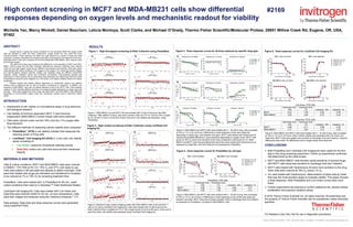
- 1. Michelle Yan, Marcy Wickett, Daniel Beacham, Leticia Montoya, Scott Clarke, and Michael O’Grady, Thermo Fisher Scientific/Molecular Probes, 29851 Willow Creek Rd, Eugene, OR, USA, 97402 RESULTS Figure 1. High-throughput screening of Killer Collection using PrestoBlue Figure 1: MDA-MB231 (A) and MCF7 (B) were treated with 10 µM of drug from the Killer Collection. After addition of drug, cells were cultured under low (1%) or normal (19%) oxygen for 24, 48 and 72 hours. At the end of each time point, cell viability was assessed using PrestoBlue. ABSTRACT Oxygen levels in typical cell culture conditions do not accurately reflect the oxygen levels cells are exposed to within the body. Furthermore, oxygen levels can vary within the tumor microenvironment. These variances can affect how cells respond to a variety of drugs and small molecules. To further understand how oxygen levels affect drug sensitivity, the response of hormone- dependent MCF7 cells were compared to hormone-independent MDA-MB231 cells, cultured under low and high oxygen. The goal of this study was to examine the differences in the sensitivity of MCF7 and MDA- MB231 cells to drugs from the Killer Collection (MicroSource Discovery Systems) when cultured under low and high oxygen levels and for different drug exposure time periods. Cells were grown using standard cell culture conditions (19% oxygen) or low oxygen (1%). Using high-content imaging and high-throughput analysis methods, viability was assessed using two different mechanistic readouts; cellular metabolic activity and membrane permeability. Post-screening analysis was performed to confirm positive hits by performing dose-responses and determining IC50 and EC50 concentrations using the same reagents as in the screen, and assess oxidative stress and cellular proliferation. Results showed that viability differed depending on mechanistic readout and platform method used to determine hits as well as duration of exposure to compound. In addition, the response of MDA-MB231 cells was not always identical to that of the MCF7 cells. Post-screening analysis of “hits” indicated different potencies of compounds tested depending on oxygen level and cell type. These data suggest that some drugs may affect MCF7 and MDA-MB231 cells differently depending on environmental oxygen levels and mechanistic readout used to determine cellular health. INTRODUCTION Assessment of cell viability is a foundational assay in drug discovery and biological research Cell viability of hormone dependent (MCF-7) and hormone independent (MDA-MB231) human breast cells were examined Cells were cultured under normal (19%) and low (1%) oxygen after drug exposure Two different methods of measuring cellular viability were compared: PrestoBlue™ (HTS) a cell viability indicator that measures the reducing power of living cells Live/Dead™ Cell Imaging Kit (HCS) is a two color cell viability assay consisting of: Live Green: measures intracellular esterase activity Dead Red: enters only cells that have lost their membrane integrity MATERIALS AND METHODS Cells & culture conditions: MCF7 and MDA-MB231 cells were cultured in DMEM + 10% FBS at 5% CO, 19% O2 and 37⁰C until ready to use. Cells were plated in 96-well plate and allowed to attach overnight. Cells were then treated with drugs (as indicated) and transferred to incubator to be cultured at 1% or 19% O2 for remaining treatment time. PrestoBlue: Cells were loaded with 1x PrestoBlue for 60 min. under culture conditions than read on a Varioskan™ Flash Multimode Reader. Live/Dead Cell Imaging Kit: Cells were loaded with Live Green and Dead Red plus Hoechst 33342 for 30 min. at room temperature. Cells were then imaged and analyzed using the Cellomics Arrayscan™ VTI. Data analysis: Data plots and dose-response curves were generated using GraphPad Prism. CONCLUSIONS Both PrestoBlue and Live/Dead Cell Imaging Kit were useful for the first step in this drug screening experiment. Follow up experiments confirmed hits determined by the initial screen. MCF7 and MDA-MB231 cells showed varied sensitivity to several drugs, with MCF7 cells being less sensitive to Gambogic Acid and Celastrol. MCF7 cells treated with Tanshionone IIA were more sensitive to this drug when cells were cultured at 19% O2 versus 1% O2. For cells treated with Dactinomycin, determination of dead cells by Dead Red was the most sensitive assay to evaluate viability. This assay showed a dose-response, while PrestoBlue and Live Green curves were more linear. Further experiments are planned to confirm additional hits, assess cellular proliferation and examine oxidative stress. High content screening in MCF7 and MDA-MB231 cells show differential responses depending on oxygen levels and mechanistic readout for viability Thermo Fisher Scientific • 5791 Van Allen Way • Carlsbad, CA 92008 • www.lifetechnologies.com Figure 2. High-content screening of Killer Collection using Live/Dead Cell Imaging Kit Figure 2: Results for high-content imaging screen with MDA-MB231 cells (A-B) and MCF-7 cells (C-D) using Live/Dead Cell Imaging Kit. Cells were treated with 10 µM drug from the Killer Collection then incubated at 1% or 19% oxygen levels for 24 and 48 hours. At the end of each time point, cell viability was assessed using Live/Dead Cell Imaging Kit. A. B. A. B. C. D. Figure 3. Dose-response curves for all three readouts by specific drug type Figure 3: MDA-MB231 and MCF7 cells were treated with 0 – 30 µM of drug, then incubated at 19% or 1% O2 for 24 hours. Differences in dose-response curves were observed depending on assay type, and between cell types. MCF7 cells treated with Tanshinone IIA showed the greatest difference in drug sensitivity depending on oxygen levels. MCF7 cells grown at 19% O2 were more sensitive to the drug compared to cells grown at 1%. MDA- MB231 and MCF7 cells showed differential responses when treated with Diactinomycin as assessed by Dead Red, and with Pristimerin as assessed by PrestoBlue. A. B. C. Figure 4. Dose-response curves for PrestoBlue by cell type Figure 4: MDA-MB231 and MCF7 cells were treated with 0 – 30 µM of drug, then incubated at 19% or 1% O2 for 24 hours. Differences in dose-response curves (IC50) was observed between cell types. MCF7 were less sensitive (greater IC50) to Gambogic Acid and Celastrol as assessed by PrestoBlue, compared to MDA-MB231 cells. Figure 5. Dose-response curves for Live/Dead Cell Imaging Kit PrestoBlue, IC50 (µM) Gambogic Acid, 19% Gambogic Acid, 1% Celastrol, 19% Celastrol, 1% MCF7 5.90 7.25 9.92 9.77 MDA-MB231 2.05 1.38 2.64 2.31 Live Green, IC50 (µM) Gambogic Acid, 19% Gambogic Acid, 1% Celastrol, 19% Celastrol, 1% MCF7 8.23 7.39 6.45 6.36 MDA-MB231 3.00 3.36 6.73 5.02 Dead Red, EC50 (µM) Gambogic Acid, 19% Gambogic Acid, 1% Celastrol, 19% Celastrol, 1% MCF7 4.68 4.71 4.06 4.32 MDA-MB231 2.71 3.64 3.14 3.35 #2169 Figure 5: MDA-MB231 and MCF7 cells were treated with 0 – 30 µM of drug, then incubated at 19% or 1% O2 for 24 hours. When cellular viability was assessed with Live Green and Dead Red, MCF7 cells were slightly less sensitive to Gambogic Acid and Celastrol. However, differences were small. No differences due to oxygen levels were observed. For Research Use Only. Not for use in diagnostic procedures. © 2016 Thermo Fisher Scientific Inc. All rights reserved. All trademarks are the property of Thermo Fisher Scientific and its subsidiaries unless otherwise specified.