This study assessed the toxicity and clearance of CdTe and CdHgTe quantum dots (QDs) in vitro and in vivo. Both CdTe and CdHgTe QDs showed concentration-dependent cytotoxicity to human breast and prostate cancer cells in vitro. CdTe QD toxicity increased over time, while CdHgTe toxicity did not. In vivo, CdTe QDs could be detected by fluorescence imaging in mice up to 18 days after subcutaneous injection, while CdHgTe fluorescence disappeared within 6 days, likely due to instability. This study provides insights into the toxicity and clearance of two types of QDs.
Atmospheric Exposure to Cr III Powder Causes Genotoxicity in Rattus Norvegicus.inventionjournals
Several chemical elements are responsible for altering the genetic integrity of living beings. The metal Cr stands out in this regard. It exists in two oxidation states, Cr VI and Cr III, and has been investigated as an important environmental and occupational contaminant. Although the former is considered carcinogenic, the latter is classified as safe, even for human use in food supplementation. However, most studies with Cr( III) have been carried out by different routes to how it is occupationally found – in the atmosphere. This study evaluated the genotoxicity of Cr(III) inhaled during 8 hours of exposure to the maximum concentration permitted by ATSDR. Fifteen male Rattus norvegicus were used in this study. There were 3 groups (n=5 per group); these were - group exposed to Cr (III) powder (S), the negative control group (NC) and the positive control group (PC). The animals were exposed to Cr aerosol particles at a flow rate of 9L/min and atmospheric concentration of 500μg/m3 for only 8 hours in this study. An increase in genotoxicity and mutagenicity in the group exposed to the metal powder was observed. These findings suggest that further studies should be carried out in order to establish safe levels of exposure to Cr III in work environments
V79C cells were derived from V79 cell line by through chronic oxidative stress that was found to be radio-resistant. These cells had demonstrated transformation–like stable changes and could be used as model system to study oxidant induced carcinogenesis. Our objective was to understand mechanism of radiation-resistance in these cells. Apoptotic cell death was inhibited in these cells as visualized microscopically by Hoechst staining and nucleosomal ladder formation in agarose gel. Release of cytochrome c in cytoplasm and Apoptosis Inducing Factor in the mitochondrial and nuclear fraction of cells were determined by Western blotting. The caspase 9 and caspase 3 activities in these cells were estimated from fluorimetric assays. These results revealed that the radiation resistance was due to inhibition of caspase-dependent apoptotic death pathways although Apoptosis Inducing Factor mediated pathway remained unaffected. These findings may aid in understanding the mechanism of radiation resistance in tumors arising from oxidative stress.
The IOSR Journal of Pharmacy (IOSRPHR) is an open access online & offline peer reviewed international journal, which publishes innovative research papers, reviews, mini-reviews, short communications and notes dealing with Pharmaceutical Sciences( Pharmaceutical Technology, Pharmaceutics, Biopharmaceutics, Pharmacokinetics, Pharmaceutical/Medicinal Chemistry, Computational Chemistry and Molecular Drug Design, Pharmacognosy & Phytochemistry, Pharmacology, Pharmaceutical Analysis, Pharmacy Practice, Clinical and Hospital Pharmacy, Cell Biology, Genomics and Proteomics, Pharmacogenomics, Bioinformatics and Biotechnology of Pharmaceutical Interest........more details on Aim & Scope).
All manuscripts are subject to rapid peer review. Those of high quality (not previously published and not under consideration for publication in another journal) will be published without delay.
High content screening in MCF7 and MDA-MB231 cells show differential response...Thermo Fisher Scientific
Oxygen levels in typical cell culture conditions do not accurately reflect the oxygen levels cells are exposed to within the body. Furthermore, oxygen levels can vary within the tumor microenvironment. These variances can affect how cells respond to a variety of drugs and small molecules. To further understand how oxygen levels affect drug sensitivity, the response of hormone-dependent MCF7 cells were compared to hormone-independent MDA-MB231 cells, cultured under low and high oxygen.
Expression of Genetically Engineered Chitinase Gene of Pyrococcus furiosusIJERDJOURNAL
ABSTRACT: Wild-type Pyrococcus furiosus is most likely unable to grow on chitin in the natural biotope due to a nucleotide insertion which separates the chitinase gene into two ORFs, whereas a genetically engineered strain with the deleted nucleotide is able to grow on chitin. In the latest studies, the recombinant enzyme activity against the crystal chitins was examined. But there are still some conflictions. In our study, to shed a light on whether the construct composed of a catalytic domain and a chitin binding domain show any activity against crystalline chitin, the construct was created in the pET 28b (+) expression vector and expressed in Escherichia coli. The chitinase with an approximately 55 kDa molecular weight was determined. The activity of the enzyme was measured spectrophotometrically. Despite the presence of enzyme activity against the colloidal chitin, no significant activity against the crystal chitin has been measured.
Pluripotent Stem Cell Markers and microRNA Expression May Correlate with Dent...asclepiuspdfs
Background: Recent evidence has demonstrated that dental pulp stem cells (DPSC) may represent a source of pluripotent progenitors capable of differentiating into many cell and tissue types. Although microRNAs are known to modulate differentiation and function in human dental tissues, much of this research has focused selectively on tooth development. The primary objective of this study was to evaluate the expression of microRNA in dental pulp stem cell isolates to compare with classical biomarkers of cellular phenotypes and pluripotency. Materials and Methods: Using eight previously isolated and characterized DPSC isolates, growth and viability were evaluated and RNA was extracted for mRNA screening. DPSC biomarker and microRNA expression were analyzed for comparison with cellular phenotypes. Results: Evaluation of the growth and proliferation rates of each cell line resulted in the categorization of DPSC isolates into rapid, intermediate, and slow doubling times, which demonstrated higher viability among the most rapidly proliferating DPSCs. Analysis of DPSC biomarkers (Oct-4, Sox-2, NANOG) revealed an association with total live cell count, while microRNA expression (miR-27, miR-218, miR-124, and miR-16) appeared to be more closely associated with cellular viability. Conclusions: Although this study was limited to a small number of DPSC isolates, these results suggest a more thorough investigation and evaluation of biomarkers and microRNA expression may be necessary to elucidate the associations and complex interconnections with DPSC viability, proliferation, differentiation, and pluripotency.
Objective: To investigate the changes in the retina due to deltamethrin toxicity and the process in cell inflammation and apoptosis.
Study Design: Sixteen Wistar albino rats were randomly divided into two groups as control (n=8) and deltamethrin (n=8) groups. Saline was given to the control group, and 0.5 mL of 5 mg/kg deltamethrin was given to the deltamethrin group for 14 days each. Blood was collected for biochemical analysis. Retinal tissue was processed for histological examination.
Results: Compared to the control group, MDA levels were high while GSH and CAT levels were low in the deltamethrin group. Histopathological analysis showed spaces between the pigment epithelium, irregularity in the delimiting membrane, degenerated ganglion, cone and bacillus cell, pyknotic nuclei, thinned inner limitation membrane, and thickened vascular wall. The control group showed FAS expression in the pigment layer limiting membranes, in the nuclei of many cone and bacillus cells, and ganglion cells in the control group sections. In the deltamethrin group, FAS expression was observed in the inner and outer limiting membranes of the pigment epithelium, cone and bacillus cells, and ganglion cell nuclei. In the control group, negative NOS expression in the pigment epithelium and outer limiting membranes, internal limitation membrane, and ganglion cells in the cone and bacillus cell nuclei were observed. In the deltamethrin group, NOS expression was positive in the pigment epithelium, cone and bacillus, and ganglion cell nuclei.
Conclusion: We suggest that deltamethrin toxicity induced apoptotic process due to increased inflammation in the retina and may cause visual impairment as a result of neural damage.
Keywords: deltamethrin, FAS, insecticides, NOS, nitric oxide synthase, retina
International Journal of Engineering and Science Invention (IJESI) is an international journal intended for professionals and researchers in all fields of computer science and electronics. IJESI publishes research articles and reviews within the whole field Engineering Science and Technology, new teaching methods, assessment, validation and the impact of new technologies and it will continue to provide information on the latest trends and developments in this ever-expanding subject. The publications of papers are selected through double peer reviewed to ensure originality, relevance, and readability. The articles published in our journal can be accessed online.
We are strong believers of innovation, and over the years we have constantly changed our technology to give cinema viewers an enthralling experience. While cinema screens have changed, so have the projects and picture quality changed. Here we highlight some of the major changes in cinema projection technology and how cinema screens have had to adapt to these changes.
descripcion completa de las hemorroides internas desea saber como eliminarlo de manera natural y sencilla entonces ingrese en el enlace:
http://tinyurl.com/mcfsjou
Airvoi's complete phone system is loaded with robust and standard featuresavencorp
Here is the presentation provides you frequently asked questions answers, manuals and guides about Avencorp Digi Inc and complete phone system ay AIRVOI. Watch the presentation and know more. Source: http://www.airvoi.com/airvoi_business_features.html
In-vitro biological activities of the free new H4L ( indole-7-thiocarbohydrazone) ligand and its Ni(II), Pd(II) , Pt(II),
Cu(II), Ag(I), Zn(II) and Cd(II) complexes are screened against two cancerous cell lines, that revealed significant
activity only for [Cu2Cl2(H4L)2(PPh3)2] after 72 h treatment by the highest tested concentrations. The Copper(I)
complex was characterized by X-ray Crystallography and the NMR spectra, whereas it has been confirmed to have
momentous cytotoxicity against ovarian, breast cancerous cell lines (Caov-3, MCF-7). The apoptosis-inducing
properties of the Cu(I) complex have been investigated through fluorescence microscopy visualization, DNA
fragmentation analysis and propidium iodide flow cytometry.
Anticancer Activity of New Di-Nuclear Copper (I) ComplexTaghreed Al-Noor
In-vitro biological activities of the free new H4L ( indole-7-thiocarbohydrazone) ligand and its Ni(II), Pd(II) , Pt(II),
Cu(II), Ag(I), Zn(II) and Cd(II) complexes are screened against two cancerous cell lines, that revealed significant
activity only for [Cu2Cl2(H4L)2(PPh3)2] after 72 h treatment by the highest tested concentrations. The Copper(I)
complex was characterized by X-ray Crystallography and the NMR spectra, whereas it has been confirmed to have
momentous cytotoxicity against ovarian, breast cancerous cell lines (Caov-3, MCF-7). The apoptosis-inducing
properties of the Cu(I) complex have been investigated through fluorescence microscopy visualization, DNA
fragmentation analysis and propidium iodide flow cytometry.
High Content Screening of automated wound healing and cytotoxicity assays in ...HCS Pharma
To find anti-cancer drugs, different cellular modifications can be looked at: cell death effect on proliferating cells either in 2D culture or on spheroids (3D culture), anti-mitotic, anti-migration/invasion, anti-angiogenesis effects… Different assays can be performed to follow these different effects. High content screening is a multi-parametric technology allowing searching for poly-effects drugs.
In this poster, several well known compounds were tested in 3 different assays: cytotoxicity in 2D culture, cytotoxicity in spheroids (3D culture) and scratch assay (also named wound healing). These 3 models were automated in 96-well plates.
Minimal criteria for defining multipotent mesenchymal stromal cells. The International Society for Cellular Therapy position statement M Dominici1, K Le Blanc2, I Mueller3, I Slaper-Cortenbach4, FC Marini5, DS Krause6, RJ Deans7, A Keating8, DJ Prockop9 and EM Horwitz10
Discovery of novel immunotherapy represents a main and intense focus of reseach in oncology. Proof of concept studies in animal represent a challenge and require a well characterized and appropriate animal models with most of the time customized approaches. Some recent developments and data generated for immune checkpoint modulators, adoptive cell transfer therapy, vaccines and bispecific T cell engagers will be presented.
Neurotoxicity assay on 2D and 3D culture using High Content Screening (HCS) t...HCS Pharma
As shown by AstraZeneca in nature reviews*, one third of safety failures along the drug discovery process is linked to CNS toxicity uncovered in clinical trials. To avoid this attrition, the potential neurotoxicity of any drug going through the blood brain barrier (BBB) needs to be assessed in the very early stages of new chemical entities (NCE) research. Neurotoxicity assays can be performed on the SH-SY5Y human cell line by using High-Content Screening (HCS) technologies. The present study was performed using classical 2D and 3D culture protocols. In this poster, 2D results and preliminary 3D culture results on multiple reference compounds are depicted.
The Role of Radiotherapy in the Treatment of Early Stage Ocular Marginal Zone...daranisaha
To evaluate the benefit of radiotherapy, compared with other treatment in ocular marginal zone lymphoma, retrospectively we analyzed our experience, with the end-points: efficacy, measured for complete response, Progression-Free Survival (PFS) and Overall Survival
ESCOZINETM is an innovative polarized, potentiated bio-active peptide extracted from the Blue Caribbean Scorpion (Rhopularus Princeps) which contains amino acids, proteins and minerals. Medolife filed a patent in 2012 for its ESCOZINETM and polarization technology (Patent # US 8,097,284 B2). The polarization technique acts as a delivery system and additionally, amplifies the highly positive
The Radiosensitivity Effect of Hydroxyurea on HT29 Cell Lineiosrphr_editor
The IOSR Journal of Pharmacy (IOSRPHR) is an open access online & offline peer reviewed international journal, which publishes innovative research papers, reviews, mini-reviews, short communications and notes dealing with Pharmaceutical Sciences( Pharmaceutical Technology, Pharmaceutics, Biopharmaceutics, Pharmacokinetics, Pharmaceutical/Medicinal Chemistry, Computational Chemistry and Molecular Drug Design, Pharmacognosy & Phytochemistry, Pharmacology, Pharmaceutical Analysis, Pharmacy Practice, Clinical and Hospital Pharmacy, Cell Biology, Genomics and Proteomics, Pharmacogenomics, Bioinformatics and Biotechnology of Pharmaceutical Interest........more details on Aim & Scope).
Identification of the Enrichment Pathways of Catalase in Multiple Primary Tumorsdaranisaha
Reactive oxygen species (ROS) has been detected in almost all cancers, which it involves many aspects of carcinogenesis and tumor progression. We previously reported a novel oncogenic role of manganese superoxide dismutase (MnSOD; SOD2) in invasive lung adenocarcinoma (LUAD) by upregulating forkhead box protein M1 (FOXM1) and matrix metalloproteinase-2 (MMP2) expression. In this study, we used a comprehensive analysis and further evaluated the effects of a hydrogen peroxide (H2O2) scavenger, catalase (CAT) in The Cancer Genome Atlas (TCGA) datasets of the different cancer types.
Identification of the Enrichment Pathways of Catalase in Multiple Primary Tumors
nihms95470
1. In vitro and In vivo Assessment of CdTe and CdHgTe Toxicity and
Clearance
Li Liu1,*, Jun Zhang2,*,#, Xing Su1, and Ralph P. Mason1,3
1 Cancer Imaging Program, Department of Radiology, The University of Texas, Southwestern
Medical Center, Dallas, TX 75390, USA
2 College of Chemistry and Chemical Engineering, Inner Mongolia University, Hohhot, Inner
Mongolia 010021, P. R. China
3 Joint Program in Biomedical Engineering UT Arlington and UT Southwestern Medical Center,
Dallas, TX 75390, USA
Abstract
Semiconductor QDs are being developed as fluorescent tags for biomedical applications such as
imaging, targeting, therapeutic carriers, drug delivery, nanomedicine, and in vitro and in vivo
biological labeling. However, potential toxicity and clearance of semiconductor QDs in biological
systems are of concerned. We have tested toxicity and clearance in vitro and in vivo (subcutaneous)
of CdTe and CdHgTe semiconductor QDs in human breast cancer MCF7 and MBA-MD-231 cells
and prostate cancer PC3 cells over a period of 40 days. Our results show that both CdTe and CdHgTe
QDs are cytotoxic to human breast and prostate cancer cells. CdHgTe ODs were cleared rapidly from
the site of injection, while CdTe were still detectable 18 days after injection, but were cleared by 30
days.
Keywords
Semiconductor QDs; CdTe; CdHgTe; Toxicity; Clearance
1. INTRODUCTION
Semiconductor QDs have been developed as fluorescence tags in biological and medical
applications such as in vitro and in vivo biological labeling, imaging,1–6 targeting,7,8
therapeutic carriers and drug delivery.9–13 To avoid QD aggregation, improve water-solubility
and biocompatibility, and exert specific surface chemistry for targeting and delivery, various
conformations of semiconductor QDs, such as core-shell structure, organic molecule-modified
geometry, and functional polymer capped structure have been developed14–16. Semiconductor
QDs are shown to have advantages of efficient luminescence, high photobleaching threshold,
flexible surface chemistry, and good water-solubility for biological and medical applications.
However, the issues of toxicity and clearance of QDs in bio-systems are of concern,17–24 as
these two issues are not fully understood and must be carefully assessed if QDs are to move
from scientific curiosity to biomedical application.
Several reports have examined the toxicity and clearance of QDs.8,18–32 One common
understanding for toxicity of cadmium-containing semiconductor QDs is that their toxicity is
#Author to whom correspondence should be addressed. cejzhang@imu.edu.cn.
* Same contribution to this work
NIH Public Access
Author Manuscript
J Biomed Nanotechnol. Author manuscript; available in PMC 2009 October 5.
Published in final edited form as:
J Biomed Nanotechnol. 2008 December 1; 4(4): 524–528. doi:10.1166/jbn.2008.018.
NIH-PAAuthorManuscriptNIH-PAAuthorManuscriptNIH-PAAuthorManuscript
2. closely related to the concentration of free Cd2+.17,21,22,28,29 Derfus et al observed that the
cytotoxicity of CdTe QDs was correlated to the liberation of free Cd2+.29 and Zhang et al
recently presented similar evidence showing that the cytotoxicity of QDs behaves in a
concentration- and size-dependent manner17. Another common assumption is that surface
capping layers of QDs synthesized under different methods may contribute to the level of
toxicity following different mechanisms such as oxidation and breakdown of nanostructures.
21,22,28 The distribution and damage of QDs to organs are highly variable, and the clearance
of QDs in bio-system also varies according to the concentration and structure of QDs.28, 33,
34.
In this paper, we assess the toxicity and clearance of semiconductor CdTe and CdHgTe QDs
in vitro in human breast and prostate cancer cells and in vivo following subcutaneous injection
in mice.
2. MATERIALS AND METHODS
2.1 Synthesis of Semiconductor CdTe and CdHgTe QDs
The preparation of CdTe QDs has been reported elsewhere.35–38 The QDs were stabilized by
thioglycolic acid (TGA) on their surfaces, enhancing water solubility, and facilitating
conjugation with ligands. The average sizes of QDs used was 6–8 nm. The procedures for
making CdTe QDs are briefly described as follows: CdTe QDs were created by the reaction
of precursors containing cadmium perchlorate hydrate [Cd(ClO4)2*H2O] and hydrogen
telluride (H2Te) through vigorous stirring. The Cd2+ solution was prepared by dissolving 731
mg of Cd(ClO4)2*H2O in 125 mL of water. TGA (0.396 mL) was then added to the solution.
0.1M NaOH solution was also added to adjust the pH to approximately 11. The solution was
then purged with nitrogen for at least 30 minutes. H2Te gas was produced by the chemical
reaction of excess aluminum telluride with 0.5 M sulfuric acid in an inert atmosphere (nitrogen)
and was combined with the above Cd2+ solution using a set-up described previously36. After
completion of the reaction a yellow solution of CdTe QD nuclei was obtained. This solution
was then refluxed at 100 °C to promote crystal growth. The QDs were extracted and stored at
4 °C in the dark. The near infrared emitting CdHgTe QDs were obtained by adding 2.5 ml of
0.1 M mercury perchlorate solution to 50 ml of CdTe QD solution to gradually form CdHgTe
QDs.
2.2 Fluorescent Imaging
Red CdTe and Infrared CdHgTe QDs with fluorescence emission peaks in the 650 nm and 900
nm ranges, respectively were used. Imaging was performed using a multispectral Maestro
Fluorescent Imaging System (Cri, Waltham, MA) with a green filter. For in vivo fluorescent
imaging experiments CdTe and CdHgTe QDs (50 μl) were injected subcutaneously into mice
at two locations. A mouse was anesthetized with 80 μl of a mixture of ketamine, xylazine, and
acepromazine. Fluorescent images were acquired immediately after injection of CdTe and
CdHgTe QDs with 100 ms exposure. CdTe and CdHgTe clearance and toxicity were monitored
over a period of 40 days.
2.3 Toxicity
Human breast cancer MCF7 and MDA-MD-231 cells and prostate cancer PC3 cells were
seeded in 96-well plates (Costar, Corning, NY) at a concentration of 1 × 10⁴ cells in 100 μl of
medium per well. Each treatment condition was assessed in groups of 8. After 24 hours, the
medium was aspirated and new medium containing the QDs were added. At the indicated time,
total cell number was determined using a crystal violet assay. Briefly, the medium was aspirated
and 1% glutaraldehyde (100 μl; Sigma, St. Louis, MO) in PBS was added for incubation for
15 minutes. After removing glutaraldehyde, 0.5% crystal violet (Sigma) was incubated for 15
Liu et al. Page 2
J Biomed Nanotechnol. Author manuscript; available in PMC 2009 October 5.
NIH-PAAuthorManuscriptNIH-PAAuthorManuscriptNIH-PAAuthorManuscript
3. minutes, and the plates were washed with water (twice) and soaked in water for 10 min before
drying at room temperature. Once dry, 100 μl of Sorenson’s solution (a solution of 9 g tri-
sodium citrate in 305 ml of distilled water with 195 ml 0.1 N HCl and 500 ml 90% ethanol)
was added to elute the crystal violet. After 30 minutes, it was read at 540 nm using an ELX800
microplate reader (Bio-Tek Instruments, Winooski, VT).
3. RESULTS AND DISCUSSION
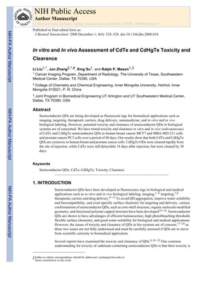
We tested the concentration and time-dependent cytotoxicity of CdTe and CdHgTe QDs in
human breast cancer MCF7 and MBA-MD-231 cells and prostate cancer PC3 cells. Both CdTe
and CdHgTe QDs showed concentration-dependent cytotoxicity (Figs. 1 and 2) for each cell
type. Intriguingly, cytotoxicity of CdTe QDs increased with exposure time, but this was not
seen for CdHgTe for any of the tumor cell lines. For 24 hrs exposure, there was minimal
cytotoxicity up to 5 μM, but it increased significantly with higher concentrations.
When CdTe and CdHgTe QDs (50 μL) were injected subcutaneously into a group of 6 mice
at two locations all survived and fluorescent images were acquired immediately and up to 40
days. Figure 3(a) shows the in vivo fluorescence spectra of CdTe and CdHgTe QDs at 650 nm
and 900 nm. Figure 3(b) indicates the change of fluorescence signals for CdTe and CdHgTe
QDs over 30 days. The fluorescent signals from CdTe QDs decayed more slowly than those
of CdHgTe QDs. CdTe QDs still showed 15% fluorescent intensity after 18 days, but signal
disappeared by 30 days. The fluorescent signals from CdHgTe QDs decayed rapidly and no
fluorescent signal was detectable by the 6th day. The first row in Figure 3(c) shows the
fluorescence images of a mouse 1, 6, 18 and 30 days following subcutaneous injection of 50
μL CdHgTe (left fluorescence spot) and CdTe (right fluorescence spot) QDs. Fluorescence
from CdTe QDs could be observed clearly at 18 days, while the fluorescence from CdHgTe
QDs persisted less than 6 days. The discrepancy in fluorescence decay may be due to the lower
chemical-stability of CdHgTe in comparison with CdTe QDs. Spectral unmixing showed the
fluorescent images individually for CdTe and CdHgTe QDs, respectively (Figure 3c, second
and third rows), Combined images of QDs overlaid on auto fluorescence (Figure 4d).
CONCLUSION
Both CdTe and CdHgTe QDs could be detected by fluorescent imaging in vitro and in vivo.
Both CdTe and CdHgTe QDs showed some cytotoxicity in human breast MCF7 and MBA-
MD-231 and prostate cancer PC3 cells at 5 μM. CdTe QD toxicity increased with exposure
time, but this was not seen in CdHgTe QDs. CdTe QDs could be observed in vivo following
SC administration in mice for up to 18 days, while the fluorescent signal in CdHgTe
disappeared within 6 days, possibly due to instability.
Acknowledgments
Supported by the Department of Energy (DE-FG02-05CH11280) and Southwestern Small Animal Imaging Research
Program (SW-SAIRP), funded in part by NCI U24 CA126608. J. Zhang would like to thank financial aid from the
NSFC (Grants No. 20601012), Inner Mongolia University “513 program” (No. 206043) and “Nanolab program”, and
the Educational Department of Inner Mongolia (NJZY07011).
References and Notes
1. Chan WCW, Nie S. Science 1998;281:2016. [PubMed: 9748158]
2. Jamieson T, Bakhshi R, Petrova D, Pocock R, Imani M, Seifalian AM. Biomaterials 2008;28:4717.
[PubMed: 17686516]
3. Hoshino A, Manabe N, Fujioka K, Suzuki K, Yasuhara M, Yamamoto K. Journal of Artificial Organs
2007;10:149. [PubMed: 17846713]
Liu et al. Page 3
J Biomed Nanotechnol. Author manuscript; available in PMC 2009 October 5.
NIH-PAAuthorManuscriptNIH-PAAuthorManuscriptNIH-PAAuthorManuscript
4. 4. Gao X, Dave SR. Bio-applications of Nanoparticles 2007;620:57.
5. Zhang J, Sun J, Liu L, Huang Y, Mason RP. Journal of Nanoscience and Nanotechnology 2008;8:1155.
[PubMed: 18468115]
6. Su J, Zhang J, Liu L, Huang Y, Mason RP. Journal of Nanoscience and Nanotechnology 2008;8:1174.
[PubMed: 18468119]
7. Gao X, Cui Y, Levenson RM, Chung LWK, Nie S. Nature Biotechnology 2004;22:969.
8. Medintz IL, Uyeda HT, Goldman ER, Mattoussi H. Nature Materials 2005;4:435.
9. Chen W. Journal of Nanoscience and Nanotechnology 2008;8:1019. [PubMed: 18468106]
10. Chen W, Zhang J. Journal of Nanoscience and Nanotechnology 2006;6:1159. [PubMed: 16736782]
11. Takeda M, Tada H, Higuchi H, Kobayashi Y, Kobayashi M, Sakurai Y, Ishida T, Ohuchi N. Breast
Cancer 2008;15:145. [PubMed: 18317884]
12. Yezhelyev MV, Qi L, O’Regan RM, Nie S, Gao X. Journal of The American Chemical Society
2008;130:9006. [PubMed: 18570415]
13. Liu W, Choi HS, Zimmer JP, Tanaka E, Frangioni JV, Bawendi MG. Journal of The American
Chemical Society 2007;129:14530. [PubMed: 17983223]
14. Guo W, Li J, Wang Y, Peng X. Journal of The American Chemical Society 2003;125:3901. [PubMed:
12656625]
15. Kim S, Bawendi MG. Journal of The American Chemical Society 2003;125:14652. [PubMed:
14640609]
16. Dubertret B, Skourides P, Norris DJ, Noireaux V, Brivanlou AH, Libchaber A. Science
2002;298:1759. [PubMed: 12459582]
17. Zhang Y, Chen W, Zhang J, Liu J, Chen G, Pope C. Journal of Nanoscience and Nanotechnology
2007;7:497. [PubMed: 17450785]
18. Male KB, Lachance B, Hrapovic S, Sunahara G, Luong JHT. Analytical Chemistry 2008;80:5480.
19. Byrne SJ, Williams Y, Davies A, Corr SA, Rakovich A, Gun’ko YK, Rakovich YR, Donegan JF,
Volkov Y. Small 2007;3:1152. [PubMed: 17534993]
20. Chang E, Thekkek N, Yu W, Colvin VL, Drezek R. Small 2006;2:1412. [PubMed: 17192996]
21. Fischer HC, Liu L, Pang K, Chan WCW. Advanced Functional Materials 2006;16:1299.
22. Fisher HC, Chan WCW. Current Opinion in Biotechnology 2007;18:565. [PubMed: 18160274]
23. Choi HS, Liu W, Misra P, Tanaka E, Zimmer JP, Ipe BI, Bawendi MG, Frangioni JV. Nature
Biotechnology 2007;25:1165.
24. Cai W, Hsu AR, Li Z, Chen X. Nanoscale Research Letters 2007;2:265.
25. Guo G, Liu W, Liang J, He Z, Xu H, Yang X. Materials Letters 2007;61:1641.
26. Han M, Gao X, Su JZ, Nie S. Nature Biotechnology 2001;19:631.
27. Ballou B, Ernst LA, Waggoner. Current Medicinal Chemistry 2005;12:795. [PubMed: 15853712]
28. Ballou B, Lagerholm BC, Ernst LA, Bruchez MP, Waggoner AS. Bioconjugation Chemistry
2004;15:79.
29. Derfus AM, Chan WCW, Bhatia SN. Nano Letters 2004;4:11.
30. Fountaine TJ, Wincovitch SM, Geho DH, Garfield SH, Pittaluga S. Modern Pathology 2006;19:1181.
[PubMed: 16778828]
31. Smith AM, Ruan G, Rhyner MN, Nie S. Annals of Biomedical Engineering 2006;34:3. [PubMed:
16450199]
32. Fu A, Gu W, Larabell C, Alivisatos AP. Current Opinion in Neurobiology 2005;15:568. [PubMed:
16150591]
33. Voura EB, Jaiswal JK, Mattoussi H, Simon SM. Nature Medicine 2004;10:993.
34. Lovric J, Bazzi HS, Cuie Y, Fortin GR, Winnik FM, Maysinger D. Journal of Molecular Medicine
2005;83:377. [PubMed: 15688234]
35. Morgan NY, English S, Chen W, Chernomordik V, Russo A, Smith PD, Gandjbakhche A. Academic
Radiology 2005;12:313. [PubMed: 15766692]
36. Gaponik N, Talapin DV, Rogach AL, Hoppe K, Shevchenko EV, Kornowski A, Eychmuller A, Weller
H. J Phys Chem B 2002;106:7177.
Liu et al. Page 4
J Biomed Nanotechnol. Author manuscript; available in PMC 2009 October 5.
NIH-PAAuthorManuscriptNIH-PAAuthorManuscriptNIH-PAAuthorManuscript
5. 37. Joly AG, Chen W, McCready DE, Malm J, Bovin J. Phys Rev B 2005;71:165304.
38. Chen W, Zhang J, Westcott SL, Joly AG, Malm J, Bovin J. J Appl Phys 2006;99:034302.
Liu et al. Page 5
J Biomed Nanotechnol. Author manuscript; available in PMC 2009 October 5.
NIH-PAAuthorManuscriptNIH-PAAuthorManuscriptNIH-PAAuthorManuscript
6. Figure 1.
Cytotoxicity of CdTe and CdHgTe QDs in breast cancer MDA-MB-231 and MCF7 Cells
Liu et al. Page 6
J Biomed Nanotechnol. Author manuscript; available in PMC 2009 October 5.
NIH-PAAuthorManuscriptNIH-PAAuthorManuscriptNIH-PAAuthorManuscript
7. Figure 2.
Cytotoxicity of CdTe and CdHgTe QDs in prostate cancer PC3 cells
Liu et al. Page 7
J Biomed Nanotechnol. Author manuscript; available in PMC 2009 October 5.
NIH-PAAuthorManuscriptNIH-PAAuthorManuscriptNIH-PAAuthorManuscript
8. Figure 3.
(a) Fluorescence spectra of CdTe and CdHgTe QDs; (b) CdTe and CdHgTe clearance in
vivo; (c) First row: Fluorescence images of grey mouse following subcutaneous injection of
50 μL CdHgTe (left fluorescent spot) and CdTe (right fluorescent spot) QDs observed at 1, 6,
18 and 30 days; Second row: Spectrally unmixed images to show CdTe; Third row: Spectrally
unmixed images showing CdHgTe QDs. Fourth row: Combined images of CdTe and CdHgTe
overlaid on autofluorescence.
Liu et al. Page 8
J Biomed Nanotechnol. Author manuscript; available in PMC 2009 October 5.
NIH-PAAuthorManuscriptNIH-PAAuthorManuscriptNIH-PAAuthorManuscript