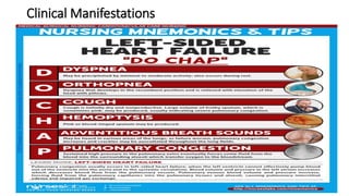
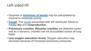
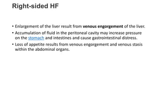
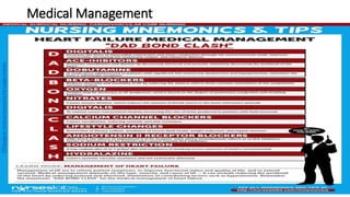
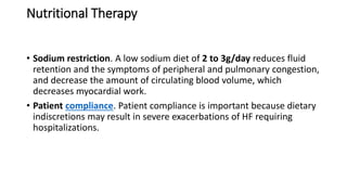
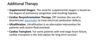
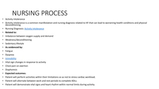
Heart Failure
DEFINITION
• Heart failure is the inability of the heart to pump sufficient blood to
meet the needs of the tissues for oxygen and nutrients.
• The term heart failure indicates myocardial disease in which there is
a problem with contraction of the heart (systolic dysfunction) or
filling of the heart (diastolic dysfunction) that may or may not cause
pulmonary or systemic congestion.
• Heart failure is most often a progressive, life-long condition that is
managed with lifestyle changes and medications to prevent episodes
of acute decompensated heart failure.
CLASSIFICATION
• Heart failure is classified into two types: left-sided heart failure and
right-sided heart failure.
Left-Sided Heart Failure
• Left-sided heart failure or left ventricular failure have different
manifestations with right-sided heart failure.
• Pulmonary congestion occurs when the left ventricle cannot
effectively pump blood out of the ventricle into the aorta and the
systemic circulation.
• Pulmonary venous blood volume and pressure increase, forcing fluid
from the pulmonary capillaries into the pulmonary tissues and alveoli,
causing pulmonary interstitial edema and impaired gas exchange.
Right-Sided Heart Failure
• When the right ventricle fails, congestion in the peripheral tissues and
the viscera predominates.
• The right side of the heart cannot eject blood and cannot
accommodate all the blood that normally returns to it from the
venous circulation.
• Increased venous pressure leads to JVD and increased capillary
hydrostatic pressure throughout the venous system.
Causes
• Systemic diseases are usually one of the most common causes of heart failure.
• Coronary artery disease. Atherosclerosis of the coronary arteries is the primary
cause of HF, and coronary artery disease is found in more than 60% of the
patients with HF.
• Ischemia. Ischemia deprives heart cells of oxygen and leads to acidosis from the
accumulation of lactic acid.
• Cardiomyopathy. HF due to cardiomyopathy is usually chronic and progressive.
• Systemic or pulmonary hypertension. Increase in afterload results from
hypertension, which increases the workload of the heart and leads to
hypertrophy of myocardial muscle fibers.
• Valvular heart disease. Blood has increasing difficulty moving forward, increasing
pressure within the heart and increasing cardiac workload.
Pathophysiology
• Heart failure results from a variety of cardiovascular conditions, including
chronic hypertension, coronary artery disease, and valvular disease.
• As HF develops, the body activates neurohormonal compensatory mechanisms.
• Systolic HF results in decreased blood volume being ejected from the ventricle.
• The sympathetic nervous system is then stimulated to
release epinephrine and norepinephrine.
• Decrease in renal perfusion causes renin release, and then promotes the
formation of angiotensin I.
• Angiotensin I is converted to angiotensin II by ACE which constricts the blood
vessels and stimulates aldosterone release that causes sodium and fluid
retention.
• There is a reduction in the contractility of the muscle fibers of the heart as the
workload increases.
• Compensation. The heart compensates for the increased workload by increasing
the thickness of the heart muscle.
Clinical Manifestations
Left-sided HF
• Dyspnea or shortness of breath may be precipitated by
minimal to moderate activity.
• Cough. The cough associated with left ventricular failure is
initially dry and nonproductive.
• Pulmonary crackles. Bibasilar crackles are detected earlier
and as it worsens, crackles can be auscultated across all lung
fields.
• Low oxygen saturation levels. Oxygen saturation may
decrease because of increased pulmonary pressures.
Right-sided HF
• Enlargement of the liver result from venous engorgement of the liver.
• Accumulation of fluid in the peritoneal cavity may increase pressure
on the stomach and intestines and cause gastrointestinal distress.
• Loss of appetite results from venous engorgement and venous stasis
within the abdominal organs.
Prevention
• Prevention of heart failure mainly lies in lifestyle management.
• Healthy diet. Avoiding intake of fatty and salty foods greatly improves
the cardiovascular health of an individual.
• Engaging in cardiovascular exercises thrice a week could keep
the cardiovascular system up and running smoothly.
• Smoking cessation. Nicotine causes vasoconstriction that increases
the pressure along the vessels.
Complications
• Many potential problems associated with HF therapy relate to the use
of diuretics.
• Hypokalemia. Excessive and repeated dieresis can lead to hypokalemia.
• Hyperkalemia. Hyperkalemia may occur with the use of ACE inhibitors,
ARBs, or spironolactone.
• Prolonged diuretic therapy might lead to hyponatremia and result
in disorientation, fatigue, apprehension, weakness, and muscle cramps.
• Dehydration and hypotension. Volume depletion from excessive fluid
loss may lead to dehydration and hypotension.
Assessment and Diagnostic Findings
• HF may go undetected until the patient presents with signs and symptoms of
pulmonary and peripheral edema.
• ECG: May show hypertrophy, axis deviation, ischemia, and damage patterns.
Dysrhythmias and ST-T segment abnormalities may be present.
• Chest x-ray: May show enlarged cardiac shadow or abnormal contour indicating
ventricular aneurysm.
• Sonograms (echocardiography, Doppler, and transesophageal echocardiography):
May reveal chamber dimensions, valvular function/structure, and ventricular
dilation and dysfunction.
• Heart scan (MUGA): Measures cardiac volume, ejection fraction, and wall motion.
• Exercise or pharmacological stress myocardial perfusion: Determines presence of
myocardial ischemia and wall motion abnormalities.
• PET scan: Sensitive test for evaluating myocardial ischemia and viability.
• Cardiac catheterization: Assesses pressures, differentiates right- versus left-sided
heart failure, and evaluates coronary artery patency.
• Liver enzymes: Elevated in liver congestion/failure.
• Digoxin and other cardiac drug levels: Determines therapeutic range.
• Bleeding and clotting times: Identifies clotting risks and therapeutic range.
• Electrolytes: May be altered due to fluid shifts, renal function, or diuretic therapy.
• Pulse oximetry: Measures oxygen saturation, especially in conjunction with COPD or
chronic HF.
• Arterial blood gases (ABGs): Reflects respiratory and acid-base status.
• BUN/creatinine: Evaluates renal perfusion and function.
• Serum albumin/transferrin: Indicates protein intake and liver function.
• Complete blood count (CBC): Assesses for anemia, polycythemia, and dilutional
changes.
• ESR: Evaluates acute inflammatory reaction.
• Thyroid studies: Determines thyroid activity as a potential precipitator of HF.
Medical Management
Pharmacologic Therapy
• ACE Inhibitors. ACE inhibitors slow the progression of HF, improve exercise
tolerance, decrease the number of hospitalizations for HF, and
promote vasodilation and diuresis by decreasing afterload and preload.
• Angiotensin II Receptor Blockers. ARBs block the conversion of angiotensin
I at the angiotensin II receptor and cause decreased blood pressure,
decreased systemic vascular resistance, and improved cardiac output.
• Beta Blockers. Beta blockers reduce the adverse effects from the constant
stimulation of the sympathetic nervous system.
• Diuretics. Diuretics are prescribed to remove excess extracellular fluid by
increasing the rate of urine produced in patients with signs and symptoms
of fluid overload.
• Calcium Channel Blockers. CCBs cause vasodilation, reducing systemic
vascular resistance but contraindicated in patients with systolic HF.
Nutritional Therapy
• Sodium restriction. A low sodium diet of 2 to 3g/day reduces fluid
retention and the symptoms of peripheral and pulmonary congestion,
and decrease the amount of circulating blood volume, which
decreases myocardial work.
• Patient compliance. Patient compliance is important because dietary
indiscretions may result in severe exacerbations of HF requiring
hospitalizations.
Additional Therapy
• Supplemental Oxygen. The need for supplemental oxygen is based on
the degree of pulmonary congestion and resulting hypoxia.
• Cardiac Resynchronization Therapy. CRT involves the use of a
biventricular pacemaker to treat electrical conduction defects.
• Ultrafiltration. Ultrafiltration is an alternative intervention for
patients with severe fluid overload.
• Cardiac Transplant. For some patients with end-stage heart failure,
cardiac transplant is the only option for long term survival.
NURSING PROCESS
• Activity Intolerance
• Activity intolerance is a common manifestation and nursing diagnosis related to HF that can lead to worsening health conditions and physical
deconditioning.
• Nursing Diagnosis: Activity Intolerance
• Related to:
• Imbalance between oxygen supply and demand
• Weakness/deconditioning
• Sedentary lifestyle
• As evidenced by:
• Fatigue
• Dyspnea
• Immobility
• Vital sign changes in response to activity
• Chest pain on exertion
• Diaphoresis
• Expected outcomes:
• Patient will perform activities within their limitations so as not to stress cardiac workload.
• Patient will alternate between work and rest periods to complete ADLs.
• Patient will demonstrate vital signs and heart rhythm within normal limits during activity.
• Assessment:
• 1. Observe cardiopulmonary response to activity.
The nurse can monitor the patient’s heart rate, oxygen saturation, and cardiac
rhythm during activity. A rise or drop in blood pressure, tachycardia, or EKG
changes can signify overexertion and help plan appropriate interventions.
• 2. Assess the patient’s perspective.
Assess the patient’s understanding of their condition and their perceived
activity limitations. The goal is to ensure the patient is not overexerting
themselves but also feels motivated to make progress with their activity
tolerance and maintain independence.
• 3. Assess the degree of debility.
Interventions can be tailored to the severity of the patient’s symptoms. Assess
the level of fatigue, weakness, and dyspnea in relation to activity and length of
exertion. The nurse may need to assist with ADLs or adjust the activities the
patient can undertake for their safety.
• 1. Provide a calm environment.
• Dyspnea from HF can result in anxiety and restlessness. Provide the patient with a cool, dimly lit space free from
clutter and stimulation. Assist the patient in taking slow, controlled breaths and provide emotional support so
they feel in control.
• 2. Encourage participation.
• Even a patient with chronic HF and severe activity intolerance can assist with care to some extent. Provide
toiletries at the bedside so the patient can brush their teeth or comb their hair. Have the patient assist with
turning themselves in bed. A patient who becomes immobile from a sedentary lifestyle is at an increased risk for
other complications such as skin breakdown, deep vein thrombosis (DVT), and pneumonia.
• 3. Teach methods to conserve energy.
• Group tasks together, sit when possible when performing ADLs, plan rest periods, promote restful sleep, do not
rush activities, and avoid activities in hot or cold temperatures.
• 4. Recommend cardiac rehabilitation.
• This is a medically supervised outpatient program that teaches a patient with a cardiac history how to reduce
their risk of heart problems through exercise, heart-healthy diets, stress reduction, and management of chronic
conditions. This is a team-based approach working with providers, nurses who specialize in cardiac care, PT and
OT, and dieticians.

HEART FAILURE.pptx

  • 1.
  • 2.
    DEFINITION • Heart failureis the inability of the heart to pump sufficient blood to meet the needs of the tissues for oxygen and nutrients. • The term heart failure indicates myocardial disease in which there is a problem with contraction of the heart (systolic dysfunction) or filling of the heart (diastolic dysfunction) that may or may not cause pulmonary or systemic congestion. • Heart failure is most often a progressive, life-long condition that is managed with lifestyle changes and medications to prevent episodes of acute decompensated heart failure.
  • 3.
    CLASSIFICATION • Heart failureis classified into two types: left-sided heart failure and right-sided heart failure.
  • 4.
    Left-Sided Heart Failure •Left-sided heart failure or left ventricular failure have different manifestations with right-sided heart failure. • Pulmonary congestion occurs when the left ventricle cannot effectively pump blood out of the ventricle into the aorta and the systemic circulation. • Pulmonary venous blood volume and pressure increase, forcing fluid from the pulmonary capillaries into the pulmonary tissues and alveoli, causing pulmonary interstitial edema and impaired gas exchange.
  • 5.
    Right-Sided Heart Failure •When the right ventricle fails, congestion in the peripheral tissues and the viscera predominates. • The right side of the heart cannot eject blood and cannot accommodate all the blood that normally returns to it from the venous circulation. • Increased venous pressure leads to JVD and increased capillary hydrostatic pressure throughout the venous system.
  • 6.
    Causes • Systemic diseasesare usually one of the most common causes of heart failure. • Coronary artery disease. Atherosclerosis of the coronary arteries is the primary cause of HF, and coronary artery disease is found in more than 60% of the patients with HF. • Ischemia. Ischemia deprives heart cells of oxygen and leads to acidosis from the accumulation of lactic acid. • Cardiomyopathy. HF due to cardiomyopathy is usually chronic and progressive. • Systemic or pulmonary hypertension. Increase in afterload results from hypertension, which increases the workload of the heart and leads to hypertrophy of myocardial muscle fibers. • Valvular heart disease. Blood has increasing difficulty moving forward, increasing pressure within the heart and increasing cardiac workload.
  • 7.
    Pathophysiology • Heart failureresults from a variety of cardiovascular conditions, including chronic hypertension, coronary artery disease, and valvular disease. • As HF develops, the body activates neurohormonal compensatory mechanisms. • Systolic HF results in decreased blood volume being ejected from the ventricle. • The sympathetic nervous system is then stimulated to release epinephrine and norepinephrine. • Decrease in renal perfusion causes renin release, and then promotes the formation of angiotensin I. • Angiotensin I is converted to angiotensin II by ACE which constricts the blood vessels and stimulates aldosterone release that causes sodium and fluid retention. • There is a reduction in the contractility of the muscle fibers of the heart as the workload increases. • Compensation. The heart compensates for the increased workload by increasing the thickness of the heart muscle.
  • 8.
  • 9.
    Left-sided HF • Dyspneaor shortness of breath may be precipitated by minimal to moderate activity. • Cough. The cough associated with left ventricular failure is initially dry and nonproductive. • Pulmonary crackles. Bibasilar crackles are detected earlier and as it worsens, crackles can be auscultated across all lung fields. • Low oxygen saturation levels. Oxygen saturation may decrease because of increased pulmonary pressures.
  • 11.
    Right-sided HF • Enlargementof the liver result from venous engorgement of the liver. • Accumulation of fluid in the peritoneal cavity may increase pressure on the stomach and intestines and cause gastrointestinal distress. • Loss of appetite results from venous engorgement and venous stasis within the abdominal organs.
  • 12.
    Prevention • Prevention ofheart failure mainly lies in lifestyle management. • Healthy diet. Avoiding intake of fatty and salty foods greatly improves the cardiovascular health of an individual. • Engaging in cardiovascular exercises thrice a week could keep the cardiovascular system up and running smoothly. • Smoking cessation. Nicotine causes vasoconstriction that increases the pressure along the vessels.
  • 13.
    Complications • Many potentialproblems associated with HF therapy relate to the use of diuretics. • Hypokalemia. Excessive and repeated dieresis can lead to hypokalemia. • Hyperkalemia. Hyperkalemia may occur with the use of ACE inhibitors, ARBs, or spironolactone. • Prolonged diuretic therapy might lead to hyponatremia and result in disorientation, fatigue, apprehension, weakness, and muscle cramps. • Dehydration and hypotension. Volume depletion from excessive fluid loss may lead to dehydration and hypotension.
  • 14.
    Assessment and DiagnosticFindings • HF may go undetected until the patient presents with signs and symptoms of pulmonary and peripheral edema. • ECG: May show hypertrophy, axis deviation, ischemia, and damage patterns. Dysrhythmias and ST-T segment abnormalities may be present. • Chest x-ray: May show enlarged cardiac shadow or abnormal contour indicating ventricular aneurysm. • Sonograms (echocardiography, Doppler, and transesophageal echocardiography): May reveal chamber dimensions, valvular function/structure, and ventricular dilation and dysfunction. • Heart scan (MUGA): Measures cardiac volume, ejection fraction, and wall motion. • Exercise or pharmacological stress myocardial perfusion: Determines presence of myocardial ischemia and wall motion abnormalities.
  • 15.
    • PET scan:Sensitive test for evaluating myocardial ischemia and viability. • Cardiac catheterization: Assesses pressures, differentiates right- versus left-sided heart failure, and evaluates coronary artery patency. • Liver enzymes: Elevated in liver congestion/failure. • Digoxin and other cardiac drug levels: Determines therapeutic range. • Bleeding and clotting times: Identifies clotting risks and therapeutic range. • Electrolytes: May be altered due to fluid shifts, renal function, or diuretic therapy. • Pulse oximetry: Measures oxygen saturation, especially in conjunction with COPD or chronic HF. • Arterial blood gases (ABGs): Reflects respiratory and acid-base status. • BUN/creatinine: Evaluates renal perfusion and function. • Serum albumin/transferrin: Indicates protein intake and liver function. • Complete blood count (CBC): Assesses for anemia, polycythemia, and dilutional changes. • ESR: Evaluates acute inflammatory reaction. • Thyroid studies: Determines thyroid activity as a potential precipitator of HF.
  • 16.
  • 17.
    Pharmacologic Therapy • ACEInhibitors. ACE inhibitors slow the progression of HF, improve exercise tolerance, decrease the number of hospitalizations for HF, and promote vasodilation and diuresis by decreasing afterload and preload. • Angiotensin II Receptor Blockers. ARBs block the conversion of angiotensin I at the angiotensin II receptor and cause decreased blood pressure, decreased systemic vascular resistance, and improved cardiac output. • Beta Blockers. Beta blockers reduce the adverse effects from the constant stimulation of the sympathetic nervous system. • Diuretics. Diuretics are prescribed to remove excess extracellular fluid by increasing the rate of urine produced in patients with signs and symptoms of fluid overload. • Calcium Channel Blockers. CCBs cause vasodilation, reducing systemic vascular resistance but contraindicated in patients with systolic HF.
  • 18.
    Nutritional Therapy • Sodiumrestriction. A low sodium diet of 2 to 3g/day reduces fluid retention and the symptoms of peripheral and pulmonary congestion, and decrease the amount of circulating blood volume, which decreases myocardial work. • Patient compliance. Patient compliance is important because dietary indiscretions may result in severe exacerbations of HF requiring hospitalizations.
  • 19.
    Additional Therapy • SupplementalOxygen. The need for supplemental oxygen is based on the degree of pulmonary congestion and resulting hypoxia. • Cardiac Resynchronization Therapy. CRT involves the use of a biventricular pacemaker to treat electrical conduction defects. • Ultrafiltration. Ultrafiltration is an alternative intervention for patients with severe fluid overload. • Cardiac Transplant. For some patients with end-stage heart failure, cardiac transplant is the only option for long term survival.
  • 20.
    NURSING PROCESS • ActivityIntolerance • Activity intolerance is a common manifestation and nursing diagnosis related to HF that can lead to worsening health conditions and physical deconditioning. • Nursing Diagnosis: Activity Intolerance • Related to: • Imbalance between oxygen supply and demand • Weakness/deconditioning • Sedentary lifestyle • As evidenced by: • Fatigue • Dyspnea • Immobility • Vital sign changes in response to activity • Chest pain on exertion • Diaphoresis • Expected outcomes: • Patient will perform activities within their limitations so as not to stress cardiac workload. • Patient will alternate between work and rest periods to complete ADLs. • Patient will demonstrate vital signs and heart rhythm within normal limits during activity.
  • 21.
    • Assessment: • 1.Observe cardiopulmonary response to activity. The nurse can monitor the patient’s heart rate, oxygen saturation, and cardiac rhythm during activity. A rise or drop in blood pressure, tachycardia, or EKG changes can signify overexertion and help plan appropriate interventions. • 2. Assess the patient’s perspective. Assess the patient’s understanding of their condition and their perceived activity limitations. The goal is to ensure the patient is not overexerting themselves but also feels motivated to make progress with their activity tolerance and maintain independence. • 3. Assess the degree of debility. Interventions can be tailored to the severity of the patient’s symptoms. Assess the level of fatigue, weakness, and dyspnea in relation to activity and length of exertion. The nurse may need to assist with ADLs or adjust the activities the patient can undertake for their safety.
  • 22.
    • 1. Providea calm environment. • Dyspnea from HF can result in anxiety and restlessness. Provide the patient with a cool, dimly lit space free from clutter and stimulation. Assist the patient in taking slow, controlled breaths and provide emotional support so they feel in control. • 2. Encourage participation. • Even a patient with chronic HF and severe activity intolerance can assist with care to some extent. Provide toiletries at the bedside so the patient can brush their teeth or comb their hair. Have the patient assist with turning themselves in bed. A patient who becomes immobile from a sedentary lifestyle is at an increased risk for other complications such as skin breakdown, deep vein thrombosis (DVT), and pneumonia. • 3. Teach methods to conserve energy. • Group tasks together, sit when possible when performing ADLs, plan rest periods, promote restful sleep, do not rush activities, and avoid activities in hot or cold temperatures. • 4. Recommend cardiac rehabilitation. • This is a medically supervised outpatient program that teaches a patient with a cardiac history how to reduce their risk of heart problems through exercise, heart-healthy diets, stress reduction, and management of chronic conditions. This is a team-based approach working with providers, nurses who specialize in cardiac care, PT and OT, and dieticians.