Download to read offline

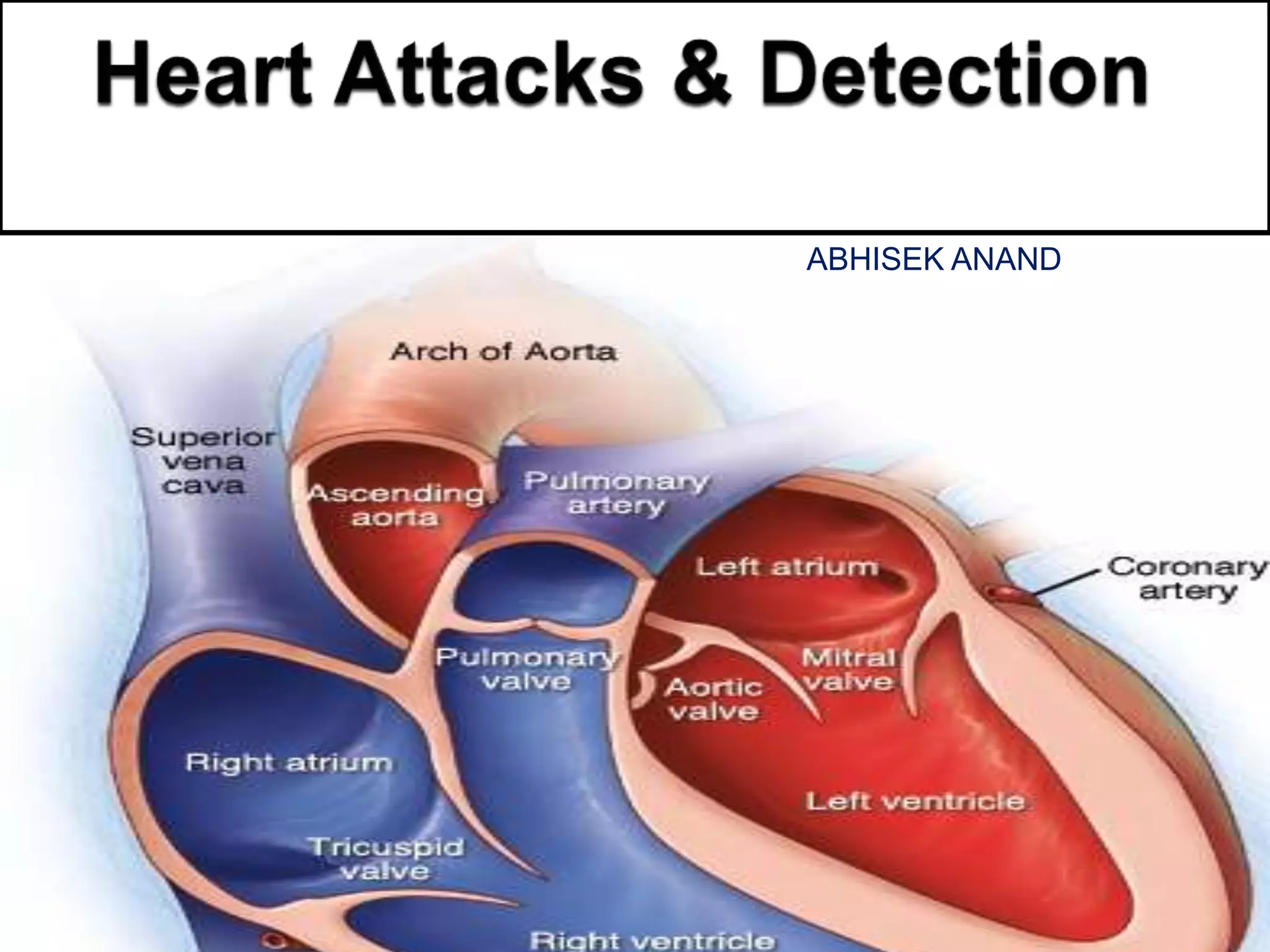
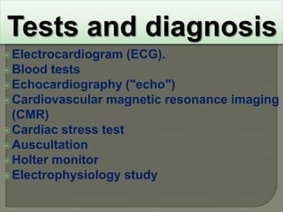
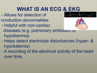
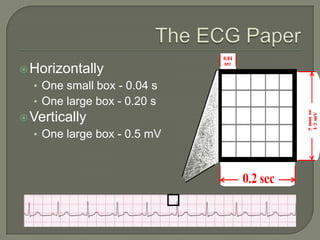
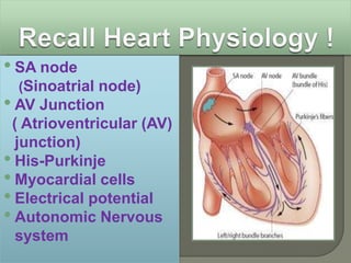

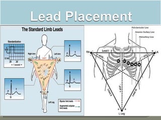
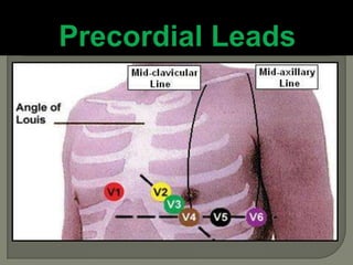
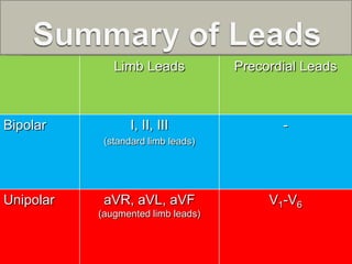

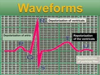
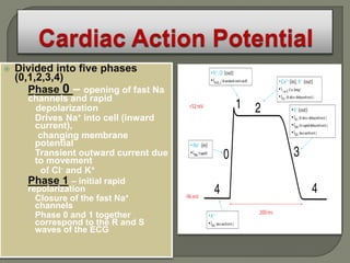
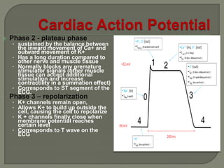
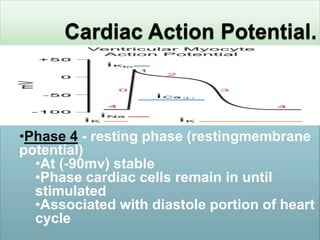
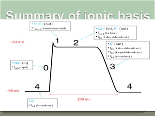
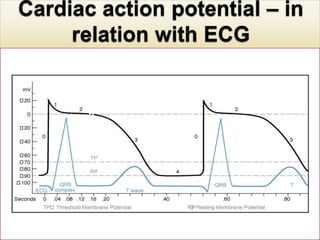
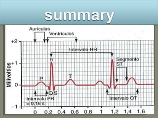
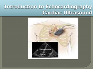
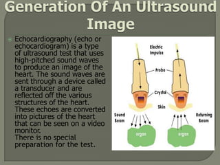
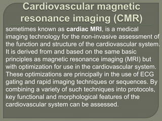
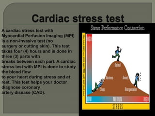

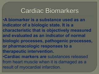


The document discusses cardiac markers, which are substances released when heart muscle is damaged during a myocardial infarction (heart attack). Some common cardiac markers measured in blood tests include troponin and CK-MB. Elevated levels of these markers can help diagnose a heart attack. The document also provides information on electrocardiograms (ECGs), echocardiograms, cardiac MRI, stress tests and other tests used to evaluate the heart.


