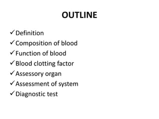
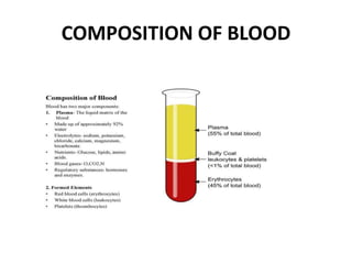
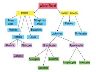
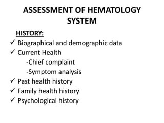
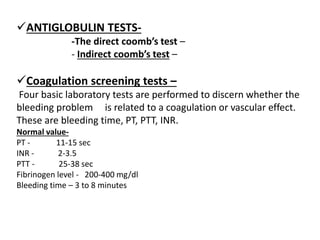
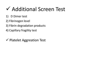
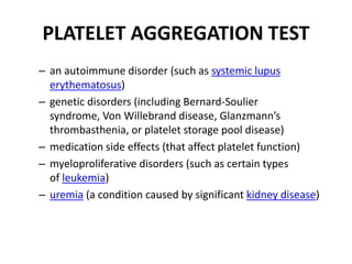
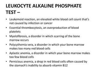
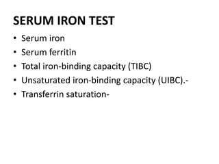
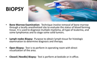
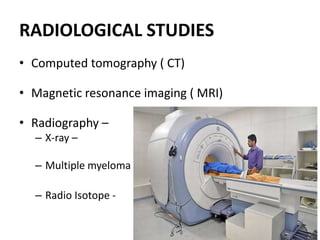
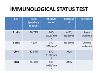
ASSESSMENT AND DIAGNOSTIC
TEST OF HEMATOLOGY SYSTEM
RAKESH KUMAR
M.Sc ( Previous)
RAKCON
25th OCT 2019
OUTLINE
Definition
Composition of blood
Function of blood
Blood clotting factor
Assessory organ
Assessment of system
Diagnostic test
INTRODUCTION
• The formation of blood cellular components-
occurs during embryonic development and
throughout adulthood to produce and replenish
the blood system. Furthermore hematopoietic
stem cells ( HSCs) can be used as a model system
for understanding tissue stem cells and their role
in ageing and oncogenesis. Hematologic
activities, such as red blood cell formation and
the clotting cascade, require a complex series of
events to allow good health andhomeostasis.
Without leukocytes to protect us, our bodies
could succumb to disease and infection
HEMATOPOIETIC SYSTEM
• Hematology is the study of blood and blood forming
tissue. This include the bone marrow, blood , spleen
and lymph system. A basic knowledge of hematology
is useful in clinical setting to evaluate the patient
ability to transport oxygen and carbon dioxide,
coagulation blood and combat infections.
Assessment of the hematology system is based on
the patient's health history , physical examination ,
and result of diagnostic studies .
COMPOSITION OF BLOOD
FUNCTION OF BLOOD
Transportation
Regulation
Protection
HEMATOPOIESIS
• Hematopoiesis is the formation of blood cellular
components. All cellular blood components are derived
from hematopoietic. In a healthy adult person,
approximately 1011–1012 new blood cells are produced
daily in order to maintain steady state levels in the
peripheral circulation.
• This stem cell is the “Mother Cell” or the originator of
all blood cells. It has the ability to self‐renew and
create progenitor stem cell lines. They are naturally
limited in number. By reviewing the chart, you can see
that all cells come from the stem cell. An attack on the
stem cell can theoretically affect all cells similarly.
Erythrocytes
Erythrocytes, or red blood cells (RBCs), originate from a
stem cell. Vitamin B12, folic acid, iron, and copper are
essential in the formation of erythrocytes. Erythropoietin
is a hormone released by the kidneys in response to
hypoxemia, which stimulates the bone marrow to produce
red blood cells. Typically, red blood cells live approximately
120 days. When the red blood cells become old and
damaged, the liver, spleen, and bone marrow cleanse
them from the blood. Increases or decreases in the red
blood cell count indicate an abnormality. Please note that
laboratory values given in this course are reference ranges
only, as values vary at different laboratories.
Normal RBC Range:
Males: 4.5 – 5.9 mil/dL
Females: 4.0 ‐ 5.2 mil/dL
Reticulocytes
The reticulocytes is immature erythrocyte. When
released from the bone marrow, red blood cells are
slightly immature and are known as reticulocytes.
Reticulocytes mature into red blood cells within a
few days. The number of reticulocytes in the blood
indicates the amount of bone marrow activity. Low
reticulocyte counts may be due to vitamin
deficiency, liver cirrhosis, or radiation therapy.
Normal Reticulocyte Range: 0.5‐2.5% of RBCs
Leukocytes
Leakocytes appear white when separated from blood . Like the
erythrocytes , leukocytes orignates from stem cells within bone marrow.
Leukocytes are typically classified as either:
Granular leukocytes
 Neutrophils
 Eosinophils
 Basophils
Non‐granular leukocytes
 Lymphocytes
 Monocytes
Normal Leukocyte Range: 4,000 ‐10,000 / uL
Platelets
Platelets are small, colorless cells that have a lifespan of seven
to ten days.
Blood Clotting: Platelets perform three major roles:
1) Decreasing the luminal size of damaged vessels to
decrease blood loss.
2) Forming blockages in injured vessels to decrease blood
loss.
3) Providing support accelerate blood coagulation through
molecules on the surface of the platelets.
Normal Platelet Range: 150,000 ‐ 450,000 /mm3
NORMAL CLOTTING MECHANISM:
• The process is important in minimizing blood loss when
various body structure are injured. Three components
contributing to normal clotting: vascular response, platelet
response and plasma clotting factors.
 Vascular response
 Platelet response
 Plasma clotting factor
ASSESSORY ORGAN
SPLEEN:
Functions:
Hematopoietic function
Filter Function
Immune Function
Storage Fuction
LYMPH SYSTEM
• The lymph system also returns excess interstitial fluid to the blood,
which is important in preventing the development of edema.
Lymph fluid is pale yellow interstitial fluid that has diffused through
lymphatic capillary walls. It circulates through a special vasculature,
much as blood moves through blood vessels. The formation of
lymph fluid increases when interstitial fluid increases, thereby
forcing more fluid into the lymph system.
• When too much interstitial fluid develops or when something
interferes with the reabsorption of lymph, lymphedema develops.
The lymphedema that may occur as a complication Of mastectomy
or lumpectomy with dissection of axillary nodes is often caused by
the obstruction of lymph flow from the removal of lymph nodes.
The lymphatic capillaries are thin-walled vessels that have an
irregular diameter. They are somewhat larger than blood capillaries
and do not contain valves. Lymphatic capillaries unite to form
lymphatic vessels that carry all lymph fluid to either the right
lymphatic duct or the thoracic duct. These large lymphatic ducts
drain into subclavian veins in the neck
LIVER
Function
 Involved in bile production
 Erythropoiesis (during fetal life and when bone
marrow production is insufficient
 Kupffer cells of the liver have reticuloendothelial
function as histiocytes; phagocytic activity and iron
storage.
 Synthesis of clotting factors, synthesis of
antithrombins
ASSESSMENT OF HEMATOLOGY
SYSTEM
HISTORY:
 Biographical and demographic data
 Current Health
-Chief complaint
-Symptom analysis
 Past health history
 Family health history
 Psychological history
HEAD TO TOE EXAMINATION
Skin
Eyes
Ears
Nose
Mouth
SYSTEM WISE ASSESSMENT
Respiratory
Cardio vascular system
Gastro intestinal tract
Genitourinary tract
Musculoskeletal system Nervous system
PHYSICAL EXAMINATION
• Inspection
• Palpation
• Percussion
• Auscultation
DIAGNOSTIC TEST
 HEMATOLOGICAL TEST
• Complete blood count –
- RBC count
- Hemoglobin
- Hematocrit
- RBC indices
- WBC count
- Platelet count.
Mean corpuscular volume (MCV)
Mean corpuscular hemoglobin (MCH)
Mean corpuscular hemoglobin concentration (MCHC)
• WBC count
• WBC differential
» Granulocytes
• Neutrophils - 55 – 70%
• Eosinophils - 1 – 4 %
• Basophils - 0.5 – 1.0 %
» Agranulocytes
• Lymphocytes - 20 – 40 %
• Monocytes - 2 – 8 %
• Reticulocyte Count
• Hemoglobin Electrophoresis
-- Hemoglobin F
-- Hemoglobin A
-- Hemoglobin C, D, E, M, and S
The normal levels of the types of
hemoglobin in adults are:
Type of hemoglobin Percentage
hemoglobin A 95% to 98%
hemoglobin A2 2% to 3%
hemoglobin F 1% to 2%
hemoglobin S 0%
hemoglobin C 0%
Perpheral Blood Smear
• Normocyte – cells of normal size and shape
• Normochromic – cells of normal color
• Anisocytes – vary from normal size
• Poikilocytes – abnormally shaped
• Microcyte – abnormally small (< 6 mm )
• Macrocyte – Abnormally large (> 9 mm )
• Hypochromic cells – pale appreance because of abnormally low HB
• Spherocytes – relatively small and round rather than biconcave
• Schistocytes- fragmented with bizarre shape
• Sickle shape – sickle shape due to presence of abnormal
hemoglobin (Hb S)
• Target cell – thin, with small amount in center
ANTIGLOBULIN TESTS-
-The direct coomb’s test –
- Indirect coomb’s test –
Coagulation screening tests –
Four basic laboratory tests are performed to discern whether the
bleeding problem is related to a coagulation or vascular effect.
These are bleeding time, PT, PTT, INR.
Normal value-
PT - 11-15 sec
INR - 2-3.5
PTT - 25-38 sec
Fibrinogen level - 200-400 mg/dl
Bleeding time – 3 to 8 minutes
 Additional Screen Test
1) D Dimer test
2) Fibrinogen level
3) Fibrin degradation products
4) Capillary fragility test
 Platelet Aggreation Test
PLATELET AGGREGATION TEST
– an autoimmune disorder (such as systemic lupus
erythematosus)
– genetic disorders (including Bernard-Soulier
syndrome, Von Willebrand disease, Glanzmann’s
thrombasthenia, or platelet storage pool disease)
– medication side effects (that affect platelet function)
– myeloproliferative disorders (such as certain types
of leukemia)
– uremia (a condition caused by significant kidney disease)
LEUKOCYTE ALKALINE PHOSPHATE
TEST –
• Leukemoid reaction, an elevated white blood cell count that’s
not caused by infection or cancer
• Essential thrombocytosis, an overproduction of blood
platelets
• Myelofibrosis, a disorder in which scarring of the bone
marrow occurs
• Polycythemia vera, a disorder in which your bone marrow
makes too many red blood cells
• Aplastic anemia, a disorder in which your bone marrow makes
too few blood cells
• Pernicious anemia, a drop in red blood cells often caused by
the stomach’s inability to absorb vitamin B12
SERUM IRON TEST
• Serum iron
• Serum ferritin
• Total iron-binding capacity (TIBC)
• Unsaturated iron-binding capacity (UIBC).-
• Transferrin saturation-
BIOPSY
• Bone Marrow Examination: Technique involve removal of bone marrow
through a locally anesthetized site to evaluate the status of blood forming
tissue. It is used to diagnose multiple myeloma, all type of leukemia, and
some lymphomas and to stage some solid tumors.
• Lymph nodes Biopsy: Purpose to obtain lymph tissue for histologic
examination to determine diagnosis and therapy.
• Open Biopsy: Test is to perform in operating room with direct
visualization of the area.
• Closed ( Needle) Biopsy: Test is perform at bedside or in office.
RADIOLOGICAL STUDIES
• Computed tomography ( CT)
• Magnetic resonance imaging ( MRI)
• Radiography –
– X-ray –
– Multiple myeloma
– Radio Isotope -
IMMUNOLOGICAL STATUS TEST
Cell Total
lymphocy
te count
Absolute
count
decrease
d
increased
T cells 56-77% 860-
1800/mm
3
AIDS,
leukemia
Acute
leukemia
B cells 7-17% 140-
370/mm3
leukemia Chronic
leukemia
CD 4 32-54% 530-
1190/mm
3
AIDS
CD 8 24-37% 430-
1060/mm
3
AIDS
IMMUNOGLOBULIN SUBTYPES
Immunoglobulin normal range in mg/dl
Ig G 550-1990
Ig M 45-145
Ig A 70-310
Ig E 0.01 – 0.04
Ig D 0- 8
• Complement assays –
Disorder that are related to a lack of normal levels of
complement components may be detected by the level of the
total serum complement (CH50) or may requiring measuring
levels of specific complement components such as c3 and c4.
SKIN TEST
• Patch test
• Scratch test
• Delayed type hypersensitivity testing
SUMMARY
• The formation of blood cellular components- occurs during
embryonic development and throughout adulthood to
produce and replenish the blood system. A basic knowledge
of hematology is useful in clinical setting to evaluate the
patient ability to transport oxygen and carbon dioxide,
coagulation blood and combat infections. we discuss the
composition and function of blood. This stem cell is the
“Mother Cell” or the originator of all blood cells. It has the
ability to self‐renew and create progenitor stem cell lines.
They are naturally limited in number. The CBC includes the
RBC count, hemoglobin, Hematocrit, RBC indices, WBC count
with and without differential and platelet count.
•
Haematological assessment

Haematological assessment

  • 2.
    ASSESSMENT AND DIAGNOSTIC TESTOF HEMATOLOGY SYSTEM RAKESH KUMAR M.Sc ( Previous) RAKCON 25th OCT 2019
  • 3.
    OUTLINE Definition Composition of blood Functionof blood Blood clotting factor Assessory organ Assessment of system Diagnostic test
  • 4.
    INTRODUCTION • The formationof blood cellular components- occurs during embryonic development and throughout adulthood to produce and replenish the blood system. Furthermore hematopoietic stem cells ( HSCs) can be used as a model system for understanding tissue stem cells and their role in ageing and oncogenesis. Hematologic activities, such as red blood cell formation and the clotting cascade, require a complex series of events to allow good health andhomeostasis. Without leukocytes to protect us, our bodies could succumb to disease and infection
  • 5.
    HEMATOPOIETIC SYSTEM • Hematologyis the study of blood and blood forming tissue. This include the bone marrow, blood , spleen and lymph system. A basic knowledge of hematology is useful in clinical setting to evaluate the patient ability to transport oxygen and carbon dioxide, coagulation blood and combat infections. Assessment of the hematology system is based on the patient's health history , physical examination , and result of diagnostic studies .
  • 6.
  • 8.
  • 9.
    HEMATOPOIESIS • Hematopoiesis isthe formation of blood cellular components. All cellular blood components are derived from hematopoietic. In a healthy adult person, approximately 1011–1012 new blood cells are produced daily in order to maintain steady state levels in the peripheral circulation. • This stem cell is the “Mother Cell” or the originator of all blood cells. It has the ability to self‐renew and create progenitor stem cell lines. They are naturally limited in number. By reviewing the chart, you can see that all cells come from the stem cell. An attack on the stem cell can theoretically affect all cells similarly.
  • 11.
    Erythrocytes Erythrocytes, or redblood cells (RBCs), originate from a stem cell. Vitamin B12, folic acid, iron, and copper are essential in the formation of erythrocytes. Erythropoietin is a hormone released by the kidneys in response to hypoxemia, which stimulates the bone marrow to produce red blood cells. Typically, red blood cells live approximately 120 days. When the red blood cells become old and damaged, the liver, spleen, and bone marrow cleanse them from the blood. Increases or decreases in the red blood cell count indicate an abnormality. Please note that laboratory values given in this course are reference ranges only, as values vary at different laboratories. Normal RBC Range: Males: 4.5 – 5.9 mil/dL Females: 4.0 ‐ 5.2 mil/dL
  • 12.
    Reticulocytes The reticulocytes isimmature erythrocyte. When released from the bone marrow, red blood cells are slightly immature and are known as reticulocytes. Reticulocytes mature into red blood cells within a few days. The number of reticulocytes in the blood indicates the amount of bone marrow activity. Low reticulocyte counts may be due to vitamin deficiency, liver cirrhosis, or radiation therapy. Normal Reticulocyte Range: 0.5‐2.5% of RBCs
  • 13.
    Leukocytes Leakocytes appear whitewhen separated from blood . Like the erythrocytes , leukocytes orignates from stem cells within bone marrow. Leukocytes are typically classified as either: Granular leukocytes  Neutrophils  Eosinophils  Basophils Non‐granular leukocytes  Lymphocytes  Monocytes Normal Leukocyte Range: 4,000 ‐10,000 / uL
  • 14.
    Platelets Platelets are small,colorless cells that have a lifespan of seven to ten days. Blood Clotting: Platelets perform three major roles: 1) Decreasing the luminal size of damaged vessels to decrease blood loss. 2) Forming blockages in injured vessels to decrease blood loss. 3) Providing support accelerate blood coagulation through molecules on the surface of the platelets. Normal Platelet Range: 150,000 ‐ 450,000 /mm3
  • 16.
    NORMAL CLOTTING MECHANISM: •The process is important in minimizing blood loss when various body structure are injured. Three components contributing to normal clotting: vascular response, platelet response and plasma clotting factors.  Vascular response  Platelet response  Plasma clotting factor
  • 17.
    ASSESSORY ORGAN SPLEEN: Functions: Hematopoietic function FilterFunction Immune Function Storage Fuction
  • 18.
    LYMPH SYSTEM • Thelymph system also returns excess interstitial fluid to the blood, which is important in preventing the development of edema. Lymph fluid is pale yellow interstitial fluid that has diffused through lymphatic capillary walls. It circulates through a special vasculature, much as blood moves through blood vessels. The formation of lymph fluid increases when interstitial fluid increases, thereby forcing more fluid into the lymph system. • When too much interstitial fluid develops or when something interferes with the reabsorption of lymph, lymphedema develops. The lymphedema that may occur as a complication Of mastectomy or lumpectomy with dissection of axillary nodes is often caused by the obstruction of lymph flow from the removal of lymph nodes. The lymphatic capillaries are thin-walled vessels that have an irregular diameter. They are somewhat larger than blood capillaries and do not contain valves. Lymphatic capillaries unite to form lymphatic vessels that carry all lymph fluid to either the right lymphatic duct or the thoracic duct. These large lymphatic ducts drain into subclavian veins in the neck
  • 19.
    LIVER Function  Involved inbile production  Erythropoiesis (during fetal life and when bone marrow production is insufficient  Kupffer cells of the liver have reticuloendothelial function as histiocytes; phagocytic activity and iron storage.  Synthesis of clotting factors, synthesis of antithrombins
  • 20.
    ASSESSMENT OF HEMATOLOGY SYSTEM HISTORY: Biographical and demographic data  Current Health -Chief complaint -Symptom analysis  Past health history  Family health history  Psychological history
  • 21.
    HEAD TO TOEEXAMINATION Skin Eyes Ears Nose Mouth
  • 22.
    SYSTEM WISE ASSESSMENT Respiratory Cardiovascular system Gastro intestinal tract Genitourinary tract Musculoskeletal system Nervous system
  • 23.
    PHYSICAL EXAMINATION • Inspection •Palpation • Percussion • Auscultation
  • 24.
    DIAGNOSTIC TEST  HEMATOLOGICALTEST • Complete blood count – - RBC count - Hemoglobin - Hematocrit - RBC indices - WBC count - Platelet count. Mean corpuscular volume (MCV) Mean corpuscular hemoglobin (MCH) Mean corpuscular hemoglobin concentration (MCHC)
  • 25.
    • WBC count •WBC differential » Granulocytes • Neutrophils - 55 – 70% • Eosinophils - 1 – 4 % • Basophils - 0.5 – 1.0 % » Agranulocytes • Lymphocytes - 20 – 40 % • Monocytes - 2 – 8 % • Reticulocyte Count • Hemoglobin Electrophoresis -- Hemoglobin F -- Hemoglobin A -- Hemoglobin C, D, E, M, and S
  • 26.
    The normal levelsof the types of hemoglobin in adults are: Type of hemoglobin Percentage hemoglobin A 95% to 98% hemoglobin A2 2% to 3% hemoglobin F 1% to 2% hemoglobin S 0% hemoglobin C 0%
  • 27.
    Perpheral Blood Smear •Normocyte – cells of normal size and shape • Normochromic – cells of normal color • Anisocytes – vary from normal size • Poikilocytes – abnormally shaped • Microcyte – abnormally small (< 6 mm ) • Macrocyte – Abnormally large (> 9 mm ) • Hypochromic cells – pale appreance because of abnormally low HB • Spherocytes – relatively small and round rather than biconcave • Schistocytes- fragmented with bizarre shape • Sickle shape – sickle shape due to presence of abnormal hemoglobin (Hb S) • Target cell – thin, with small amount in center
  • 28.
    ANTIGLOBULIN TESTS- -The directcoomb’s test – - Indirect coomb’s test – Coagulation screening tests – Four basic laboratory tests are performed to discern whether the bleeding problem is related to a coagulation or vascular effect. These are bleeding time, PT, PTT, INR. Normal value- PT - 11-15 sec INR - 2-3.5 PTT - 25-38 sec Fibrinogen level - 200-400 mg/dl Bleeding time – 3 to 8 minutes
  • 29.
     Additional ScreenTest 1) D Dimer test 2) Fibrinogen level 3) Fibrin degradation products 4) Capillary fragility test  Platelet Aggreation Test
  • 30.
    PLATELET AGGREGATION TEST –an autoimmune disorder (such as systemic lupus erythematosus) – genetic disorders (including Bernard-Soulier syndrome, Von Willebrand disease, Glanzmann’s thrombasthenia, or platelet storage pool disease) – medication side effects (that affect platelet function) – myeloproliferative disorders (such as certain types of leukemia) – uremia (a condition caused by significant kidney disease)
  • 31.
    LEUKOCYTE ALKALINE PHOSPHATE TEST– • Leukemoid reaction, an elevated white blood cell count that’s not caused by infection or cancer • Essential thrombocytosis, an overproduction of blood platelets • Myelofibrosis, a disorder in which scarring of the bone marrow occurs • Polycythemia vera, a disorder in which your bone marrow makes too many red blood cells • Aplastic anemia, a disorder in which your bone marrow makes too few blood cells • Pernicious anemia, a drop in red blood cells often caused by the stomach’s inability to absorb vitamin B12
  • 32.
    SERUM IRON TEST •Serum iron • Serum ferritin • Total iron-binding capacity (TIBC) • Unsaturated iron-binding capacity (UIBC).- • Transferrin saturation-
  • 33.
    BIOPSY • Bone MarrowExamination: Technique involve removal of bone marrow through a locally anesthetized site to evaluate the status of blood forming tissue. It is used to diagnose multiple myeloma, all type of leukemia, and some lymphomas and to stage some solid tumors. • Lymph nodes Biopsy: Purpose to obtain lymph tissue for histologic examination to determine diagnosis and therapy. • Open Biopsy: Test is to perform in operating room with direct visualization of the area. • Closed ( Needle) Biopsy: Test is perform at bedside or in office.
  • 34.
    RADIOLOGICAL STUDIES • Computedtomography ( CT) • Magnetic resonance imaging ( MRI) • Radiography – – X-ray – – Multiple myeloma – Radio Isotope -
  • 35.
    IMMUNOLOGICAL STATUS TEST CellTotal lymphocy te count Absolute count decrease d increased T cells 56-77% 860- 1800/mm 3 AIDS, leukemia Acute leukemia B cells 7-17% 140- 370/mm3 leukemia Chronic leukemia CD 4 32-54% 530- 1190/mm 3 AIDS CD 8 24-37% 430- 1060/mm 3 AIDS
  • 36.
    IMMUNOGLOBULIN SUBTYPES Immunoglobulin normalrange in mg/dl Ig G 550-1990 Ig M 45-145 Ig A 70-310 Ig E 0.01 – 0.04 Ig D 0- 8
  • 37.
    • Complement assays– Disorder that are related to a lack of normal levels of complement components may be detected by the level of the total serum complement (CH50) or may requiring measuring levels of specific complement components such as c3 and c4.
  • 38.
    SKIN TEST • Patchtest • Scratch test • Delayed type hypersensitivity testing
  • 39.
    SUMMARY • The formationof blood cellular components- occurs during embryonic development and throughout adulthood to produce and replenish the blood system. A basic knowledge of hematology is useful in clinical setting to evaluate the patient ability to transport oxygen and carbon dioxide, coagulation blood and combat infections. we discuss the composition and function of blood. This stem cell is the “Mother Cell” or the originator of all blood cells. It has the ability to self‐renew and create progenitor stem cell lines. They are naturally limited in number. The CBC includes the RBC count, hemoglobin, Hematocrit, RBC indices, WBC count with and without differential and platelet count. •