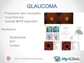
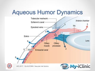
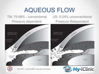
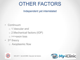
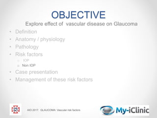
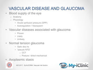
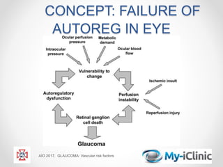
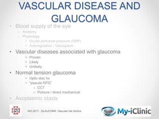
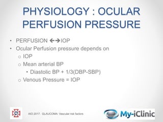
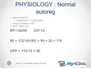
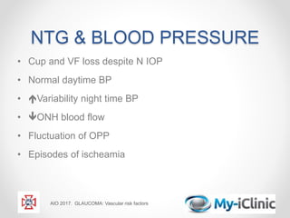
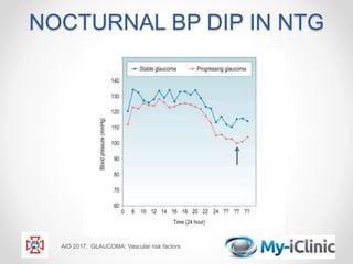
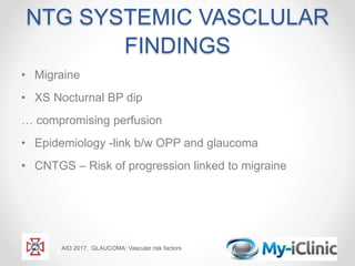
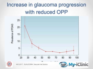
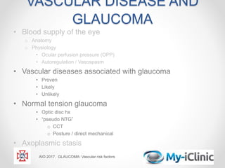
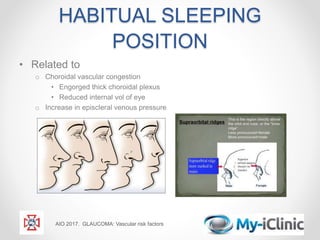
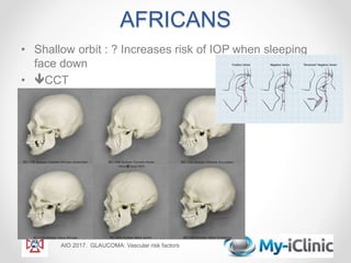
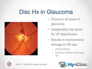
GLAUCOMA
Vascular Risk Factors
in Aetiology & Progression
Ms Bola Odufuwa
AIO 2017. GLAUCOMA: Vascular risk factors
OBJECTIVE
Explore effect of vascular disease on Glaucoma
• Definition
• Anatomy / physiology
• Pathology
• Risk factors
o IOP
o Non IOP
• Case presentation
• Management of these risk factors
AIO 2017. GLAUCOMA: Vascular risk factors
INTERACTIVE SELF
ASSESMENT
Systemic hypertension is a risk factor for glaucoma?
AIO 2017. GLAUCOMA: Vascular risk factors
•YES •NO
GLAUCOMA
• Progressive optic neuropathy
• Visual field loss
• Typically IOP dependent
Mechanism
Multifactorial
BUT
Unclear
AIO 2017. GLAUCOMA: Vascular risk factors
GLAUCOMA – unclear
variables
• Extensively studied Includes
o Optic nerve head structure
o Ocular wall properties
• Including CCT
o Systemic blood flow alterations
o Diabetes
o Refractive error
o Posture
o Migraine
o Etc….etc...etc
AIO 2017. GLAUCOMA: Vascular risk factors
OBJECTIVE
Explore effect of vascular disease on Glaucoma
• Definition
• Anatomy / physiology
• Pathology
• Risk factors
o IOP
o Non IOP
• Case presentation
• Management of these risk factors
AIO 2017. GLAUCOMA: Vascular risk factors
SIMPLISTIC
AIO 2017. GLAUCOMA: Vascular risk factors
Aqueous Humor Dynamics
AIO 2017. GLAUCOMA: Vascular risk factors
AQUEOUS FLOW
TM: 75-99% - conventional
Pressure dependent
US: 0-24% unconventional
Pressure INdependent
AIO 2017. GLAUCOMA: Vascular risk factors
ITS NOT ALL IOP
Evidently
o IOP  =  risk of damage by 2/3
o Improves prognosis
o Does NOT stop progression
Existence of OHT and NTG
o Further proof of other factors
• Pathogenesis
• Rendering eyes more sensitive to IOP
AIO 2017. GLAUCOMA: Vascular risk factors
ITS NOT ALL IOP
.. So what else
NTG and POAG
– identical clinical appearance
• Arcuate field loss
• Matches RNFL damage
• 2ndry change
o PP Heamorrhage ??
o Posterior deviation of lamina cribrosa
o Optic nerve head pit
AIO 2017. GLAUCOMA: Vascular risk factors
OTHER FACTORS
Independent yet interrelated
• Continuum
o 1.Vascular and
o 2.Mechanical factors (IOP)
o >>>axon loss
• 3rd theory
o Axoplasmic flow
AIO 2017. GLAUCOMA: Vascular risk factors
Conventional theories
o PRESSURE (Muller)
GON is a direct consequence of elevated IOP damaging lamina cribrosa and neural
axons
“Pressure plays larger role in POAG than NTG”
o VASCULAR (von Jaeger)
GON consequence of insufficient blood supply to ONH including inc IOP as a cause
of reduced OBF
AIO 2017. GLAUCOMA: Vascular risk factors
o AXOPLASMIC transfer blockage …
RGC death by apoptosis..?
Obstruciton partyly 2o to IOP
depended blood flow changes
OBJECTIVE
Explore effect of vascular disease on Glaucoma
• Definition
• Anatomy / physiology
• Pathology
• Risk factors
o IOP
o Non IOP
• Case presentation
• Management of these risk factors
AIO 2017. GLAUCOMA: Vascular risk factors
VASCULAR DISEASE AND GLAUCOMA
• Blood supply of the eye
o Anatomy
o Physiology
• Ocular perfusion pressure (OPP)
• Autoregulation / Vasospasm
• Vascular diseases associated with glaucoma
• Proven
• Likely
• Unlikely
• Normal tension glaucoma
• Optic disc hx
• “pseudo NTG”
o CCT
o Posture / direct mechanical
• Axoplasmic stasis
AIO 2017. GLAUCOMA: Vascular risk factors
BLOOD SUPPLY OF THE EYE
AIO 2017. GLAUCOMA: Vascular risk factors
GLAUCOMA: OCULAR
VASCULAR FINDINGS
• Impaired ONH perfusion
• Impaired periph retinal perfusion
• Optic disc Hx
• Local constriction retinal arteries
• Conj peri-limbal aneurysm
• 2X Crescent Choroidal PPA
AIO 2017. GLAUCOMA: Vascular risk factors
 OCULAR BLOOD FLOW
IN GLAUCOMA
•  Local resistance
o ?Structural
o ?Functional
•  Ocular perfusion pressure
•  Blood viscosity
AIO 2017. GLAUCOMA: Vascular risk factors
 Local resistance
?Structural
•  OCULAR BLOOD FLOW
IN GLAUCOMA
• Atherosclerosis
• HBP
• Diabetes
• Obesity
• Smoking
• Carotid stenosis
AIO 2017. GLAUCOMA: Vascular risk factors
 Altered blood flow
?Functional
• AUTOREGULATION
o Physiological response to
• OPP changes
• Tissue metabolic demands
DRIVEN BY
o Intrinsic capacity of organ to maintain blood flow
o Maintain nutrient supply
AIO 2017. GLAUCOMA: Vascular risk factors
CONCEPT: FAILURE OF
AUTOREG IN EYE
AIO 2017. GLAUCOMA: Vascular risk factors
VASCULAR DISEASE AND
GLAUCOMA
• Blood supply of the eye
o Anatomy
o Physiology
• Ocular perfusion pressure (OPP)
• Autoregulation / Vasospasm
• Vascular diseases associated with glaucoma
• Proven
• Likely
• Unlikely
• Normal tension glaucoma
• Optic disc hx
• “pseudo NTG”
o CCT
o Posture / direct mechanical
• Axoplasmic stasis
AIO 2017. GLAUCOMA: Vascular risk factors
PHYSIOLOGY : OCULAR
PERFUSION PRESSURE
• PERFUSION IOP
• Ocular Perfusion pressure depends on
o IOP
o Mean arterial BP
• Diastolic BP + 1/3(DBP-SBP)
o Venous Pressure = IOP
AIO 2017. GLAUCOMA: Vascular risk factors
PHYSIOLOGY : Normal
autoreg
o Mean arterial BP
• Diastolic BP + 1/3(DBP-SBP)
o Venous Pressure = IOP
o OPP = MAP - OP
BP:140/90 IOP 14
90 + 1/3(140-90) = 90 + 20 = 110
OPP = 110-14 = 96
AIO 2017. GLAUCOMA: Vascular risk factors
PHYSIOLOGY: Altered
Autoregulation
o Mean arterial BP
• Diastolic BP + 1/3(DBP-SBP)
o Venous Pressure = IOP
o OPP = MAP - OP
BP:80/60; IOP 20
60 + 1/3(80-60) = 60 + 7 = 67
OPP = 67-20 = 47
AIO 2017. GLAUCOMA: Vascular risk factors
NTG & BLOOD PRESSURE
• Cup and VF loss despite N IOP
• Normal daytime BP
• Variability night time BP
• ONH blood flow
• Fluctuation of OPP
• Episodes of ischeamia
AIO 2017. GLAUCOMA: Vascular risk factors
NOCTURNAL BP DIP IN NTG
AIO 2017. GLAUCOMA: Vascular risk factors
NTG SYSTEMIC VASCLULAR
FINDINGS
• Migraine
• XS Nocturnal BP dip
… compromising perfusion
• Epidemiology -link b/w OPP and glaucoma
• CNTGS – Risk of progression linked to migraine
AIO 2017. GLAUCOMA: Vascular risk factors
Increase in glaucoma progression
with reduced OPP
AIO 2017. GLAUCOMA: Vascular risk factors
VASCULAR DISEASE AND
GLAUCOMA
• Blood supply of the eye
o Anatomy
o Physiology
• Ocular perfusion pressure (OPP)
• Autoregulation / Vasospasm
• Vascular diseases associated with glaucoma
• Proven
• Likely
• Unlikely
• Normal tension glaucoma
• Optic disc hx
• “pseudo NTG”
o CCT
o Posture / direct mechanical
• Axoplasmic stasis
AIO 2017. GLAUCOMA: Vascular risk factors
DIABETES SYSTEMIC
EFFECT
•  Anomaly of OBF
•  Vasc leakage
•  Blood viscosity
• Capillary sheathing
• Capillary non perfusion
• Tissue oxygenation
AIO 2017. GLAUCOMA: Vascular risk factors
DIABETES OCULAR EFFECT
• Altered dynamics
• Aqueous flare
• Iris NV
AIO 2017. GLAUCOMA: Vascular risk factors
INCREASED BLOOD
VISCOSITY
AIO 2017. GLAUCOMA: Vascular risk factors
OTHER CONDITIONS
• Vasospasm /autoreg /NTG
• Nocturnal hypotension
• Atherosclerosis /cholesterol
• Diabetes
• Habitual sleeping position
o Face down
• Axoplasmic flow
• Autoimmunity
• Alzheimers
• “Combination” e.g Africans
AIO 2017. GLAUCOMA: Vascular risk factors
POSITION
• IOP higher
o in recumbent than upright position
o Lower Lateral decubitus position
o Up tp 4mmHg in normals
o Further 2mmHg in OHT
o Avg 40mmhg! Prone spinal surgery
o Acta Ophthalmol. 2015 Nov;93(7) Yoo C et al
o Anesthesiology2001; 95: 1351–1355.Cheng et al
AIO 2017. GLAUCOMA: Vascular risk factors
HABITUAL SLEEPING
POSITION
• Related to
o Choroidal vascular congestion
• Engorged thick choroidal plexus
• Reduced internal vol of eye
o Increase in episcleral venous pressure
AIO 2017. GLAUCOMA: Vascular risk factors
AFRICANS
• Shallow orbit : ? Increases risk of IOP when sleeping
face down
• CCT
AIO 2017. GLAUCOMA: Vascular risk factors
AUTOIMMUNITY
• Higher serum conc. AutoAb in GON
• NTG greater
o Antibodies to heat shock protein
o Fodrin
o ?markers for injury rather than causative
• FODRIN also greater in
o ALZHEIMERS
o vascular / neurodegenerative condition
AIO 2017. GLAUCOMA: Vascular risk factors
IT MAY NOT BE GLAUCOMA!
ALWAYS CONSIDER DIFFERENTILS
AIO 2017. GLAUCOMA: Vascular risk factors
DIFFERANTIAL Dx – disc
pallor
• NAION
• AION
• MOD – HIGH MYOPIA
• r/o intermittent <le closure
• Burnt out CRAO ( RNFL defect)
AIO 2017. GLAUCOMA: Vascular risk factors
Disc Hx in Glaucoma
• Occurs in all types of
glaucoma
• Independent risk factor
for VF deterioration
• Results in mechamnical
damage to OD esp
o Lamina cribrosa
o Trauma to capillaries that supply
OD
AIO 2017. GLAUCOMA: Vascular risk factors
DIFFERENTIAL Dx – disc hx
• Splinter hx
• PVD
• Systemic hypertension
• Diabetes
• Collaterals
AIO 2017. GLAUCOMA: Vascular risk factors
‘3rd theory’ RETROGRADE
AXOPLASMIC FLOW
• Essential for survival of RGC
• ??IOP dependent blood flow changes
• >Obstruction of flow
• Interruption >> RGC Death
AIO 2017. GLAUCOMA: Vascular risk factors
PRINCIPLES OF Rx
• 30%  in IOP irrespective of mech
o Medical
o AVOID :
non selective adrenergic ( vasoconstrictive)
*PM* use of Β blocker (nocturnal hypotension)
o Neuroprotection
o α 2 selective ( Brimodidine )
o NMDA, Memantine
o Protection from apoptosis
o Laser
o Surgical
AIO 2017. GLAUCOMA: Vascular risk factors
PRINCIPLES OF Rx II
• Review medicaiton
• Check BP
o Day
o Night
o 24 hour monitoring
• Habitual sleeping posture
• Pseudo NTG
o CCT
• Consider differentials
AIO 2017. GLAUCOMA: Vascular risk factors
CASE PRESENTATION
• 72 yo
• Male
• Nigerian
• Accident prone
• Gen health
o Various allergies
o Antihypertensive
o Antihistamine
AIO 2017. GLAUCOMA: Vascular risk factors
iop 19mmHg
AIO 2017. GLAUCOMA: Vascular risk factors
IOP 14-
16mmHg
BP 120/70
Plan: surgery
AIO 2017. GLAUCOMA: Vascular risk factors
Further investigations
• MRI scan
• Refer to Cardiology
o 24 hour BP monitoring
• Medication review
OUTCOME
• Nocturnal hypotension (on antihypertensives)
• Steroid for nasal congestion
AIO 2017. GLAUCOMA: Vascular risk factors
Management
• Stopped antihypertensive
• Non steroidal for allergic rhinitis
• OUTCOME – no progression on VF and OCT at last
visit!
AIO 2017. GLAUCOMA: Vascular risk factors
SUMMARY
Clinical risk factors
• NOT TREATABLE
• Advancing age – most important
• Refractive error
• Optic disc size/structure?
• TREATABLE
• IOP
• Vascular factors
AIO 2017. GLAUCOMA: Vascular risk factors
SUMMARY
Vascular risk factors
• Hypotension
• Vasospasm/Migraine/Raynauds
• Cigarette smoking
• Hypertension
• Diabetes
• Hypercholesterolaemia
• Atherosclerosis
• Sticky red cells
o Vasculitis
o Hypercoagulability status
• Associated with vascular dementia
• Sleep posture
• ?Shallow orbit
AIO 2017. GLAUCOMA: Vascular risk factors
THANK YOU!
AIO 2017. GLAUCOMA: Vascular risk factors
?QUESTIONS
There is a higher incidence of migraine in patients with
Normal tension glaucoma?
AIO 2017. GLAUCOMA: Vascular risk factors
• YE
S
• NO

Glaucoma vascular risk factors

  • 1.
    GLAUCOMA Vascular Risk Factors inAetiology & Progression Ms Bola Odufuwa AIO 2017. GLAUCOMA: Vascular risk factors
  • 2.
    OBJECTIVE Explore effect ofvascular disease on Glaucoma • Definition • Anatomy / physiology • Pathology • Risk factors o IOP o Non IOP • Case presentation • Management of these risk factors AIO 2017. GLAUCOMA: Vascular risk factors
  • 3.
    INTERACTIVE SELF ASSESMENT Systemic hypertensionis a risk factor for glaucoma? AIO 2017. GLAUCOMA: Vascular risk factors •YES •NO
  • 4.
    GLAUCOMA • Progressive opticneuropathy • Visual field loss • Typically IOP dependent Mechanism Multifactorial BUT Unclear AIO 2017. GLAUCOMA: Vascular risk factors
  • 5.
    GLAUCOMA – unclear variables •Extensively studied Includes o Optic nerve head structure o Ocular wall properties • Including CCT o Systemic blood flow alterations o Diabetes o Refractive error o Posture o Migraine o Etc….etc...etc AIO 2017. GLAUCOMA: Vascular risk factors
  • 6.
    OBJECTIVE Explore effect ofvascular disease on Glaucoma • Definition • Anatomy / physiology • Pathology • Risk factors o IOP o Non IOP • Case presentation • Management of these risk factors AIO 2017. GLAUCOMA: Vascular risk factors
  • 7.
    SIMPLISTIC AIO 2017. GLAUCOMA:Vascular risk factors
  • 8.
    Aqueous Humor Dynamics AIO2017. GLAUCOMA: Vascular risk factors
  • 9.
    AQUEOUS FLOW TM: 75-99%- conventional Pressure dependent US: 0-24% unconventional Pressure INdependent AIO 2017. GLAUCOMA: Vascular risk factors
  • 10.
    ITS NOT ALLIOP Evidently o IOP  =  risk of damage by 2/3 o Improves prognosis o Does NOT stop progression Existence of OHT and NTG o Further proof of other factors • Pathogenesis • Rendering eyes more sensitive to IOP AIO 2017. GLAUCOMA: Vascular risk factors
  • 11.
    ITS NOT ALLIOP .. So what else NTG and POAG – identical clinical appearance • Arcuate field loss • Matches RNFL damage • 2ndry change o PP Heamorrhage ?? o Posterior deviation of lamina cribrosa o Optic nerve head pit AIO 2017. GLAUCOMA: Vascular risk factors
  • 12.
    OTHER FACTORS Independent yetinterrelated • Continuum o 1.Vascular and o 2.Mechanical factors (IOP) o >>>axon loss • 3rd theory o Axoplasmic flow AIO 2017. GLAUCOMA: Vascular risk factors
  • 13.
    Conventional theories o PRESSURE(Muller) GON is a direct consequence of elevated IOP damaging lamina cribrosa and neural axons “Pressure plays larger role in POAG than NTG” o VASCULAR (von Jaeger) GON consequence of insufficient blood supply to ONH including inc IOP as a cause of reduced OBF AIO 2017. GLAUCOMA: Vascular risk factors o AXOPLASMIC transfer blockage … RGC death by apoptosis..? Obstruciton partyly 2o to IOP depended blood flow changes
  • 14.
    OBJECTIVE Explore effect ofvascular disease on Glaucoma • Definition • Anatomy / physiology • Pathology • Risk factors o IOP o Non IOP • Case presentation • Management of these risk factors AIO 2017. GLAUCOMA: Vascular risk factors
  • 15.
    VASCULAR DISEASE ANDGLAUCOMA • Blood supply of the eye o Anatomy o Physiology • Ocular perfusion pressure (OPP) • Autoregulation / Vasospasm • Vascular diseases associated with glaucoma • Proven • Likely • Unlikely • Normal tension glaucoma • Optic disc hx • “pseudo NTG” o CCT o Posture / direct mechanical • Axoplasmic stasis AIO 2017. GLAUCOMA: Vascular risk factors
  • 16.
    BLOOD SUPPLY OFTHE EYE AIO 2017. GLAUCOMA: Vascular risk factors
  • 17.
    GLAUCOMA: OCULAR VASCULAR FINDINGS •Impaired ONH perfusion • Impaired periph retinal perfusion • Optic disc Hx • Local constriction retinal arteries • Conj peri-limbal aneurysm • 2X Crescent Choroidal PPA AIO 2017. GLAUCOMA: Vascular risk factors
  • 18.
     OCULAR BLOODFLOW IN GLAUCOMA •  Local resistance o ?Structural o ?Functional •  Ocular perfusion pressure •  Blood viscosity AIO 2017. GLAUCOMA: Vascular risk factors
  • 19.
     Local resistance ?Structural • OCULAR BLOOD FLOW IN GLAUCOMA • Atherosclerosis • HBP • Diabetes • Obesity • Smoking • Carotid stenosis AIO 2017. GLAUCOMA: Vascular risk factors
  • 20.
     Altered bloodflow ?Functional • AUTOREGULATION o Physiological response to • OPP changes • Tissue metabolic demands DRIVEN BY o Intrinsic capacity of organ to maintain blood flow o Maintain nutrient supply AIO 2017. GLAUCOMA: Vascular risk factors
  • 21.
    CONCEPT: FAILURE OF AUTOREGIN EYE AIO 2017. GLAUCOMA: Vascular risk factors
  • 22.
    VASCULAR DISEASE AND GLAUCOMA •Blood supply of the eye o Anatomy o Physiology • Ocular perfusion pressure (OPP) • Autoregulation / Vasospasm • Vascular diseases associated with glaucoma • Proven • Likely • Unlikely • Normal tension glaucoma • Optic disc hx • “pseudo NTG” o CCT o Posture / direct mechanical • Axoplasmic stasis AIO 2017. GLAUCOMA: Vascular risk factors
  • 23.
    PHYSIOLOGY : OCULAR PERFUSIONPRESSURE • PERFUSION IOP • Ocular Perfusion pressure depends on o IOP o Mean arterial BP • Diastolic BP + 1/3(DBP-SBP) o Venous Pressure = IOP AIO 2017. GLAUCOMA: Vascular risk factors
  • 24.
    PHYSIOLOGY : Normal autoreg oMean arterial BP • Diastolic BP + 1/3(DBP-SBP) o Venous Pressure = IOP o OPP = MAP - OP BP:140/90 IOP 14 90 + 1/3(140-90) = 90 + 20 = 110 OPP = 110-14 = 96 AIO 2017. GLAUCOMA: Vascular risk factors
  • 25.
    PHYSIOLOGY: Altered Autoregulation o Meanarterial BP • Diastolic BP + 1/3(DBP-SBP) o Venous Pressure = IOP o OPP = MAP - OP BP:80/60; IOP 20 60 + 1/3(80-60) = 60 + 7 = 67 OPP = 67-20 = 47 AIO 2017. GLAUCOMA: Vascular risk factors
  • 26.
    NTG & BLOODPRESSURE • Cup and VF loss despite N IOP • Normal daytime BP • Variability night time BP • ONH blood flow • Fluctuation of OPP • Episodes of ischeamia AIO 2017. GLAUCOMA: Vascular risk factors
  • 27.
    NOCTURNAL BP DIPIN NTG AIO 2017. GLAUCOMA: Vascular risk factors
  • 28.
    NTG SYSTEMIC VASCLULAR FINDINGS •Migraine • XS Nocturnal BP dip … compromising perfusion • Epidemiology -link b/w OPP and glaucoma • CNTGS – Risk of progression linked to migraine AIO 2017. GLAUCOMA: Vascular risk factors
  • 29.
    Increase in glaucomaprogression with reduced OPP AIO 2017. GLAUCOMA: Vascular risk factors
  • 30.
    VASCULAR DISEASE AND GLAUCOMA •Blood supply of the eye o Anatomy o Physiology • Ocular perfusion pressure (OPP) • Autoregulation / Vasospasm • Vascular diseases associated with glaucoma • Proven • Likely • Unlikely • Normal tension glaucoma • Optic disc hx • “pseudo NTG” o CCT o Posture / direct mechanical • Axoplasmic stasis AIO 2017. GLAUCOMA: Vascular risk factors
  • 31.
    DIABETES SYSTEMIC EFFECT • Anomaly of OBF •  Vasc leakage •  Blood viscosity • Capillary sheathing • Capillary non perfusion • Tissue oxygenation AIO 2017. GLAUCOMA: Vascular risk factors
  • 32.
    DIABETES OCULAR EFFECT •Altered dynamics • Aqueous flare • Iris NV AIO 2017. GLAUCOMA: Vascular risk factors
  • 33.
    INCREASED BLOOD VISCOSITY AIO 2017.GLAUCOMA: Vascular risk factors
  • 34.
    OTHER CONDITIONS • Vasospasm/autoreg /NTG • Nocturnal hypotension • Atherosclerosis /cholesterol • Diabetes • Habitual sleeping position o Face down • Axoplasmic flow • Autoimmunity • Alzheimers • “Combination” e.g Africans AIO 2017. GLAUCOMA: Vascular risk factors
  • 35.
    POSITION • IOP higher oin recumbent than upright position o Lower Lateral decubitus position o Up tp 4mmHg in normals o Further 2mmHg in OHT o Avg 40mmhg! Prone spinal surgery o Acta Ophthalmol. 2015 Nov;93(7) Yoo C et al o Anesthesiology2001; 95: 1351–1355.Cheng et al AIO 2017. GLAUCOMA: Vascular risk factors
  • 36.
    HABITUAL SLEEPING POSITION • Relatedto o Choroidal vascular congestion • Engorged thick choroidal plexus • Reduced internal vol of eye o Increase in episcleral venous pressure AIO 2017. GLAUCOMA: Vascular risk factors
  • 37.
    AFRICANS • Shallow orbit: ? Increases risk of IOP when sleeping face down • CCT AIO 2017. GLAUCOMA: Vascular risk factors
  • 38.
    AUTOIMMUNITY • Higher serumconc. AutoAb in GON • NTG greater o Antibodies to heat shock protein o Fodrin o ?markers for injury rather than causative • FODRIN also greater in o ALZHEIMERS o vascular / neurodegenerative condition AIO 2017. GLAUCOMA: Vascular risk factors
  • 39.
    IT MAY NOTBE GLAUCOMA! ALWAYS CONSIDER DIFFERENTILS AIO 2017. GLAUCOMA: Vascular risk factors
  • 40.
    DIFFERANTIAL Dx –disc pallor • NAION • AION • MOD – HIGH MYOPIA • r/o intermittent <le closure • Burnt out CRAO ( RNFL defect) AIO 2017. GLAUCOMA: Vascular risk factors
  • 41.
    Disc Hx inGlaucoma • Occurs in all types of glaucoma • Independent risk factor for VF deterioration • Results in mechamnical damage to OD esp o Lamina cribrosa o Trauma to capillaries that supply OD AIO 2017. GLAUCOMA: Vascular risk factors
  • 42.
    DIFFERENTIAL Dx –disc hx • Splinter hx • PVD • Systemic hypertension • Diabetes • Collaterals AIO 2017. GLAUCOMA: Vascular risk factors
  • 43.
    ‘3rd theory’ RETROGRADE AXOPLASMICFLOW • Essential for survival of RGC • ??IOP dependent blood flow changes • >Obstruction of flow • Interruption >> RGC Death AIO 2017. GLAUCOMA: Vascular risk factors
  • 44.
    PRINCIPLES OF Rx •30%  in IOP irrespective of mech o Medical o AVOID : non selective adrenergic ( vasoconstrictive) *PM* use of Β blocker (nocturnal hypotension) o Neuroprotection o α 2 selective ( Brimodidine ) o NMDA, Memantine o Protection from apoptosis o Laser o Surgical AIO 2017. GLAUCOMA: Vascular risk factors
  • 45.
    PRINCIPLES OF RxII • Review medicaiton • Check BP o Day o Night o 24 hour monitoring • Habitual sleeping posture • Pseudo NTG o CCT • Consider differentials AIO 2017. GLAUCOMA: Vascular risk factors
  • 46.
    CASE PRESENTATION • 72yo • Male • Nigerian • Accident prone • Gen health o Various allergies o Antihypertensive o Antihistamine AIO 2017. GLAUCOMA: Vascular risk factors
  • 47.
    iop 19mmHg AIO 2017.GLAUCOMA: Vascular risk factors
  • 48.
    IOP 14- 16mmHg BP 120/70 Plan:surgery AIO 2017. GLAUCOMA: Vascular risk factors
  • 49.
    Further investigations • MRIscan • Refer to Cardiology o 24 hour BP monitoring • Medication review OUTCOME • Nocturnal hypotension (on antihypertensives) • Steroid for nasal congestion AIO 2017. GLAUCOMA: Vascular risk factors
  • 50.
    Management • Stopped antihypertensive •Non steroidal for allergic rhinitis • OUTCOME – no progression on VF and OCT at last visit! AIO 2017. GLAUCOMA: Vascular risk factors
  • 51.
    SUMMARY Clinical risk factors •NOT TREATABLE • Advancing age – most important • Refractive error • Optic disc size/structure? • TREATABLE • IOP • Vascular factors AIO 2017. GLAUCOMA: Vascular risk factors
  • 52.
    SUMMARY Vascular risk factors •Hypotension • Vasospasm/Migraine/Raynauds • Cigarette smoking • Hypertension • Diabetes • Hypercholesterolaemia • Atherosclerosis • Sticky red cells o Vasculitis o Hypercoagulability status • Associated with vascular dementia • Sleep posture • ?Shallow orbit AIO 2017. GLAUCOMA: Vascular risk factors
  • 53.
    THANK YOU! AIO 2017.GLAUCOMA: Vascular risk factors
  • 54.
    ?QUESTIONS There is ahigher incidence of migraine in patients with Normal tension glaucoma? AIO 2017. GLAUCOMA: Vascular risk factors • YE S • NO

Editor's Notes

  • #2 Vascular risk and causal factors in the development and progression of Glaucoma   OBJECTIVE: To invite delegates to consider their pre existing knowledge of any of any disease modifying changes other than IOP that patients can make to affect the outcome of the disease and re-evaluate the same at the end of the talk. Hint: lifestyle changes Treatments
  • #5 Although clinical picture of glaucomas is well described ( above) the exact mechanism leading to the specific damage is not yet clear. It is generally accepted that the mechanism of damage is multifactoral. It is well established that the most commonly associated risk factor associated with GON is inc IOP Numerous other variables in development and progresson have been identified Vascular risk factors extensively studied include: Systemic blood flow alterations Diabetes Reduced ocular blood flow Vasospasm
  • #6 Numerous other variables in development and progresson have been identified Vascular risk factors extensively studied include: Systemic blood flow alterations Diabetes Reduced ocular blood flow Vasospasm
  • #8 – early embryonic stage, in order to achieve maintain transparency of lens and cornea vessels to these tissues regress and disappear. AQUOUS REQD for nutrition of lens and cornea
  • #9 COPIED FROM : Volume 1 GLAUCOMA ( T SHARAAWAY et al >> redrawn from Figures 1 and 3 of Toris CB. Aqueous humor dynamics I, measurement methods and animal studies. The eye’s aqueous humor. Secreted in ciliary process into the posterior chamber (1) flows across the vitreous cavity (2) or through the pupil into the anterior chamber (3). circulates around the anterior chamber and eventually drains into the anterior chamber angle (4). Aqueous humor drains from the anterior chamber angle via two routes, the trabecular meshwork, Schlemm’s canal, and episcleral veins (5), or the uveoscleral outflow route. The latter route starts with the ciliary muscl e. From there, fluid may flow in many directions, including: across the sclera (6), within the supraciliary and suprachoroidal spaces (7), through emissarial canals (8), into uveal vessels (9) and vortex veins (not drawn), and possibly into ciliary processes (10) where it could be secreted again.
  • #11 increased intraocular pressure is neither sufficient nor necessary for development of primary open angle glaucoma, although it is a major risk factor. Many studies have proven IOP as risk factor and shown benefit of IOP reduction . But though IOP reduction improves prognosis, it does not stop progression. Also, the existence of NTG on one hand and OHT on the other proves that IOP is not the only risk factor – but other risk factors may be direclty or indirectly involved in the development and progression of GON
  • #12 The loss of visual field usually occurs in arcuate fashion which matches the location of damage within the retinal nerve fiber and the optic nerve head. These clinical observations provide compelling evidence that the pathophysiological events initiating RGC loss occur at the optic nerve head. Secondary changes such as hemorrhages in the peripapillary retinal nerve fiber layer, posterior deviation of the lamina cribrosa, or loss of cribrosal tissue with the development of optic nerve head pits are all associated with the exacerbation of vision loss.
  • #13 In the past, the pathophysiological processes that result in axon loss have been considered separately as either mechanical or vascular factors in the initiation and propagation of retinal ganglion cell death. A more contemporary view is that they can be treated as a continuum in which each factor contributes to the damage of retinal ganglion cell axons. Therefore, rather than being mutually exclusive it now seems likely that that vascular and mechanical factors combine to cause axon loss. Our understanding of the way in which these forces can result in neural damage has advanced considerably in the last decade. It has highlighted the importance of determining not just the gross organization of the optic nerve head and lamina cribrosa, but also the behaviors of its various cellular elements under stress.
  • #14 Significant interrelationship between IOP and blood flow factors
  • #21 Autoregulation of blood flow is described as the intrinsic capacity of an organ to maintain its blood flow despite changes in local vascular parameters (Haggendal et al 1969). Within the eye, autoregulation is defined as local vascular constriction or dilation causing vascular resistance to reciprocally increase or decrease, thereby maintaining a constant nutrient supply in response to perfusion pressure changes (Harris et al 1998). Challenges to normal ocular blood flow include increased IOP, fluctuating blood pressure, a resultant decrease in ocular perfusion pressure, and/or a rise in local tissue metabolic demands. Failure of stable blood flow regulation may lead to ischemic damage of the optic nerve and/or retinal ganglion cells, which likely contributes to further impairment in vascular regulation. A cascade of events may occur, which then leads to retinal ganglion cell death and structural changes of the optic nerve head (ONH) (Flammer et al 2001) consistent with OAG pathophysiolog
  • #22 Autoregulation of blood flow is described as the intrinsic capacity of an organ to maintain its blood flow despite changes in local vascular parameters (Haggendal et al 1969). Within the eye, autoregulation is defined as local vascular constriction or dilation causing vascular resistance to reciprocally increase or decrease, thereby maintaining a constant nutrient supply in response to perfusion pressure changes (Harris et al 1998). Challenges to normal ocular blood flow include increased IOP, fluctuating blood pressure, a resultant decrease in ocular perfusion pressure, and/or a rise in local tissue metabolic demands. Failure of stable blood flow regulation may lead to ischemic damage of the optic nerve and/or retinal ganglion cells, which likely contributes to further impairment in vascular regulation. A cascade of events may occur, which then leads to retinal ganglion cell death and structural changes of the optic nerve head (ONH) (Flammer et al 2001) consistent with OAG pathophysiolog
  • #28 NTG patients were reported to have a clearly increased prevalence of systemic hypotension. Lower systemic blood pressure, both systolic and diastolic, was also found, particularly during the night, in patients with progressive glaucoma compared to stable patients (Fig. 8.2).34 This association between glaucomatous damage and low blood pressure has been confirmed by several authors.41–43Consequently, there is little doubt that low blood pressure is an essential risk factor as is increased IOP
  • #29  But: Caution – difficulty in interpreting the various OBF studies Various ages ( young/old) Early/ late glaucoma Alteraning in BF in vrioud parts of hte eye with and without provocaiton tests Influnce of addidional factors : vasispasm, BP, vasoactive agents
  • #34 Increased Blood Viscosity Finally, the contribution of hypercoagulability states to GON has been investigated by several authors. Drance et al.1 found a relative hyperviscosity in NTG patients, though this was not confirmed by subsequent publications.44,45 A study by O’Brien et al.46 reported activation of coagulation cascades and fibrinolysis pathways in untreated OAG compared with controls. Hamard et al.,47 using a laser Doppler velocimeter, found decreased blood flow in NTG and also increased red cell aggregability. It can be concluded that although as yet there is no consistent evidence as to the presence of an abnormal rheology in NTG, the presence of abnormalities should be considered in each NTG patient.  
  • #44 Retrograde axoplasmic transport is essential for the delivery of many substances necessary for the survival of the retinal ganglion cell (RGC) bodies. Interruption of this process could trigger pathways that lead to RGC death via apoptosis.15 Obstruction of axoplasmic transport at the level of the lamina cribrosa in response to elevated IOP has been demonstrated in the primate glaucoma model using radioactive tracers.16,17 It is not yet known to what degree obstruction of axoplasmic flow might be influenced by IOP-dependent blood flow changes in the optic nerve. .  
  • #52 Our knowledge of the clinical risk factors for the development of glaucoma has been important in shaping our views of the events that occur in the optic nerve head in this disease. Advancing age remains the single most important (but untreatable) risk factor. Of the treatable risk factors, elevated intraocular pressure is the most easily identified, though it is clear that refractive error and optic disc size also play a role. In addition, there is strong evidence that the blood supply of the optic nerve head influences retinal ganglion cell survival. It is now clear that these risk factors do not act in isolation; the challenge has been the development of a framework in which they can interact to generate vision loss.