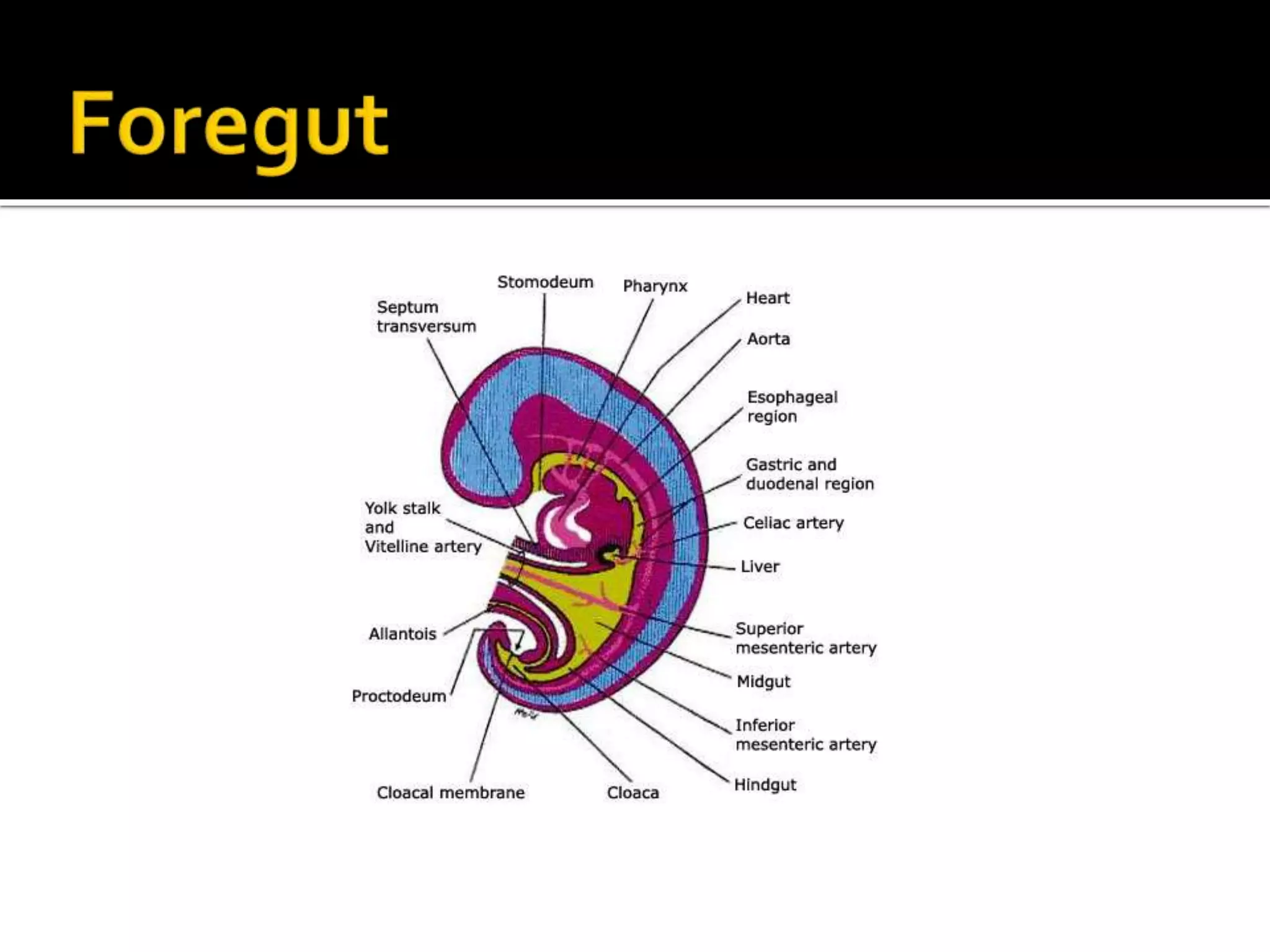
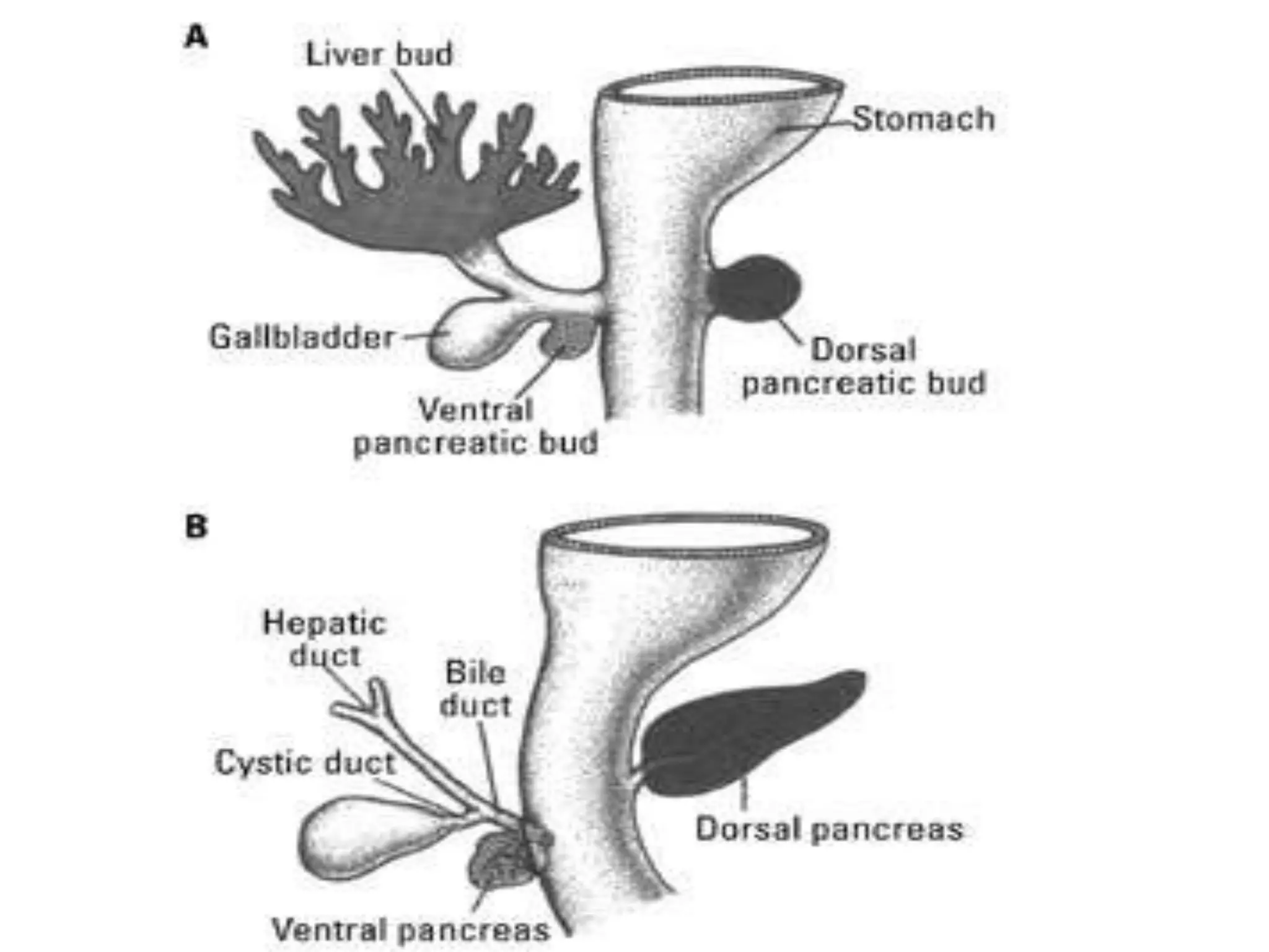
 The primordial gut at the beginning
of the 4th week is closed at the
cranial end by the oropharangeal
membrane and the caudal end by
the cloacal membrane.The
endoderm of the primordial gut
gives rise to most of the epithelium
and glands of the digestive tract.
The primordial gut is broken down
into 3 parts:The foregut, midgut,
and hindgut.
 Oral cavity, pharynx, tongue, tonsils, salivary
glands, and respiratory system.
 The Esophagus
 The Stomach
 The duodenum
 The liver and pancreas
 The esophagus develops inferiorly to the pharynx.
 It is initially short, but elongates rapidly reaching its
relative length by week 7.
 During development, the endodermal lining of the
esophagus proliferates rapidly and obliterates the
lumen.
 Recanalization occurs by the end of the embryonic
period.
 The stomach develops from the foregut starting at the 4th
week.
 It begins as a dilation of the gut tube and during
development it shifts position by both moving caudally and
rotating.
 As this occurs the left wall of the stomach will become the
anterior surface and the right side of the gut tube will
become the posterior surface of the stomach.
 Also, the dorsal border of the stomach grows faster than the
ventral border forming the greater curvature.
 Develops from the caudal part of the foregut, cranial part
of the midgut and the splanchnic mesenchyme of the
primordial gut.
 Forms a C – shaped loop that projects ventrally.
 Lumen of the duodenum becomes progressively smaller
and is temporarily obliterated and becomes recanalized
at the end of the embryonic period.
 Supplied by the celiac trunk and the superior mesenteric
artery.
 Arise as a ventral outgrowth from the caudal part of the foregut.
 Hepatic diverticulum divides into two parts.
-The larger cranial part becomes the liver
-The small caudal part becomes the gallbladder.
-The stalk of the diverticulum becomes the cystic duct.
 Hepatic cords from the hepatic diverticulum grow into the septum
transversum.
 Hepatic cords arrange themselves around the vitelline and umbillical veins,
which course through the septum transversum and form the hepatic
sinusoids.
 Initially the left and right lobes of the liver are the same size; the
right lobe later enlarges.
 Arises from the dorsal and ventral pancreatic buds
of endodermal cells which arise from the caudal part
of the foregut.
 The dorsal pancreatic bud appears first, growing rapidly.
 The ventral pancreatic bud develops near the entry of the
bile duct into the duodenum.
 As the duodenum rotates to the right the ventral
pancreatic bud is carried dorsally with the bile duct.
It soon lies posterior to the dorsal pancreatic bud
and later fuses with it.
 Derived from mesenchymal cells located between the
layers of the dorsal mesogastrium.
 Starts developing during the fifth week.
 Lobulated in the fetus. Lobules disappear before birth.
 The spleen has a dorsal attachment of the splenorenal
ligament, due to the rotation of the stomach
 Functions as a hematopoetic center until late fetal life.
 Accessory spleen(s) may develop. Occurs in about 10%
of people.
 The midgut develops into the following
structures:
 Small Intestine (excluding the proximal
duodenum)
 Cecum and appendix,
 Ascending Colon
 Proximal part of theTransverse Colon (Right 1/2 to
2/3)
All of the derivatives are supplied by the Superior
Messenteric Artery.
• Vitelline duct also known as the omphalomesenteric duct, is a long
narrow tube that joins the yolk sac to the midgut lumen of the
developing fetus
 At the beginning
of the sixth week
 The hindgut gives rise to:
 Left 1/3-1/2 of the transverse colon
 Descending colon
 Sigmoid colon
 Rectum
 Superior part of anal canal
 Epithelium of urinary bladder
 Urethra
 All hindgut derivatives are supplied by inferior
mesenteric artery
 Cloaca is the caudal end of the hindgut and is
lined with endoderm.
 At the end of the cloaca there is a cloacal
membrane which separates the endoderm
from the surface ectoderm of proctodeum or
anal pit at 4 weeks
GI presentation embryo

GI presentation embryo

  • 2.
     The primordialgut at the beginning of the 4th week is closed at the cranial end by the oropharangeal membrane and the caudal end by the cloacal membrane.The endoderm of the primordial gut gives rise to most of the epithelium and glands of the digestive tract. The primordial gut is broken down into 3 parts:The foregut, midgut, and hindgut.
  • 3.
     Oral cavity,pharynx, tongue, tonsils, salivary glands, and respiratory system.  The Esophagus  The Stomach  The duodenum  The liver and pancreas
  • 4.
     The esophagusdevelops inferiorly to the pharynx.  It is initially short, but elongates rapidly reaching its relative length by week 7.  During development, the endodermal lining of the esophagus proliferates rapidly and obliterates the lumen.  Recanalization occurs by the end of the embryonic period.
  • 5.
     The stomachdevelops from the foregut starting at the 4th week.  It begins as a dilation of the gut tube and during development it shifts position by both moving caudally and rotating.  As this occurs the left wall of the stomach will become the anterior surface and the right side of the gut tube will become the posterior surface of the stomach.  Also, the dorsal border of the stomach grows faster than the ventral border forming the greater curvature.
  • 7.
     Develops fromthe caudal part of the foregut, cranial part of the midgut and the splanchnic mesenchyme of the primordial gut.  Forms a C – shaped loop that projects ventrally.  Lumen of the duodenum becomes progressively smaller and is temporarily obliterated and becomes recanalized at the end of the embryonic period.  Supplied by the celiac trunk and the superior mesenteric artery.
  • 8.
     Arise asa ventral outgrowth from the caudal part of the foregut.  Hepatic diverticulum divides into two parts. -The larger cranial part becomes the liver -The small caudal part becomes the gallbladder. -The stalk of the diverticulum becomes the cystic duct.  Hepatic cords from the hepatic diverticulum grow into the septum transversum.  Hepatic cords arrange themselves around the vitelline and umbillical veins, which course through the septum transversum and form the hepatic sinusoids.  Initially the left and right lobes of the liver are the same size; the right lobe later enlarges.
  • 9.
     Arises fromthe dorsal and ventral pancreatic buds of endodermal cells which arise from the caudal part of the foregut.  The dorsal pancreatic bud appears first, growing rapidly.  The ventral pancreatic bud develops near the entry of the bile duct into the duodenum.  As the duodenum rotates to the right the ventral pancreatic bud is carried dorsally with the bile duct. It soon lies posterior to the dorsal pancreatic bud and later fuses with it.
  • 11.
     Derived frommesenchymal cells located between the layers of the dorsal mesogastrium.  Starts developing during the fifth week.  Lobulated in the fetus. Lobules disappear before birth.  The spleen has a dorsal attachment of the splenorenal ligament, due to the rotation of the stomach  Functions as a hematopoetic center until late fetal life.  Accessory spleen(s) may develop. Occurs in about 10% of people.
  • 13.
     The midgutdevelops into the following structures:  Small Intestine (excluding the proximal duodenum)  Cecum and appendix,  Ascending Colon  Proximal part of theTransverse Colon (Right 1/2 to 2/3) All of the derivatives are supplied by the Superior Messenteric Artery.
  • 14.
    • Vitelline ductalso known as the omphalomesenteric duct, is a long narrow tube that joins the yolk sac to the midgut lumen of the developing fetus
  • 15.
     At thebeginning of the sixth week
  • 17.
     The hindgutgives rise to:  Left 1/3-1/2 of the transverse colon  Descending colon  Sigmoid colon  Rectum  Superior part of anal canal  Epithelium of urinary bladder  Urethra  All hindgut derivatives are supplied by inferior mesenteric artery
  • 18.
     Cloaca isthe caudal end of the hindgut and is lined with endoderm.  At the end of the cloaca there is a cloacal membrane which separates the endoderm from the surface ectoderm of proctodeum or anal pit at 4 weeks