what is strabismus ?
what are different type of verticle squint ?
what is A pattern = means a relative convergence in upgaze & relative divergence in down gaze with minimum difference of 10 PD between upgaze & downgaze .
what is V pattern strabismus ?
it is relative divergence in upgaze & relative convergence in downgaze with minimum difference of 15 PD between upgaze & downgaze …
what is alphabate pattern strabismus ?
PBCT is more sensitive than Krimsky test ?
the measurement of squint in upgae 25 degree & in downgaze 33-35 degree i.e PBCT ( PRISM BAR COVER TEST )
what is Lamba pattern strabismus ?
what is X pattern strabismus ?
what is delta pattern ?
This document discusses exodeviation, also known as divergent strabismus, where the visual axes diverge outward instead of converging. There are two main types - concomitant and incomitant. Exodeviation can be primary, such as infantile exotropia which begins in the first 6 months of life, or intermittent exotropia where the eyes diverge intermittently. Secondary exodeviation includes sensory exotropia caused by visual impairment, and consecutive exotropia following correction of esotropia. Treatment depends on the type and may include correcting refractive errors, vision therapy, prism therapy, or surgery. The angle of exodeviation is measured using tests
This document discusses the diagnosis and management of superior oblique palsy. It begins by describing the anatomy and function of the superior oblique muscle. Superior oblique palsy can result in hypertropia, excyclotorsion, and esotropia that are greater in certain gazes. Causes may be congenital or acquired from trauma or vascular issues. Diagnosis involves evaluating eye movements, diplopia, and head tilt. Non-surgical treatment includes patching or prisms while surgery involves weakening the antagonist inferior oblique muscle or tucking the superior oblique tendon. The goal of treatment is to expand the field of single vision while minimizing complications.
This document discusses various types of vertical eye movement disorders including: principles of cyclovertical deviations; the Park 3 step test for diagnosing superior oblique palsy; classifications of incomitant and comitant vertical deviations including dissociated vertical deviation, inferior oblique overaction, and superior oblique palsy; diagnostic tests and treatments for these conditions; and references for further information.
- Microtropia is a small-angle, usually unilateral, strabismus of 10Δ or less. It is the most common form of abnormal binocular single vision.
- Characteristics include a foveal suppression scotoma in the deviating eye, reduced visual acuity in that eye, and anisometropia in nearly all cases. Microtropia can be classified as primary or secondary.
- Management aims to obtain the best possible visual acuity in each eye through refractive correction, occlusion therapy, and restoration of binocular single vision if needed for an associated strabismus.
The document discusses retinal correspondence and abnormal retinal correspondence (ARC) in patients with strabismus. It defines normal retinal correspondence as when stimulation of corresponding retinal points produces single vision, while ARC is when non-corresponding points produce single vision. ARC can be harmonious, unharmonious, or paradoxical depending on its relationship to the objective angle of deviation. Several clinical tests are described to assess retinal correspondence, including Bagolini's striated glasses test, red filter test, synoptophore, and Worth's four dot test. Occlusion therapy is mentioned as a treatment to prevent worsening of ARC and promote normal correspondence.
Convergence insufficiency is the inability to maintain binocular convergence without undue effort. It is the most common cause of eyestrain. It can be caused by refractive errors, presbyopia, muscle imbalances, or other factors like wide pupil distance. Clinical features include eyestrain in desk workers and blurred near vision. Diagnosis involves measuring near point of convergence over 10cm and difficulty maintaining 30 degrees of convergence. Treatment includes optical correction, orthoptic exercises to improve near point convergence and fusional vergence, relaxation exercises, and prism therapy. Surgical treatment is a last resort.
This document discusses contact lenses, including their classification based on anatomical position, material, and mode of wear. It covers topics such as contact lens design, related terms, ideal materials, indications for use, contraindications, and optics. Rigid and soft contact lenses are described. Factors that affect contact lens fit and comfort are also summarized.
Fitting an Astigmatic Patient is really a challenging.Though fitting a Toric Cornea is another challenge in CL Dispensing practice.This Slide will give you a basic considerations in RGP Toric lens.
This document provides information about corneal topography and keratometry. It defines the cornea and its dimensions. It describes the historical evolution of keratometry from its first description in 1619 to modern computerized corneal topography systems. The document explains the principles, procedures, techniques, and applications of keratometry and corneal topography in evaluating the cornea. It also discusses the limitations and assumptions of keratometry measurements.
The synoptophore is an instrument used to objectively and subjectively measure eye alignment and binocular vision. It consists of two tubes that each present a different image to each eye. Alignment of the tubes can be adjusted horizontally, vertically, and torsionally to measure any deviation. Simultaneous perception, fusion, and stereoscopic targets are used to assess the grade of binocular single vision from grade 1 to grade 3. Objective measurement can be done by neutralization or corneal reflection. Fusional amplitudes and retinal correspondence can also be evaluated. The synoptophore is useful for diagnosing strabismus and binocular vision disorders.
Eccentric fixation occurs when an amblyopic eye fixes on a point other than the fovea. It is important to diagnose as it impacts visual acuity and treatment. Eccentric fixation can be evaluated using several tests including the corneal light reflex test, ophthalmoscopy, after image transfer, and perimetry. Treatment may involve occlusion of the good eye combined with use of a red filter over the amblyopic eye to encourage central fixation. Careful monitoring of fixation behavior is important for guiding amblyopia treatment.
This document discusses various options for correcting presbyopia with contact lenses, including bifocal, monovision, and multifocal lenses. Bifocal lenses have simultaneous vision designs like concentric, aspheric, and diffractive lenses or alternating/translating designs. Monovision fits one eye for distance and one for near. Factors in fitting presbyopic contact lenses include visual requirements, occupation, binocularity, medication, and tear film status. Fitting requires assessing balance of distance and near vision and allowing adaptation time, with patient education crucial. Presbyopia correction with contact lenses provides an alternative to bifocal glasses but requires careful patient screening and management of expectations.
Vertical Deviations
Hyperphoria is a latent, or hidden, upward deviation of one eye. It is often associated with other conditions like horizontal misalignments or incomitant, or non-equal, muscle deviations. Primary hyperphoria of less than 3 degrees is generally due to slight anatomical differences between the eyes. Symptoms can be relieved through refractive correction, orthoptic treatment with prisms, or in rare cases surgery. Dissociated vertical deviation is a rare anomaly where the covered eye moves upward behind the cover regardless of which eye is covered.
Nystagmus assessments and management mehediMehedi Hasan
Nystagmus is involuntary eye movement that can cause vision problems. It has many potential causes including issues with the eye, brain, inner ear or genetic conditions. There are different types classified by timing, direction of eye movement, and other factors. Evaluation involves assessing symptoms, medical history and characterizing the nystagmus through observation. Management may include optical corrections, vision therapy, medical treatments, or rarely surgery to improve eye alignment and vision.
Esotropia , classification , diagnosis and managementDrAzmat Ali
This document provides information on various types of esotropia (convergent strabismus), including:
- Accommodative esotropia caused by uncorrected hyperopia or a high AC/A ratio
- Partially accommodative esotropia with both accommodative and non-accommodative components
- Non-accommodative esotropia including convergence excess, cyclical esotropia, and acquired forms
- Esotropia associated with high myopia or nystagmus is also discussed
Treatment options including refractive correction, orthoptic exercises, prisms, botulinum toxin, and surgery are mentioned for different types of esotropia.
Pattern strabismus occurs when there is a change in the magnitude of horizontal deviation between up and down gaze. The most common types are A pattern (convergence in up gaze) and V pattern (divergence in up gaze). Pattern strabismus can be caused by abnormalities of vertical or horizontal muscle action, anatomical anomalies, disorders of muscle innervation, or anomalous muscle insertions. Evaluation involves measuring the deviation in different gazes using cover-uncover testing and Hess screening. Management may involve adaptation or surgery tailored to the specific pattern, which aims to improve alignment and binocular function.
This document discusses tinted lenses, which are lenses that have been dyed with organic dyes or metallic oxides to give them color. Tinted lenses can be used in glasses or contact lenses and serve purposes like fashion, visual comfort, improving visibility and contrast, and protecting the eye from harmful radiation. The document outlines different types of tints like bright yellows and browns that provide UV protection and darker greens and grays for sunglasses. It also discusses reasons for using tinted lenses such as for light sensitivity, contrast enhancement, reducing migraines, and reading comfort. Finally, it lists some ocular conditions where tinted lenses may be prescribed, such as age-related macular degeneration and cataracts
This document discusses frame adjustment and quality checking. It describes 7 off-face adjustments including x-ing, temple spread, pantoscopic angle, temple fold angle, pad angles, face form, and 4-point touch. It also discusses 7 on-face adjustments including horizontal alignment, vertex distance, frame height, segment height, temple bend, pad contact, and skin/lash clearance. Key details are provided about properly adjusting specific angles and alignments during the fitting process.
Fitting soft contact lenses requires considering many patient-specific factors to achieve excellent vision and ocular health. A proper fit involves selecting the correct total diameter, base curve, thickness, and material based on the patient's prescription, corneal shape, lifestyle, and health. Trial lenses are used to evaluate fit parameters like coverage, centration, movement, comfort, and vision to optimize on-eye performance while avoiding issues like tightness or looseness that could impact ocular health or vision. The goal is to find a lens that provides optimum vision and good comfort without causing any ocular insult.
This document discusses eccentric fixation (EF), a condition where an eye fails to fixate with the fovea and instead fixates at another retinal point. It describes several theories for the cause of EF, including suppression, anomalous correspondence, motor, and sensory motor theories. It outlines methods for investigating EF, such as ophthalmoscopy and visuscopy. Treatment options discussed include occlusion therapy and pleoptic treatment to encourage foveal fixation, though EF is often difficult to fully correct once established. The document also discusses microtropia, a small-angle strabismus associated with EF and amblyopia.
Binocular vision refers to the coordinated use of both eyes to produce a single mental impression. It develops through childhood as the visual and motor mechanisms mature. Theories of binocular vision aim to explain how a single visual percept is formed from two retinal images. Key components include retinal correspondence, the horopter curve, and Panum's fusional area. Abnormalities can disrupt binocular vision and cause issues like diplopia or suppression. Clinical tests evaluate retinal correspondence, stereopsis, and fusional abilities to assess binocular function.
Measurement of optical center of ophthalmic lensesGauriChaudhary7
The document discusses optical centers of lenses and their importance. It defines an optical center as the point where light rays pass through the lens without deviation. Determining the proper position of the optical center is important to avoid prismatic effects and ensure proper refraction. The document outlines several manual methods for locating the optical center, including using a light source to align the lens images. It also discusses how decentering a lens can induce or reduce prismatic effects depending on the lens power and direction of decentration.
1. Intermittent exotropia is the most common type of exodeviation, usually beginning in early childhood.
2. It involves periods where the eyes are aligned (phoric phase) and misaligned outward (tropic phase), with control deteriorating over time.
3. Surgical treatment is recommended if control is worsening, as indicated by an increasing tropic phase, loss of fusion, increased deviation angle, or suppression development. Non-surgical management can be tried for small angles or young ages but is often ineffective long-term.
The document discusses pantoscopic tilt, which is when the bottom of eyeglass frames are angled toward the cheeks. It describes how proper pantoscopic tilt helps maximize the amount of bridge surface resting on the nose. The document also mentions retroscopic tilt, when the bottom of frames is angled away from the cheeks, and orthoscopic tilt, when frames have no angle. Additionally, it explains how lens tilt improves how glasses look and function for patients, and depends on ear and nose bridge heights, requiring frames to be properly adjusted for individual wearers before measurements.
This document discusses soft toric contact lenses for correcting astigmatism. It defines astigmatism and describes various types. It explains that toric lenses contain a cylindrical component to correct astigmatism unlike standard soft lenses. The document outlines several designs and methods for stabilizing toric soft contact lenses, including prism ballast, dynamic stabilization, and reverse prism designs. It provides steps for fitting toric lenses including diagnosis, trial lenses, and assessing lens rotation to finalize the axis. Examples of toric lens prescriptions and assessments of fit are also summarized.
1. This document discusses various types of vertical strabismus and cyclo deviations, classifying them as comitant or incomitant. Comitant deviations occur with horizontal deviations, while incomitant include paretic, restrictive, and dissociated vertical deviations.
2. Incomitant vertical deviations include apparent oblique muscle dysfunction, paretic deviations caused by muscle palsies, and restrictive deviations. Dissociated vertical deviation is also discussed in detail.
3. Treatment depends on the type of vertical deviation and may include orthoptics, prism therapy, or surgical correction such as weakening or strengthening procedures on the oblique muscles.
Ghostdogg productions presents browns syndrome online version 2-1Alistair Hamilton
A general overview and summary of what I think is one of the most intriguing forms of Pediatric Strabismus seen to date. (Note: this presentation was initially made before Alphabet Pattern Strabismus so the transitions are a little off.) Hope you enjoy
This document provides information on examining a case of strabismus. It begins with the author's disclosure statement and then is divided into sections on history, examination, equipment, motor status, sensory status, measurement of deviation, and special tests. The history section covers items like visual acuity, eye involvement, and medical history. Examination assesses areas such as head posture, ocular deviation, eye movements, and binocular vision status. Various tests and equipment used are also outlined.
This document discusses esotropia, or convergent strabismus, where the eyes turn inward. It defines strabismus and the different types of eye turns that can occur. It specifically focuses on esotropia, describing the different types including accommodative esotropia caused by hyperopia, non-accommodative esotropia from other causes, and infantile esotropia presenting in the first 6 months of life. It discusses the evaluation and management of esotropia, including correcting refractive errors, treating amblyopia with patching, and considering surgery if needed to align the eyes. The goal of treatment is to improve vision in both eyes and maximize binocular vision.
The document discusses patterns of strabismus, specifically the A pattern and V pattern. The A pattern involves relative convergence on upgaze and divergence on downgaze, while the V pattern is the opposite with relative divergence on upgaze and convergence on downgaze. Variants include the X, Y, lambda, and diamond patterns. The etiology of these patterns involves dysfunction of the horizontal, vertical, or oblique eye muscles. Clinical features may include anomalous head posture, amblyopia, and abnormal retinal correspondence. Diagnosis involves measuring alignment in upgaze and downgaze while preventing accommodation.
This document discusses various pattern deviations that can occur in horizontal strabismus. It describes V and A patterns, which are differences in horizontal deviation between upgaze and downgaze. V patterns have more divergence in upgaze, while A patterns have more divergence in downgaze. These can be caused by oblique muscle dysfunction or variations in orbital anatomy. Rare patterns like X, Y and lambda are also mentioned. Diagnostic testing and surgical management options depending on the underlying etiology are provided.
This document defines and classifies different types of squint, including:
- Latent squint (heterophoria), where the eyes have a tendency to deviate but remain aligned with fusion.
- Manifest squint (heterotropia), where there is a visible misalignment of the eyes. This includes concomitant squint, where the deviation is the same in all directions of gaze, and paralytic squint, where the deviation varies with gaze direction.
- The causes, signs, and treatments of different types of squint are described, including exercises, optical correction, amblyopia therapy
This document discusses superior oblique palsy:
1. The superior oblique nerve passes through the midbrain, decussates at the anterior medullary velum, and travels between arteries in the cavernous sinus and superior orbital fissure outside the annulus of Zinn.
2. Superior oblique palsy can be congenital or acquired. Common acquired causes include trauma. Presentation includes vertical and torsional diplopia that worsens with head tilt. Evaluation involves testing like Hess screen and three-step test.
3. Management depends on degree of deviation and may include weakening the ipsilateral inferior oblique or recessing it, with contralateral inferior rect
This document summarizes information about the anatomy and physiology of the eye, eye movements, amblyopia, and strabismus. It describes the three layers of the eye (fibrous, vascular, and neural), the extraocular muscles that control eye movement, and the cranial nerves involved (3rd, 4th, 6th). It defines amblyopia as reduced vision in one eye due to lack of coordination between the eyes and brain. Strabismus is described as misalignment of the eyes that can be constant or intermittent. Treatment options for amblyopia and strabismus include glasses, patching, and sometimes surgery to correct muscle imbalance.
This document discusses strabismus (misalignment of the eyes) including:
1. Definitions of strabismus, heterophoria (latent squint), and heterotropia (manifest squint).
2. Types of strabismus including esotropia (inward turning), exotropia (outward turning), and vertical deviations.
3. Extraocular muscles that control eye movement and their nerve supply.
4. Medical and surgical treatments for strabismus including occlusion therapy, prisms, botulinum toxin injections, and different surgical procedures.
This document discusses the evaluation of ocular examination in cataract patients. It covers various components of the eye exam including head posture, facial asymmetry, visual axis, ocular movements, eyelids, conjunctiva and more. Abnormal head postures can indicate strabismus while facial asymmetry may be due to dental, skeletal or muscular issues. Tests like cover tests are used to evaluate the visual axis and detect strabismus. Ocular movements involve evaluations of ductions, versions, vergences and following diagnostic positions of gaze. The eyelids, lashes, glands and tumors are also examined for abnormalities.
Cyclovertical anomalies refer to vertical deviations of the eyes involving the cyclovertical muscles. There are various types including comitant and incomitant vertical deviations. Patterns of strabismus like V, A, X, Y patterns are classified based on how the horizontal deviation changes between upgaze and downgaze. Conditions like superior oblique overaction, palsy, inferior oblique overaction are described. Dissociated vertical deviation is an innervational disorder seen in infantile strabismus and involves predominately vertical movements of one eye drifting upward and outward. Surgical management of these conditions aims to weaken muscles or transpose them.
EVALUATION OF A SQUINT PATIENT (4).pptxMalvikaSuresh
This document summarizes the evaluation process for a patient with strabismus (misaligned eyes). It discusses examining the patient's history, visual acuity, refractive error, sensory status, and motor status. Sensory tests evaluate binocularity and eye alignment, including tests for fusion, suppression, diplopia, and stereopsis. The motor examination measures the size and nature of any eye misalignment. The goal is to determine the cause of strabismus, assess binocularity, measure the deviation, diagnose amblyopia, and develop a treatment plan.
Binocular vision allows for single, three-dimensional vision using both eyes. It provides advantages like better visual acuity and depth perception. Binocular vision requires muscle balancing, retinal correspondence, and fusion of the two retinal images. Disturbances can include strabismus, which is an ocular misalignment. Strabismus is classified and treatments involve exercises, prisms, or surgery to improve alignment and binocularity.
This document defines and describes occipito-posterior position, which occurs when the occiput of the fetal head is positioned posteriorly in the birth canal. It has an incidence of about 10% and can cause dystocia if the occiput does not anteriorly rotate. Diagnosis is made through abdominal palpation, auscultation, and vaginal examination to locate the fetal position. Ultrasound may also be used. Spontaneous rotation usually occurs, but if not the baby's head will flex, extend, and rotate through the birth canal to deliver face first before fully rotating to an occiput anterior position.
This document provides information about infantile esotropia, including its definition, clinical features, etiology, differential diagnosis, and management. Infantile esotropia is defined as a large-angle esotropia present before 6 months of age. It is the most common form of strabismus. Clinical features include alternating or crossed fixation, apparent limitation of abduction, and associated motor abnormalities like inferior oblique overaction and dissociated vertical deviation. Treatment involves correcting refractive errors, amblyopia therapy, observation for small angles, and early surgical correction typically between 6 months to 2 years of age.
Motor adaptation in paretic and nonparetic strabismuskopila kafle
This document discusses motor adaptation in paresis and non-paresis strabismus. It begins by defining strabismus and describing the different types. It then discusses the consequences of strabismus and the different ways motor adaptation occurs, including through changes in muscle tone, compensatory head posture, and blind spot mechanisms. It describes how motor adaptation occurs differently in incomitant versus comitant strabismus. The document goes on to discuss various cranial nerve palsies and how they result in specific eye positions and compensatory head postures. It also covers special restrictive disorders like Duane's retraction syndrome.
This document summarizes key aspects of sensory evaluation of squint or strabismus. It begins by describing normal binocular development and vision, including the development of binocular fusion and stereopsis in infants. It then discusses abnormal binocular vision including sensory adaptations like suppression, anomalous retinal correspondence, and eccentric fixation. Finally, it outlines several tests used to evaluate the sensory system in strabismus, including visual acuity tests, Worth four-dot test, Bagolini striated glasses, 4 prism base out test, synaptophore, and after-image tests.
This document summarizes key concepts related to strabismus and eye movement examination. It defines strabismus as misalignment of the visual axis and describes various types of phorias and tropias. Objective tests for strabismus are outlined like the cover-uncover and prism bar cover tests. Details are provided about extraocular muscle function and innervation. Grading of binocular vision and tests for suppression are also summarized. The document covers important topics in a comprehensive yet concise manner.
Similar to Ghostdogg productions present ~ alphabet pattern strabismus online version (20)
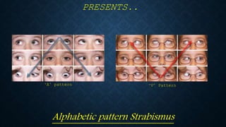
Muktapishti is a traditional Ayurvedic preparation made from Shoditha Mukta (Purified Pearl), is believed to help regulate thyroid function and reduce symptoms of hyperthyroidism due to its cooling and balancing properties. Clinical evidence on its efficacy remains limited, necessitating further research to validate its therapeutic benefits.
One health condition that is becoming more common day by day is diabetes.
According to research conducted by the National Family Health Survey of India, diabetic cases show a projection which might increase to 10.4% by 2030.
Travel vaccination in Manchester offers comprehensive immunization services for individuals planning international trips. Expert healthcare providers administer vaccines tailored to your destination, ensuring you stay protected against various diseases. Conveniently located clinics and flexible appointment options make it easy to get the necessary shots before your journey. Stay healthy and travel with confidence by getting vaccinated in Manchester. Visit us: www.nxhealthcare.co.uk
Does Over-Masturbation Contribute to Chronic Prostatitis.pptxwalterHu5
In some case, your chronic prostatitis may be related to over-masturbation. Generally, natural medicine Diuretic and Anti-inflammatory Pill can help mee get a cure.
These lecture slides, by Dr Sidra Arshad, offer a quick overview of the physiological basis of a normal electrocardiogram.
Learning objectives:
1. Define an electrocardiogram (ECG) and electrocardiography
2. Describe how dipoles generated by the heart produce the waveforms of the ECG
3. Describe the components of a normal electrocardiogram of a typical bipolar lead (limb II)
4. Differentiate between intervals and segments
5. Enlist some common indications for obtaining an ECG
6. Describe the flow of current around the heart during the cardiac cycle
7. Discuss the placement and polarity of the leads of electrocardiograph
8. Describe the normal electrocardiograms recorded from the limb leads and explain the physiological basis of the different records that are obtained
9. Define mean electrical vector (axis) of the heart and give the normal range
10. Define the mean QRS vector
11. Describe the axes of leads (hexagonal reference system)
12. Comprehend the vectorial analysis of the normal ECG
13. Determine the mean electrical axis of the ventricular QRS and appreciate the mean axis deviation
14. Explain the concepts of current of injury, J point, and their significance
Study Resources:
1. Chapter 11, Guyton and Hall Textbook of Medical Physiology, 14th edition
2. Chapter 9, Human Physiology - From Cells to Systems, Lauralee Sherwood, 9th edition
3. Chapter 29, Ganong’s Review of Medical Physiology, 26th edition
4. Electrocardiogram, StatPearls - https://www.ncbi.nlm.nih.gov/books/NBK549803/
5. ECG in Medical Practice by ABM Abdullah, 4th edition
6. Chapter 3, Cardiology Explained, https://www.ncbi.nlm.nih.gov/books/NBK2214/
7. ECG Basics, http://www.nataliescasebook.com/tag/e-c-g-basics
Our backs are like superheroes, holding us up and helping us move around. But sometimes, even superheroes can get hurt. That’s where slip discs come in.
2. DEDICATED TO...
• Dr. Burton J. Kushner, MD.
• Your interest in this subject of Binocular vision & Strabismus is what has kept me interested and enjoying doing what I
do...
A. J. Hamilton
3. HISTORY OF AV PATTERN
STRABISMUS
• Originally described as “an abnormal variation in the angle of horizontal strabismus in
vertical gaze” by Alexander Duane in 1897.
• Later labelled as “Horizontal strabismus with associated vertical elements” from 1896-1956.
• First official series of papers with emphasis on the measurement of horizontal deviation in
vertical gaze was penned by Uretts-Zavalia in 1948, in his papers “Abducción en la elevación”
and “Paralisis bilateral congenita del musculo oblicuo inferior”. Attention was also made to
the point that overaction and subsequently underaction of the oblique muscles were in fact
associated with increased and decreased convergence or divergence.
• By the late 1950’s, the syndrome was officially given the title of ‘A’ or ‘V’ Pattern strabismus,
by Dr. D. G. Albert and F. D. Costenbader in 1955 and 1957 respectively, owing to both ‘A’ and
‘V’ patterns being the most commonly noticed forms. The name was then shortened to ‘A-V
Pattern Strabismus’ as it stands today.
• The syndrome as a whole falls under the Strabismus sub-group of ‘Complex strabismus’ or
‘comitant horizontal/vertical deviations in Strabismus’
4. INCIDENCE OF AV PATTERN STRABISMUS
• 15-25% of cases of pediatric strabismus occur with A/V Pattern Strabismus as a subsidiary symptom
associated with most common forms of Strabismus: Brown’s Syndrome, Duane’s retraction Syndrome, or
any association with any form of over/underaction of the Superior/Inferior obliques
• A/V Pattern Strabismus is the most common form of Infantile Strabismus.
• While ‘A’ Pattern is perhaps the most common, ‘V’ Pattern in either esotropia or exotropia is quite
prevalent in both pediatric and adult strabismus categories.
• Other rarer forms such as ‘X’ pattern, Lambda pattern, and ‘Y’ pattern strabismus have also been
documented and seen; Lambda pattern being a sub-group of ‘A’ pattern, while ‘Y’ pattern is more closely
associated with ‘V’ pattern. ‘X’ pattern to date remains a group of its own, since so little is still unknown
of this sub-type.
• These special forms of incomitance in vertical gaze consist of nothing more than modifications of the
classic A or V patterns; therefore,they should not be regarded as separate entities.
6. ‘A’ PATTERN STRABISMUS
‘A’ PATTERN ESOTROPIA
• An A pattern is present when a horizontal deviation shows a more convergent (less divergent)
alignment in upgaze compared with downgaze.
• An ‘A’ pattern is considered significant if the difference between upgaze and downgaze is ≥10 Δ.
• Patients with ‘A’ pattern esotropia will show on clinical presentation:
• An increase of the esotropia in midline upgazes, and a decrease of the esotropia on
midline downgazes
• The eyes will be mostly straight in Primary gaze and direct downgaze
• Patients will adopt a chin-up head posture to compensate for single binocular vision.
• An ‘A’ Pattern is often associated with:
• Primary superior oblique overaction
• Inferior oblique underaction/palsy with subsequent superior oblique overaction.
• Inferior rectus underaction
7. ‘A’ PATTERN ESOTROPIA
This 6-year-old girl presents
with a evident ‘A’ pattern
Esotropia associated with a
right superior oblique
overaction, visible as an
overshoot of the right eye on
adduction. Noticeable is the
child’s assumed chin-up head
tilt to the left to compensate
for single binocular vision in
primary gaze. Also evident is
the convergence of the eyes
on direct upgaze, hence an ‘A’
pattern.
8. ‘A’ PATTERN STRABISMUS
‘A’ PATTERN EXOTROPIA
• In cases of an ‘A’ pattern exotropia, the deviation of the eyes is more pronounced as the
eyes move more towards midline downgaze rather than midline upgaze.
• There is an evident divergence of the eyes in direct downgaze, with the eyes ‘splaying
outwards’.
• Like in ‘A’ pattern esotropia, a patient’s eyes in primary and upgaze will often look
straight, and work together closely to normal. ‘A’ pattern exotropia is much more
prevalent in midline downgazes, rather than upgaze. This misalignment is often not
noticed by parent’s whose children have this form of strabismus.
• Similar again to an ‘A’ pattern esotropia, patients will often assume an abnormal head
posture, except that it will be a chin-down, head turn, again allowing the patient to
achieve single binocular vision in primary gaze.
9. Mongoloid feature
- On upgaze, the eyes
converge inward
-In Primary position
eyes are slightly
exotropic
- On downgaze, the
eyes diverge outward
“A” PATTERN EXOTROPIA:
10. ‘A’ PATTERN EXOTROPIA
This 20-year-old girl shows
a visible ‘A’ pattern
exotropia associated with
overaction of her right
superior oblique, most
evident in down and left,
and down and right gazes.
In primary gaze she adopts
a chin-down head turn
away from the affected
eye, in order for both her
eyes to fuse in single
binocular gaze. In direct
downgaze there is a visible
‘outward splaying’ of both
eyes. The exotropic
deviation is minimal to
normal in all upgazes.
11. ‘V’ PATTERN STRABISMUS
‘V’ PATTERN ESOTROPIA
• First described by Alexander Duane in 1897 when he described a patient with
bilateral superior oblique palsy
• Patients with a ‘V’ pattern esotropia, as similar to ‘A’ pattern, will show a definite
increase of horizontal deviation more so in downgaze, with a corresponding decrease
of the deviation in upgaze.
• The inferior oblique is an abductor also, and secondary or primary overaction of
that muscle will result in a relatively less convergent or more divergent position in
upward gaze, producing a V pattern.
13. ‘V’ PATTERN ESOTROPIA
This 6 ½ year old boy shows a
very apparent ‘V’ pattern
esotropia, made evident by the
extreme chin-down head
posture, which he has adopted
in order to fuse in primary
position. Evident also is the
apparent ‘upshoot’ of his left eye
on adduction, and apparent
‘overshoot’ on elevation in
adduction, causing both his eyes
to ‘splay outwards’, also evident
on direct upgaze. This is a
result of an evident overation of
his left inferior oblique. On
direct downgaze, his eyes seem
to work in tandem almost
normally.
14. ‘V’ PATTERN STRABISMUS
‘V’ PATTERN EXOTROPIA
• In cases of a ‘V’ pattern exotropia, the deviation of the eyes is more pronounced as the
eyes move more towards midline upgaze rather than midline downgaze.
• There is an evident divergence of the eyes in direct upgaze, with both eyes “splaying
outwards”.
• Overaction of the Inferior oblique can be quite apparent in an evident “upshoot” of the
globe on adduction.
• Similar again to an ‘A’ pattern esotropia, patients will often adopt an abnormal head
posture, except that it will be a chin-up head turn. This allows the patient to achieve
single binocular vision in primary gaze. Parents of children with a ‘V’ pattern will often
notice that their child will occasionally ‘tilt’ their head upwards, to bring their eyes to an
downward position in order to achieve binocular vision in primary gaze.
15. ‘V’ PATTERN EXOTROPIA
This 7-year-old girl presents
with an evident ‘V’ pattern
esotropia, clearly shown by
the chin-up head posture
which she has adopted in
order to obtain single
binocular vision in primary
gaze. Evident is the
significant overaction of her
left inferior oblique in the
form of a gross overshoot of
her left eye in adduction and
elevation in adduction. Also
significant is the quite
evident “outwards splaying’
of both her eyes in direct
upgaze. Note the horizontal
deviation is significantly less
noticeable in downgazes, and
more so in upgazes, hence a
‘V’ pattern.
17. ‘A’ PATTERN “LAMBDA” STRABISMUS
This young girl shows a
definitive λ pattern, a variant
of an ‘A’ pattern exotropia,
associated with a bilateral
superior oblique overaction. As
with an ‘A’ pattern exotropia,
she adopts a chin-down head
posture in primary. Note that
the divergence is only
manifested most in downgaze
to her right. The divergence is
evident by the ‘outward
splaying’ of both her eyes on
downgaze right, and direct
downgaze. As with an ‘A’
pattern exotropia, there is a
definite convergence of her eyes
on direct upgaze.
18. ‘X’ PATTERN STRABISMUS
• Patients with ‘X’ pattern often present with a visible exotropia that is only present
or evident in direct upward and downward gaze.
• They often have little to no horizontal deviation on either adduction, or abduction,
with only minimal deviation in primary position.
• Most patients will not adopt an abnormal head posture, but will ‘raise’ or ‘dip’ their
head, depending on which direction they are needing to visualize, be it upgaze or
downgaze, in order to bring both eyes into single binocular vision.
19. ‘X’ PATTERN STRABISMUS
This boy presents with an ‘X’
pattern strabismus, made evident
by that there is a definite
divergence of both eyes, shown by
the hallmark ‘outward splaying’ of
both eyes on both upgaze and
downgaze. This is heightened by
the fact that he ‘dips’ his head
down to bring his eyes “up” on
upgaze, and ‘raises’ his head to
bring his eyes ‘Downwards” on
downgaze. What makes this
pattern difficult to distinguish is
that there is little horizontal
deviation on either adduction or
abduction, and that he doesn’t
adopt any abnormal head posture
in primary.
20. ‘Y’ PATTERN STRABISMUS
• Like ‘X’ pattern, patients with this form of AV pattern strabismus will only display
the deviation of the eye in all gazes above midline gaze.
• There is little to no deviation in either primary position or direct downgaze.
• This pattern is often hard to distinguish from a bilateral Inferior oblique overaction,
but has been documented in the form of ‘Pseudo Y pattern associated with Inferior
Oblique overaction’.
• The most adopted theory as to the cause for a ‘Y’ pattern is an abberant innervation
of the Lateral Rectus in up gaze.
21. ‘Y’ PATTERN ANTI ELEVATION SYNDROME
• Occurs as a complication of the inferior oblique anterior transposition. This
procedure changes the Inferior oblique from being an elevator, which restricts
elevation in adduction.
• Typically, there is marked fundus extortion associated with this sub-pattern of ‘Y’
pattern strabismus.
• Treatment for this usually involves a retroplacement of the anteriorly placed
Inferior oblique, thus reverting it to a standard recession.
22. ‘Y’ PATTERN ANTI-ELEVATION
SYNDROME
This teenage boy shows an Anti-elevation pattern following bilateral IO anterior
transposition. On looking to his left, what looks like an overelevation of his right eye in
abduction, is actually a secondary deviation due to fixation duress. His right (adducted) eye
is ‘stuck’ in an elevated and adducted position, due to the IO now having become an anti-
elevator., thus restricting its normal rotation
23. PSEUDO-INFERIOR OBLIQUE OVERACTION
ASSOCIATED WITH ‘Y’ PATTERN
STRABISMUS
• Described by Dr. Burton J Kushner, Pseudo-inferior oblique overaction occurs in an unusual
manner in which patients with this strabismus pattern will demonstrate the general features of a
‘Y’ pattern, but with the addition of an over elevation of the adducting eye in the field of the inferior
oblique muscle, resulting in a ‘Y’ pattern with a large evident exotropia in all up gazes.
• When compared to a true inferior oblique overaction it is very difficult to distinguish on
presentation. It is only by noting the rapid ‘overshoot’ of the adducting eye as it reaches maximum
elevation from direct adduction. This rapid overshoot of the adducting eye is best seen in still
frames taken from a film of the patient as they look from direct adduction to direct elevation.
• A hypertropia only develops after the eyes have passed beyond midline gaze. This hypertropia then
reverts to a gross abduction of the adducting eye once it has reached maximum elevation in
adduction.
24. PSEUDO ‘Y’ PATTERN STRABISMUS
A) This teenage girl shows a pseudo-inferior
oblique overaction with ‘Y’ pattern exotropia. When
viewed in the field of the inferior obliques, it is
indistinguishable from a true inferior oblique
overaction. In primary and midline gazes there is
no overaction of the inferior oblique. B) It is only by
breaking down the frame rate to 0.1 second
intervals from direct adduction to upgaze in
adduction, can one see that there is no ‘outward
splaying” of the eyes until she has reached
maximum elevation in adduction.
B
25. GRADING OF OBLIQUE
MUSCLE OVERACTION
• Grading of oblique muscle overaction in practice is more of measuring the amount
by visual interpretation rather than exact science. Most surgeons use a grading
scale of 1+ to 4+ overaction. For inferior oblique grading, 1+ overaction means only
slight overaction or over elevation in adduction. Grade 4+ means the most
overelevation possible. Grades 2+ and 3+ overaction are the two gradations between
those extremes.
27. CAUSES OF AV PATTERN
STRABISMUS
In Strabismic documentation and presentation, there are three main groups of causes
of AV Pattern Strabismus. These are:
• Oblique muscle dysfunction
• Orbital abnormality, and
• Abduction with a large exodeviation
28. MANAGEMENT AND TREATMENT OF AV
PATTERN STRABISMUS
• Of the four main forms of Alphabet pattern
strabismus (‘A’, ‘V’, ‘X’ & ‘Y’) each have certain
methods of treatment. Some correlate with
others while some require more extensive
corrective measures.
29. AV PATTERN STRABISMUS WITH
ORBITAL ABNORMALITY
This young boy has Crouzon’s disease with an evident ‘V’ pattern associated with a craniofacial
abnormality. Note the ‘upshoot’ and ‘downshoot’ of both eyes as he looks to both his left, in
abduction, and right, in adduction. The arrow mark above his left eye is the surgeon’s mark
indicating a left ‘V’ pattern exotropia.
30. Pre Treatment Evaluation
Detailed History
Assessment of BCVA
– Cycloplegic Refraction and correction
– Measurement of angle of deviation in all the 9
positions of gaze for near and far, with and without
optical correction
Uniocular and binocular motility with particular
attention to the oblique muscle dysfunction
MANAGEMENT
31. TREATMENT
• Factors for treatment of A V pattern strabismus are as follows:
• When binocular vision is disturbed, as in A exotropia and V esotropia,
and the treatment is surgery.
• An increase in deviation in downgaze (with A exotropia and V
esotropia) may cause discomfort during reading or in near work.
• Patients with large A or V patterns usually also have significant
corresponding oblique muscle dysfunctions.
32. TREATMENT
• Bilateral superior oblique weakening can collapse large A patterns up 30−40 PD or
more. Bilateral IO weakening is not expected to collapse as large a pattern.
• Bilateral inferior oblique weakening can collapse large V patterns when significant
IO overaction is present.
• In patients with V pattern and IOOA in association with congenital esotropia
complex, consideration should be given to performing IO anterior transposition,
even if DVD is not obvious.
• The A and V patterns can be diminished by horizontal offsets of the vertical rectus
muscles. temporal
33. TREATMENT
TRANSPORTATION OF THE INFERIOR RECTI
• Transposition of the inferior recti will expand the closed end of the V pattern by
weakening the adduction vector of the inferior recti in downgaze. Transposing the
SRs nasally will help close the open end of the V pattern in an exotropic patient and
transposing the IRs nasally will help close an A-pattern exotropia.
M a l e
34. TRANSPOSITION OF THE RECTI TO CORRECT
AV PATTERN STRABISMUS
•
This boy initially presented with a ‘V’ pattern exotropia which is evident by the exotropia of his left eye in
primary, and the ‘outward splaying’ of both eyes in direct upgaze. In the second 9-nine gaze diagnostic
versions, we see him again following a bilateral lateral rectus recession. Now his eyes are straight in
primary position, and there is no evidence of any horizontal deviation in upgaze.
35. IN SUMMARY
• Treatment of AV Patterns is relatively summed up in this crucial treatment goal:
Weaken the Overacting oblique muscles to alleviate the horizontal deviation!!!
• IF, there is no oblique dysfunction, treatment can consist of bilateral or unilateral
surgery on transpositioning of the horizontal recti muscles.
• Regarded as the most recognizable form of Infantile strabismus.
• Evidence of any type of horizontal deviation is particularly demonstrated in either direct
upgaze, or direct downgaze.
• ‘A’ & ‘V’ pattern strabismus accounts for 15-25% of most horizontal strabismus anomalies
to date.
36.
37. REFERENCES
Online references:
• The Strabismus Minute: http://www.cybersight.org
• Strabismus Summary series: http://www.geocities.ws/sapatney/webavst.htm
• McGill University Pediatric & Adult Strabismus:
http://www2.medicine.mcgill.ca/strabismus/atlas/patients/atlasNav/atlas.php
• A Pattern Esotropia & Exotropia/V pattern Esotropia & Exotropia:
https://emedicine.medscape.com/article/1199714-overview
& https://emedicine.medscape.com/article/1199825-overview
• Pattern Strabismus - Grand Round lectures by Dr. Robert O. Hoffman, John A. Moran Eye
care centre, Utah: https://www.youtube.com/watch?v=APjGJ_soPG4
38. REFERENCES CON’T
Paper references:
• Duane A. Isolated paralysis of the ocular muscles. Arch Ophthalmol,1897; 26: 317–34.
• Albert DG. Personal Communication. In Parks MM. Annual review: Strabismus. Arch
Ophthalmol 1957; 58: 136–60.
• Costenbader FD. The “A” and “V” patterns in strabismus. Trans Am Acad Ophthalmol
Otolaryngol, 1964; 68: 354–86.
• Sharat, S. Parija, S. A-V Pattern Strabismus- A Simplified Approach. Orissa Journal of
Ophthalmol 2010; 40-43.
• Kushner BJ. Pseudo inferior oblique overaction associated with ‘Y’ and ‘V’ patterns.
Ophthalmology 1991; 98: 1500–5.
39. REFERENCES CON’T
Textbook references:
• Rosenbaum, Arthur L., Santiago, Alvina P. Clinical Strabismus management. Principles and Surgical techniques.,
The Curtis Centre, Philadelphia, Philadelphia, Pennsylvania. W.B. Saunders ,1999.
• von Noorden, Gunter K. MD, Campos, Emilio C. Binocular Vision and Motility – Theory and management of
Strabismus, 6th edition. Baylor College of Medicine, Houston, Texas, Mosby 2002
• Ellis, George S. At the Crossings – Pediatric Ophthalmology and Strabismus, New Orleans Academy of
Ophthalmology, 2004.
• Taylor, David MD, Hoyt, Creig S. Pediatric Ophthalmology and Strabismus 4th edition. Elsevier Saunders 2005
• Coats, David K., Olitsky, Scott E. Strabismus Surgery and it’s complications, Springer 2007
• Kanski, Jack J., Bowling, Brad. Clinical Ophthalmology – A Systematic approach, 7th edition. ExpertConsult,
Elsevier Saunders, 2011
• Kushner, Burton J. Strabismus – Practical pearls you won’t find in Textbooks. Springer 2017.