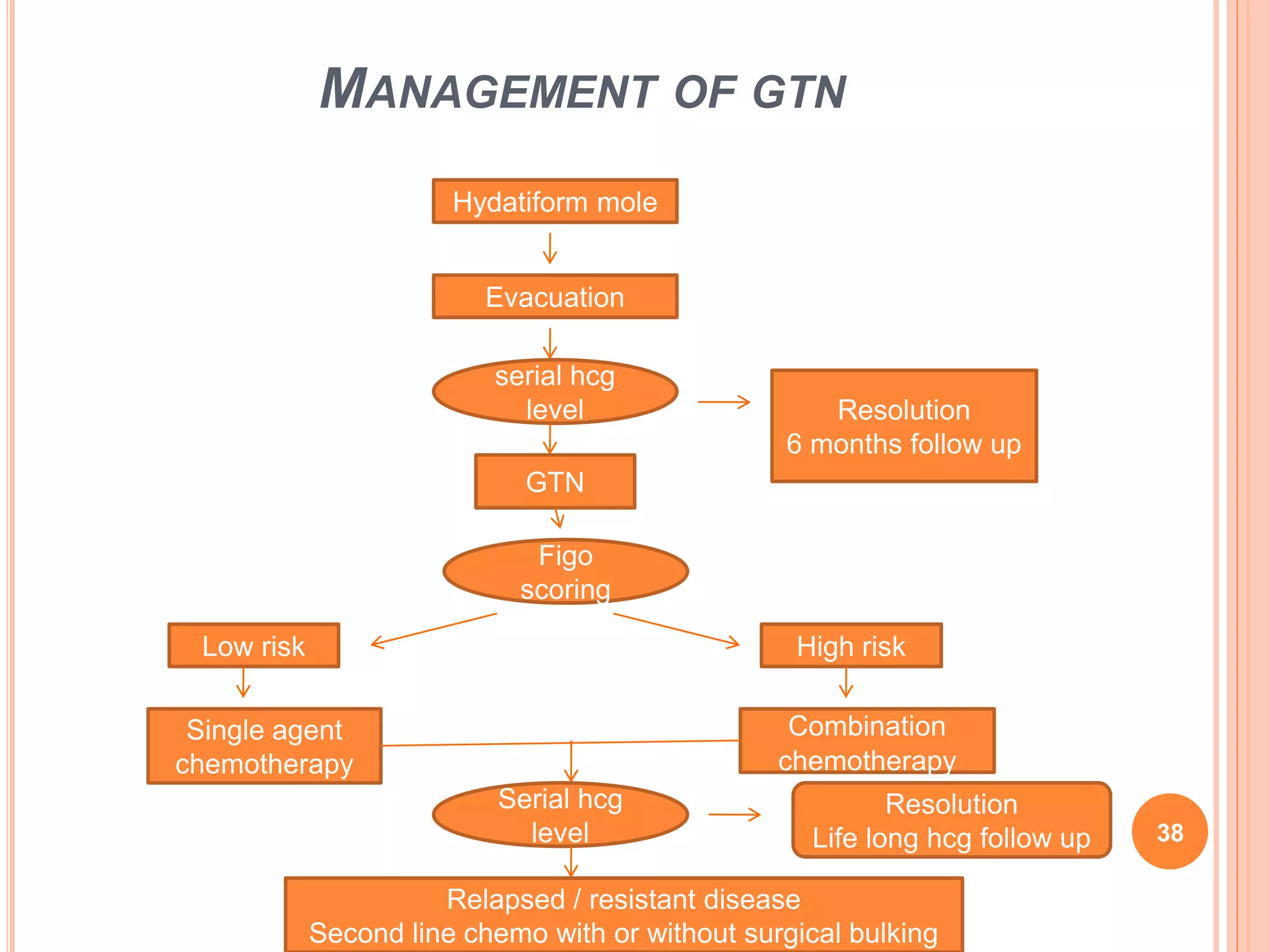
GESTATIONAL TROPHOBLASTIC
DISEASE PART - 2
1
GESTATIONAL TROPHOBLASTIC
NEOPLASIA
 Accounts < 1% of female reproductive system cancers
 Develops after a molar pregnancy , a live birth or an
abortion/ectopic pregnancy
 Includes :
1. INVASIVE MOLE
2. CHORIOCARCINOMA
3. PLACENTAL SITE TROPHOBLASTIC TUMOUR
4. EPITHELOID TROPHOBLASTIC TUMOUR
2
EPIDEMIOLOGY FOR CHORIOCARCINOMA
 Incidence after live birth – 1 in 50,000 pregnancies
COUNTRY INCIDENCE
EUROPE / NORTH AMERICA 1 in 40,000 pregnancies
1 in 40 hydatiform moles
SOUTHEAST ASIA 9.2 / 40,000 pregnancies
JAPAN 3.2 / 40,000 pregnancies
3
INVASIVE MOLE (CHORIOADENOMA DESTRUENS)
 Always develop from hydatiform mole and not normal pregnancy
 Arise from myometrial invasion ( direct tissue or venous channels)
 Complete mole – 15% 15 % metastasize to lung and vagina
Partial mole – 3 to 4%
 Rest ,
- perforate myometrium :intraperitoneal bleed
- erode uterine vessels : vaginal hemorrhage
- necrotic tumour involve uterine wall :
nidus for bacterial infection
 Microscopic : presence of hydropic villi with trophoblasts extending to
myometrium
 Diagnosed clinically and treated with chemotherapy
4
GESTATIONAL NON GESTATIONAL
• Gross : dark or red blackish mass with
hemorrhage
•Seen in both genders
• Microscopic :
- abnormal trophoblastic hyperplasia
- absence of chorionic villi
- hemorrhage and necrosis invading
myometrium
• Develop in ovaries , testicles, chest and
abdomen
• Vascular invasion resulting in spread to
distant sites
• Choriocarcinoma mixed with other types of
cancer forming mixed germ cell tumor
• Lung (80%) , vagina( 30%), pelvis (20%) ,
liver (10%), brain (10%) and others
( spleen , kidney , GIT)
• Less responsive to chemotherapy and less
favorable prognosis
5
6
PLACENTAL SITE TROPHOBLASTIC TUMOUR
 Extremely rare , slow growing , rarely metastasize
 Mostly follow non molar pregnancy but
seen with hydatiform mole and abortion
 Arise from placental implantation site
 Predominantly of mononuclear intermediate
trophoblasts without chorionic villi infiltrating
myometrial fibres
 Confined to uterus with propensity for lymphatic and late lung, brain
metastasis
 Low levels of hCG compared to tumor burden
 Immunohistochemical staining reveals diffuse presence of cytokeratin and
HPL whereas hcg is only focal
7
EPITHELOID TROPHOBLASTIC TUMOUR
 Rare variant of GTD
 Behaves like PSTT
 Most often occurs after term pregnancy
but can take several years to occur
 Used to be called as atypical choriocarcinoma
 Can be found growing in cervix , confused with cervical cancer
 Develops from neoplastic transformation of chorionic type
intermediate trophoblasts
8
CLINICAL FEATURES
 POST MOLAR GTN ( invasive mole or choriocarcinoma)
SYMPTOMS SIGNS
• Irregular vaginal bleeding • Enlarged irregular uterus
• Infection causing vaginal
discharge, pelvic cramps
and fever
• Persistent B/L ovarian
enlargement
• Rarely, intra abdominal
bleeding with severe pain
9
 METASTASIZE
 Diagnosis of choriocarcinoma should be considered in any
women of reproductive age group presenting with these acute
events
AREA INVOLVED SYMPTOMS
• Pulmonary • Chest pain, cough , hemoptysis, dyspnea
• Vaginal lesions • Fornices or suburetheral region
extremely vascular
• Liver • Epigastric pain or right upper quadrant pain,
Jaundice, hemoptysis
• Brain • Headache,seizures, hemiplegia
10
11
 Non molar GTN
- Choriocarcinoma has no characteristic symptoms or sign
( either related to uterus or metastatic sites)
 PSTT & ETT : irregular uterine bleeding
: symmetric uterine enlargement
: symptoms related to metastatic sites
12
INDICATION OF CHEMOTHERAPY IN GTD
1. Plateaued or rising hCG after evacuation
- Plateau Four or more values over a period of 3 weeks or longer
( 1,7,14, 21 )
- Rise (>10%) during three weekly consecutive measurements or longer during
a period of 2 weeks or more (1,7,14)
2. Elevated hcg 6 months after evacuation even if still falling
3. Histological evidence of choriocarcinoma
4. Evidence of metastasis in brain , liver or GIT , or radiological opacities
>2 cm on chest Xray
5. Pulmonary, vulval or vaginal metastases unless hcg falling
6. Heavy vaginal bleeding or evidence of GI or intraperitoneal
hemorrhage
7. Serum hcg >20,000 IU/L more than 4 weeks after evacuation
( risk of uterine perforation)
13
DIAGNOSIS
 History
 Pelvic examination
 Serum ßhcg level
 Pelvic doppler ultrasound
 Chest X-ray
OTHERS
 CT , MRI
 Blood investigations : CBC, LFT, RFT, PS, TFT
14
1) History
- Age
- Symptoms
- Duration
- Antecedent pregnancy ( molar or non molar)
- Interval from antecedent pregnancy
- Prior treatment ( evacuation or chemotherapy with single or
multiple agent)
2) Examination
- Pallor
- R/S ( RR or rales on auscultation)
- CNS ( level of consciousness)
- P/A ( uterine enlargement, organomegaly , tenderness, guarding )
- P/S ( bleeding , vaginal/ suburetheral nodules)
- Bimanual ( uterine size and theca lutein cyst)
15
3 ) Pelvic Doppler USG
- volume of uterus measured
- theca lutein cyst and uterine involvement
- molar tissue has low resistance circulation( AV shunting)
- low pulsatile index of uterine artery ( poor prognosis and
predictor of chemotherapy response)
16
4 ) ß hcg
 exists in 2 forms
- ß hcg regular : normal pregnancy
- hcg – H : produced by invasive cytotrophoblast
ß hcg hcg - H
Production Syncytiotrophoblast Invasive cytotrophoblast
Present - Full normal pregnancy
- All forms of GTD
- Implantation phase of normal
pregnancy
- In GTN
Method of action Endocrine Autocrine
Role Maintain progesterone
production
Promotes growth and invasion
of trophoblasts
17
5) CHEST XRAY
- Snowstorm appearance
- Discrete rounded densities
- Pleural effusion
- Embolic pattern caused by
pulmonary occlusion
 Negative pelvic examination
and chest Xray – metastasis of
other site uncommon
 If chest X-ray positive :
- CT chest – to find out micrometastasis ( 40%)
- MRI brain – early cerebral lesion , MRI pelvis
- CT abdomen
- Check CSF hcg level, CSF / serum > 1:60 – cerebral metastasis
18
PATHOLOGIC DIAGNOSIS
1. Curettage
 Repeat curettage avoided
- To prevent risk of hemorrhage, perforation or infection
- Increase risk of need of chemotherapy ( hcg > 5000 IU/L)
- Unless excessive uterine bleeding and intracavitary , should be
avoided
2. Biopsy of metastatic lesion
( dangerous for vaginal lesion – profuse bleeding)
3. Examination of hysterectomy specimen and placenta
19
FIGO ANATOMIC STAGING SYSTEM FOR GTN
 Stage 1 : patients have persistently elevated hcg level and tumor
confined to the uterine corpus
 Stage 2 : patients have metastasis to genital tract
( adnexa, vagina , broad ligament)
 Stage 3 : patients have pulmonary metastases with or without
uterine, vaginal, or pelvic involvement
 Stage 4 : patients have advanced disease and involvement of the
brain , liver , kidneys or GIT 20
PROGNOSTIC SCORING AND STAGING
 Prognostic score is central to management of GTN
 Predicts likelihood of developing resistance to single agent
chemotherapy
 Two categories : low risk (0-6)
: high risk (≥7)
 Predicts likelihood of cure
- Low risk: 100%
- High risk : 95%
 Adverse Prognostic factors in high risk group:
- Longer duration from antecedent pregnancy
- Presence of liver metastases
- Combined liver and brain metastasis ( 10% survival)
21
0 1 2 4
 Age (yrs) ≤39 >39
 Antecedent pregnancy Hydatiform
mole
Abortion Term
 Interval b/w end of
antecedent pregnancy
and start of
chemotherapy
( months)
<4 4-6 7-12 >12
 Pretreatment hcg
(IU/L)
<10³ 10³ - 104 104 - 105 >105
 ABO groups O or A B or AB
 Largest tumor,
including uterine (cm)
<3 3-5 >5
 Site of metastases spleen ,
kidney
GIT Brain , Liver
 Number of metastases 1-3 4-8 >8
 Prior chemotherapy 1 drug ≥2 drugs
Low risk:<7 and High risk : >7
22
TREATMENT
 MEDICAL
 SURGICAL
 RADIATION
23
LOW RISK DISEASE
 Includes : stage 1
: stage 2 and 3 , score <7
 single agent chemotherapy with Methotrexate or Actinomycin D
 Fertility desired : single agent chemotherapy resistant ( 20%)
sequential single agent
local uterine resection combination resistant (10%)
24
 If fertility not desired
- Hysterectomy + adjuvant single agent chemotherapy
- Adjuvant chemo : 1) to prevent dissemination of tumor cells
2) maintain cytotoxic level in case of dissemination
3) treat occult metastases @ the time of surgery
 Chemotherapy continued until hcg normal and at least further 6 weeks
RESISTANCE IN STAGE 1 RESISTANCE IN STAGE 2 -3
• Presence of metastases • Age >35 years
• Clinicopathologic diagnosis of
choriocarcinoma
• FIGO score > 7
• Pretreatment hcg > 50,000 IU/L • Pretreatment hcg > 100,000 IU/l
• Non molar antecedent
pregnancy
• Large vaginal metastases
25
CHEMOTHERAPY REGIMENS FOR LOW RISK
 Methotrexate and folinic acid – cycle repeated every 14 days
 Remission rate – 70 to 90%
METHOTREXATE FOLINIC ACID
• 1mg/kg IM d 1,3,5,7 • 0.1 mg/kg IM d 2,4,6,8
• 50 mg IM every 48 hrs for total 4
doses
• 15 mg orally 30 hr after each injection
of methotrexate
26
HIGH RISK DISEASE
 Includes : stage 4
: stage 2-3 , score ≥7
 Initial treatment with multiagent chemotherapy with or without adjuvant
surgery or radiation therapy
 Chemotherapy continued for at least 6 weeks after the first normal hcg
YEAR REGIMEN
•1970s • MAC ( MTX , Act- D, cyclophosphamide) regimen
• early 1980s • CHAMOCA ( Cyclophosphamide , Hydroxyurea , Act-D ,
MTX-FA , Vincristine , doxorubicin) regimen
•1980s • EMA-CO( Etoposide, high dose MTX-FA , Act-D,
Cyclophosphamide , Vincristine) regimen
27
CHEMOTHERAPY REGIMEN FOR HIGH RISK
DAY DRUG DOSING
1 Etoposide
Actinomycin D
MTX
100mg/m² IV over 30 min
0.5mg IVP
100mg/m² IVP , then
200mg/m² in 500 ml over
12h
2 Etoposide
Actinomycin D
FA
100mg/m² IV over 30 min
0.5mg IVP
15 mg IM or PO every
12 h for 4 doses starting
24 h after MTX
8 Cyclophosphamide
vincristine
600mg/m² IV
1 mg/m² IVP
EVERY 2 WEEKS
28
RESIDUAL POST TREATMENT MASSES
 After chemotherapy , patients are reimaged
 Pelvic USG and any abnormal investigation at diagnosis
 In USG, presence of uterine vascular malformations are normal and no
intervention needed unless active bleeding
 UAE prevents need of hysterectomy
 Residual brain lesion needs irradiation:
- Whole brain irradiation ( 3000 cGy in 200 cGy fractions) , or
- Surgical excision with stereotactic irradiation with simultaneous
systemic chemotherapy ( EMA –CO with MTX 1mg/m² and FA every 12
hr for 3 days starting 32 hrs after MTX infusion )
- Alternative to brain irradiation, intrathecal MTX with high dose IV MTX
29
 Residual masses at other sites - not resected and follow up
by standard hcg surveillance
 Liver metastasis – if resistant to combination chemo
: hepatic arterial infusion of chemo
: hepatic resection
: hepatic arterial embolisation
 Complete serological response necessary for cure but
complete radiological response is not
30
RESISTANT AND RELAPSED HIGH RISK DISEASE
 Relapsed – rising hcg after complete serological response
to EMA/CO ( at least 6 weeks of normal hcg
post chemo)
 Resistant/ refractory – rising or plateau hcg while on
EMA /CO
 FDG – PET ( fluorodeoxyglucose) can be used to find sites of
disease
 Treatment – surgical resection with chemotherapy or
salvage chemotherapy alone 31
 Most effective 2nd line regimen
– EMA/EP ( EMA and etoposide plus cisplatin – omits 2nd day dose of
etoposide and ACT-D)
 Less toxic , alternative
– fortnightly regimen of paclitaxel + cisplatin
alternating with paclitaxel + etoposide
 Other drugs – vinblastine, bleomycin, ifosfamide, 5-FU
 High dose chemotherapy with peripheral stem cell transplantation
 New chemotherapy drugs – molecular targeted therapies 32
33
SIDE EFFECTS
COMMON SPECIFIC
•Hair loss •Methotrexate : diarrhea, sore throat, stomatitis,
conjunctivitis, abdominal and chest pain , liver damage
-Hair loss and blood effects not seen
•Mouth sores •Actinomycin – D : severe nausea and vomiting, hair loss,
blood effects
•Loss of appetite •Etoposide : leukemia, breast cancer, colon cancer
•Nausea and vomiting •Cyclophosphamide and ifosfamide : nausea and hair
loss, bladder irritation
•Low blood counts •Bleomycin : lung problems
•Advancing age of
menopause :
1 year – single agent
3 yrs – multi agent
•Vincristine and cisplatin : neuropathy
•hearing loss and kidney damage ( persists)
34
PSTTS AND ETTS
 Chemoresistant with lymphatic spread
 Standard treatment – hysterectomy with lymph node clearance and
ovarian preservation
 In metastasis, EMA/EP with or without surgical resection
 Most imp prognostic factor : interval from causative pregnancy
- > 4 yrs post pregnancy( high mortality)
- < 4 yrs post pregnancy ( high survival)
 5 reported cases of fertility sparing surgery
- One : successful pregnancy
- Two: long term remission but no term pregnancy
- Two : need for hysterectomy for relapse
35
FOLLOW UP AFTER CHEMOTHERAPY
 Relapse risk : 3% with maximum in 1st year
 Measure serum hCG weekly after course of chemotherapy
3 consecutive week normal value
low risk high risk
monthly for an year monthly for 2 years
- Physical exam every 3-6 months for first year and then 6
monthly further 37
MANAGEMENT OF GTN
Hydatiform mole
Evacuation
serial hcg
level Resolution
6 months follow up
GTN
Figo
scoring
Low risk High risk
Single agent
chemotherapy
Combination
chemotherapy
Serial hcg
level
Resolution
Life long hcg follow up
Relapsed / resistant disease
Second line chemo with or without surgical bulking
38
CONTRACEPTION AND FUTURE PREGNANCY
 Contraception should be maintained during treatment and for 1 year
after completion of chemotherapy, preferably oral contraceptives
 Helps in uninterrupted hcg follow up
 Permit elimination of mature ova that may have been damaged by
exposure to cytotoxic drugs
 Most women resume ovarian function after chemotherapy
 Had successful pregnancies without increase congenital anomalies but
high risk of abortions and still birth
39
THANK YOU
40

Gestational trophoblastic disease part 2-1 - copy

  • 1.
  • 2.
    GESTATIONAL TROPHOBLASTIC NEOPLASIA  Accounts< 1% of female reproductive system cancers  Develops after a molar pregnancy , a live birth or an abortion/ectopic pregnancy  Includes : 1. INVASIVE MOLE 2. CHORIOCARCINOMA 3. PLACENTAL SITE TROPHOBLASTIC TUMOUR 4. EPITHELOID TROPHOBLASTIC TUMOUR 2
  • 3.
    EPIDEMIOLOGY FOR CHORIOCARCINOMA Incidence after live birth – 1 in 50,000 pregnancies COUNTRY INCIDENCE EUROPE / NORTH AMERICA 1 in 40,000 pregnancies 1 in 40 hydatiform moles SOUTHEAST ASIA 9.2 / 40,000 pregnancies JAPAN 3.2 / 40,000 pregnancies 3
  • 4.
    INVASIVE MOLE (CHORIOADENOMADESTRUENS)  Always develop from hydatiform mole and not normal pregnancy  Arise from myometrial invasion ( direct tissue or venous channels)  Complete mole – 15% 15 % metastasize to lung and vagina Partial mole – 3 to 4%  Rest , - perforate myometrium :intraperitoneal bleed - erode uterine vessels : vaginal hemorrhage - necrotic tumour involve uterine wall : nidus for bacterial infection  Microscopic : presence of hydropic villi with trophoblasts extending to myometrium  Diagnosed clinically and treated with chemotherapy 4
  • 5.
    GESTATIONAL NON GESTATIONAL •Gross : dark or red blackish mass with hemorrhage •Seen in both genders • Microscopic : - abnormal trophoblastic hyperplasia - absence of chorionic villi - hemorrhage and necrosis invading myometrium • Develop in ovaries , testicles, chest and abdomen • Vascular invasion resulting in spread to distant sites • Choriocarcinoma mixed with other types of cancer forming mixed germ cell tumor • Lung (80%) , vagina( 30%), pelvis (20%) , liver (10%), brain (10%) and others ( spleen , kidney , GIT) • Less responsive to chemotherapy and less favorable prognosis 5
  • 6.
  • 7.
    PLACENTAL SITE TROPHOBLASTICTUMOUR  Extremely rare , slow growing , rarely metastasize  Mostly follow non molar pregnancy but seen with hydatiform mole and abortion  Arise from placental implantation site  Predominantly of mononuclear intermediate trophoblasts without chorionic villi infiltrating myometrial fibres  Confined to uterus with propensity for lymphatic and late lung, brain metastasis  Low levels of hCG compared to tumor burden  Immunohistochemical staining reveals diffuse presence of cytokeratin and HPL whereas hcg is only focal 7
  • 8.
    EPITHELOID TROPHOBLASTIC TUMOUR Rare variant of GTD  Behaves like PSTT  Most often occurs after term pregnancy but can take several years to occur  Used to be called as atypical choriocarcinoma  Can be found growing in cervix , confused with cervical cancer  Develops from neoplastic transformation of chorionic type intermediate trophoblasts 8
  • 9.
    CLINICAL FEATURES  POSTMOLAR GTN ( invasive mole or choriocarcinoma) SYMPTOMS SIGNS • Irregular vaginal bleeding • Enlarged irregular uterus • Infection causing vaginal discharge, pelvic cramps and fever • Persistent B/L ovarian enlargement • Rarely, intra abdominal bleeding with severe pain 9
  • 10.
     METASTASIZE  Diagnosisof choriocarcinoma should be considered in any women of reproductive age group presenting with these acute events AREA INVOLVED SYMPTOMS • Pulmonary • Chest pain, cough , hemoptysis, dyspnea • Vaginal lesions • Fornices or suburetheral region extremely vascular • Liver • Epigastric pain or right upper quadrant pain, Jaundice, hemoptysis • Brain • Headache,seizures, hemiplegia 10
  • 11.
  • 12.
     Non molarGTN - Choriocarcinoma has no characteristic symptoms or sign ( either related to uterus or metastatic sites)  PSTT & ETT : irregular uterine bleeding : symmetric uterine enlargement : symptoms related to metastatic sites 12
  • 13.
    INDICATION OF CHEMOTHERAPYIN GTD 1. Plateaued or rising hCG after evacuation - Plateau Four or more values over a period of 3 weeks or longer ( 1,7,14, 21 ) - Rise (>10%) during three weekly consecutive measurements or longer during a period of 2 weeks or more (1,7,14) 2. Elevated hcg 6 months after evacuation even if still falling 3. Histological evidence of choriocarcinoma 4. Evidence of metastasis in brain , liver or GIT , or radiological opacities >2 cm on chest Xray 5. Pulmonary, vulval or vaginal metastases unless hcg falling 6. Heavy vaginal bleeding or evidence of GI or intraperitoneal hemorrhage 7. Serum hcg >20,000 IU/L more than 4 weeks after evacuation ( risk of uterine perforation) 13
  • 14.
    DIAGNOSIS  History  Pelvicexamination  Serum ßhcg level  Pelvic doppler ultrasound  Chest X-ray OTHERS  CT , MRI  Blood investigations : CBC, LFT, RFT, PS, TFT 14
  • 15.
    1) History - Age -Symptoms - Duration - Antecedent pregnancy ( molar or non molar) - Interval from antecedent pregnancy - Prior treatment ( evacuation or chemotherapy with single or multiple agent) 2) Examination - Pallor - R/S ( RR or rales on auscultation) - CNS ( level of consciousness) - P/A ( uterine enlargement, organomegaly , tenderness, guarding ) - P/S ( bleeding , vaginal/ suburetheral nodules) - Bimanual ( uterine size and theca lutein cyst) 15
  • 16.
    3 ) PelvicDoppler USG - volume of uterus measured - theca lutein cyst and uterine involvement - molar tissue has low resistance circulation( AV shunting) - low pulsatile index of uterine artery ( poor prognosis and predictor of chemotherapy response) 16
  • 17.
    4 ) ßhcg  exists in 2 forms - ß hcg regular : normal pregnancy - hcg – H : produced by invasive cytotrophoblast ß hcg hcg - H Production Syncytiotrophoblast Invasive cytotrophoblast Present - Full normal pregnancy - All forms of GTD - Implantation phase of normal pregnancy - In GTN Method of action Endocrine Autocrine Role Maintain progesterone production Promotes growth and invasion of trophoblasts 17
  • 18.
    5) CHEST XRAY -Snowstorm appearance - Discrete rounded densities - Pleural effusion - Embolic pattern caused by pulmonary occlusion  Negative pelvic examination and chest Xray – metastasis of other site uncommon  If chest X-ray positive : - CT chest – to find out micrometastasis ( 40%) - MRI brain – early cerebral lesion , MRI pelvis - CT abdomen - Check CSF hcg level, CSF / serum > 1:60 – cerebral metastasis 18
  • 19.
    PATHOLOGIC DIAGNOSIS 1. Curettage Repeat curettage avoided - To prevent risk of hemorrhage, perforation or infection - Increase risk of need of chemotherapy ( hcg > 5000 IU/L) - Unless excessive uterine bleeding and intracavitary , should be avoided 2. Biopsy of metastatic lesion ( dangerous for vaginal lesion – profuse bleeding) 3. Examination of hysterectomy specimen and placenta 19
  • 20.
    FIGO ANATOMIC STAGINGSYSTEM FOR GTN  Stage 1 : patients have persistently elevated hcg level and tumor confined to the uterine corpus  Stage 2 : patients have metastasis to genital tract ( adnexa, vagina , broad ligament)  Stage 3 : patients have pulmonary metastases with or without uterine, vaginal, or pelvic involvement  Stage 4 : patients have advanced disease and involvement of the brain , liver , kidneys or GIT 20
  • 21.
    PROGNOSTIC SCORING ANDSTAGING  Prognostic score is central to management of GTN  Predicts likelihood of developing resistance to single agent chemotherapy  Two categories : low risk (0-6) : high risk (≥7)  Predicts likelihood of cure - Low risk: 100% - High risk : 95%  Adverse Prognostic factors in high risk group: - Longer duration from antecedent pregnancy - Presence of liver metastases - Combined liver and brain metastasis ( 10% survival) 21
  • 22.
    0 1 24  Age (yrs) ≤39 >39  Antecedent pregnancy Hydatiform mole Abortion Term  Interval b/w end of antecedent pregnancy and start of chemotherapy ( months) <4 4-6 7-12 >12  Pretreatment hcg (IU/L) <10³ 10³ - 104 104 - 105 >105  ABO groups O or A B or AB  Largest tumor, including uterine (cm) <3 3-5 >5  Site of metastases spleen , kidney GIT Brain , Liver  Number of metastases 1-3 4-8 >8  Prior chemotherapy 1 drug ≥2 drugs Low risk:<7 and High risk : >7 22
  • 23.
  • 24.
    LOW RISK DISEASE Includes : stage 1 : stage 2 and 3 , score <7  single agent chemotherapy with Methotrexate or Actinomycin D  Fertility desired : single agent chemotherapy resistant ( 20%) sequential single agent local uterine resection combination resistant (10%) 24
  • 25.
     If fertilitynot desired - Hysterectomy + adjuvant single agent chemotherapy - Adjuvant chemo : 1) to prevent dissemination of tumor cells 2) maintain cytotoxic level in case of dissemination 3) treat occult metastases @ the time of surgery  Chemotherapy continued until hcg normal and at least further 6 weeks RESISTANCE IN STAGE 1 RESISTANCE IN STAGE 2 -3 • Presence of metastases • Age >35 years • Clinicopathologic diagnosis of choriocarcinoma • FIGO score > 7 • Pretreatment hcg > 50,000 IU/L • Pretreatment hcg > 100,000 IU/l • Non molar antecedent pregnancy • Large vaginal metastases 25
  • 26.
    CHEMOTHERAPY REGIMENS FORLOW RISK  Methotrexate and folinic acid – cycle repeated every 14 days  Remission rate – 70 to 90% METHOTREXATE FOLINIC ACID • 1mg/kg IM d 1,3,5,7 • 0.1 mg/kg IM d 2,4,6,8 • 50 mg IM every 48 hrs for total 4 doses • 15 mg orally 30 hr after each injection of methotrexate 26
  • 27.
    HIGH RISK DISEASE Includes : stage 4 : stage 2-3 , score ≥7  Initial treatment with multiagent chemotherapy with or without adjuvant surgery or radiation therapy  Chemotherapy continued for at least 6 weeks after the first normal hcg YEAR REGIMEN •1970s • MAC ( MTX , Act- D, cyclophosphamide) regimen • early 1980s • CHAMOCA ( Cyclophosphamide , Hydroxyurea , Act-D , MTX-FA , Vincristine , doxorubicin) regimen •1980s • EMA-CO( Etoposide, high dose MTX-FA , Act-D, Cyclophosphamide , Vincristine) regimen 27
  • 28.
    CHEMOTHERAPY REGIMEN FORHIGH RISK DAY DRUG DOSING 1 Etoposide Actinomycin D MTX 100mg/m² IV over 30 min 0.5mg IVP 100mg/m² IVP , then 200mg/m² in 500 ml over 12h 2 Etoposide Actinomycin D FA 100mg/m² IV over 30 min 0.5mg IVP 15 mg IM or PO every 12 h for 4 doses starting 24 h after MTX 8 Cyclophosphamide vincristine 600mg/m² IV 1 mg/m² IVP EVERY 2 WEEKS 28
  • 29.
    RESIDUAL POST TREATMENTMASSES  After chemotherapy , patients are reimaged  Pelvic USG and any abnormal investigation at diagnosis  In USG, presence of uterine vascular malformations are normal and no intervention needed unless active bleeding  UAE prevents need of hysterectomy  Residual brain lesion needs irradiation: - Whole brain irradiation ( 3000 cGy in 200 cGy fractions) , or - Surgical excision with stereotactic irradiation with simultaneous systemic chemotherapy ( EMA –CO with MTX 1mg/m² and FA every 12 hr for 3 days starting 32 hrs after MTX infusion ) - Alternative to brain irradiation, intrathecal MTX with high dose IV MTX 29
  • 30.
     Residual massesat other sites - not resected and follow up by standard hcg surveillance  Liver metastasis – if resistant to combination chemo : hepatic arterial infusion of chemo : hepatic resection : hepatic arterial embolisation  Complete serological response necessary for cure but complete radiological response is not 30
  • 31.
    RESISTANT AND RELAPSEDHIGH RISK DISEASE  Relapsed – rising hcg after complete serological response to EMA/CO ( at least 6 weeks of normal hcg post chemo)  Resistant/ refractory – rising or plateau hcg while on EMA /CO  FDG – PET ( fluorodeoxyglucose) can be used to find sites of disease  Treatment – surgical resection with chemotherapy or salvage chemotherapy alone 31
  • 32.
     Most effective2nd line regimen – EMA/EP ( EMA and etoposide plus cisplatin – omits 2nd day dose of etoposide and ACT-D)  Less toxic , alternative – fortnightly regimen of paclitaxel + cisplatin alternating with paclitaxel + etoposide  Other drugs – vinblastine, bleomycin, ifosfamide, 5-FU  High dose chemotherapy with peripheral stem cell transplantation  New chemotherapy drugs – molecular targeted therapies 32
  • 33.
  • 34.
    SIDE EFFECTS COMMON SPECIFIC •Hairloss •Methotrexate : diarrhea, sore throat, stomatitis, conjunctivitis, abdominal and chest pain , liver damage -Hair loss and blood effects not seen •Mouth sores •Actinomycin – D : severe nausea and vomiting, hair loss, blood effects •Loss of appetite •Etoposide : leukemia, breast cancer, colon cancer •Nausea and vomiting •Cyclophosphamide and ifosfamide : nausea and hair loss, bladder irritation •Low blood counts •Bleomycin : lung problems •Advancing age of menopause : 1 year – single agent 3 yrs – multi agent •Vincristine and cisplatin : neuropathy •hearing loss and kidney damage ( persists) 34
  • 35.
    PSTTS AND ETTS Chemoresistant with lymphatic spread  Standard treatment – hysterectomy with lymph node clearance and ovarian preservation  In metastasis, EMA/EP with or without surgical resection  Most imp prognostic factor : interval from causative pregnancy - > 4 yrs post pregnancy( high mortality) - < 4 yrs post pregnancy ( high survival)  5 reported cases of fertility sparing surgery - One : successful pregnancy - Two: long term remission but no term pregnancy - Two : need for hysterectomy for relapse 35
  • 36.
    FOLLOW UP AFTERCHEMOTHERAPY  Relapse risk : 3% with maximum in 1st year  Measure serum hCG weekly after course of chemotherapy 3 consecutive week normal value low risk high risk monthly for an year monthly for 2 years - Physical exam every 3-6 months for first year and then 6 monthly further 37
  • 37.
    MANAGEMENT OF GTN Hydatiformmole Evacuation serial hcg level Resolution 6 months follow up GTN Figo scoring Low risk High risk Single agent chemotherapy Combination chemotherapy Serial hcg level Resolution Life long hcg follow up Relapsed / resistant disease Second line chemo with or without surgical bulking 38
  • 38.
    CONTRACEPTION AND FUTUREPREGNANCY  Contraception should be maintained during treatment and for 1 year after completion of chemotherapy, preferably oral contraceptives  Helps in uninterrupted hcg follow up  Permit elimination of mature ova that may have been damaged by exposure to cytotoxic drugs  Most women resume ovarian function after chemotherapy  Had successful pregnancies without increase congenital anomalies but high risk of abortions and still birth 39
  • 39.