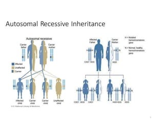
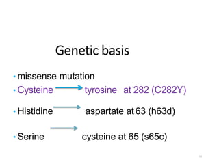
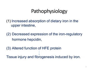
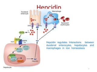
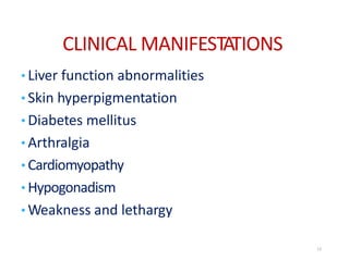
GENETICS
UNIT – IV
GENETIC CONDITIONS OF ADOLESCENTS
AND ADULTS
TOPIC : Genetic Haemochromatosis
PRESENTED BY
Mrs. SOUMYA SUBRAMANI, M.Sc.(N)
LECTURER, MSN DEPARTMENT
CON- SRIPMS, COIMBATORE.
1
SYNONYMS
• HEREDITARY HEMOCHROMATOSIS
• PRIMARY HEMOCHROMATOSIS
GENETIC HEMOCHROMATOSIS
2
Introduction
• Genetic/ Hereditary hemochromatosis is an inherited
predisposition to absorb excess iron (Fe) from the diet
• Total body iron may reach up to 20-60g, normal body
iron is 3-4g
• Mutations in the HFE gene are the most common
cause of adult onset iron overload.
• In some predisposed individuals, excessive iron
absorption and subsequent storage in various organs
(i.e. liver, pancreas, heart, joints and pituitary)
eventually lead to cellular injury
• If untreated, over time this can cause irreversible
tissue/organ damage and shorten life expectancy
3
Definition
• Genetic Hemochromatosis is autosomal recessive
genetic disorder that results from abnormal
accumulation of iron in parenchymal organs such
as the liver, pancreas, and heart leading to organ
toxicity.
4
Autosomal Recessive Inheritance
5
Epidemiology
• Hereditary hemochromatosis (HH) remains the
most common genetic disorder in Caucasians.
• Women typically presented approximately 10
years later than men
• Female: male- 1 : 10
6
CLASSIFICATION
• Primary hemochromatosis
• Secondary hemochromatosis
Types of hemochromatosis Gene mutated
Type 1 HFE
Type 2 Either HFE 2or HAMP
Type 3 TFR2
Type 4 SLC40A1
7
Genetic of Hemochromatosis
• The HAMP, HFE, HFE2,SLC40A and TFR2 are the genes
that regulate iron metabolism, i.e., absorption, transport and
storage.
• Mutations in the above mentioned genes will increase the
iron absorption and its subsequent transport and distribution.
8
HFE gene
• HFE gene on chromosome 6
• Involved in iron homeostasis by interacting with
transferrin receptor in basolateral membrane of
intestinal epithelial cells
• HFE protein normally limits amount of iron uptake by
gut and regulates amount of iron stored in the tissues
• A defect in uptake of transferrin associated iron may lead
to upregulation of enterocyte iron specific divalent metal
transporters and excessive iron absorption. 9
Genetic basis
• missense mutation
• Cysteine tyrosine at 282 (C282Y)
• Histidine aspartate at 63 (h63d)
• Serine cysteine at 65 (s65c)
10
Pathophysiology
(1) Increased absorption of dietary iron in the
upper intestine,
(2) Decreased expression of the iron-regulatory
hormone hepcidin,
(3) Altered function of HFE protein
Tissue injury and fibrogenesis induced by iron.
11
Hepcidin
Hepcidin regulates Interactions between
duodenal enterocytes, hepatocytes and
macrophages in iron homeostasis
12
CLINICAL MANIFESTATIONS
• Liver function abnormalities
• Skin hyperpigmentation
• Diabetes mellitus
• Arthralgia
• Cardiomyopathy
• Hypogonadism
• Weakness and lethargy
13
Hepatic manifestations
• Liver is usually the first organ to be affected
• Hepatomegaly in >95% of symptomatic
patients.
• Cirrhosis, Portal hypertension and esophageal
varices.
• Hepatocellular carcinoma develops in about
30% of patients with cirrhosis.
• Incidence increases with age, common in men,
almost exclusively in cirrhotic patients
14
Skin
• Skin pigmentation – Landen- grey (bronze)
skin pigmentation, Results from increased
melanin and iron in the dermis.
• Pigmentation usually is generalized,
More pronounced on;
• The face, neck,
• Extensor aspects of the lower forearms
• Dorsa of the hands, lower legs,
• Genital regions,
15
Diabetes
• Diabetes mellitus occurs in about 65%
• More likely to develop in those with a family history of
diabetes
• Insulin resistance is more common in
association with hemochromatosis
16
Arthropathy
• Arthropathy develops in 25–50% of
symptomatic patients.
• Chondrocalcinosis occurs secondary to deposition of
Calcium pyrophosphate.
17
Calcium pyrophosphate
deposition disease, atypical
osteoarthritis involving the
2nd & 3rd MCP joints & KJ
18
Cardiac manifestations
• Presenting manifestation in about
15%.
• Most common manifestation is
congestive heart failure.
• Cardiac arrhythmias & Varying
degrees of AV block.
• premature supraventricular
beats, tachyarrhythmias
• Atrial flutter
• Atrial fibrillation
19
Hypogonadism
•Occurs in both sexes.
•Impairment of hypothalamic-pituitary function by iron
deposition
• Loss of libido
• Impotence
• Amenorrhea
• Testicular atrophy
• Gynecomastia
• sparse body hair
20
Screening for HH
• High risk groups;
• Family history of HH
• Those with suspected organ involvement
• Those with chance detection of biochemical
and/or radiological abnormalities
• Optimal timing for screening family members is
between the ages of 18 and 30,
21
Diagnosis
1. Transferrin saturation —
• If transferrin saturation >45%
• the presence of the C282Y and H63D mutation
confirm the diagnosis of hemochromatosis
2.Plasma ferritin — normal 40 to 200 ng/ml
• >200 mcg/L in premenopausal women
• > 300 mcg/L in men and postmenopausal women
indicate primary iron overload
• Serum ferritin levels have an additional value as a
predictor of advanced fibrosis and cirrhosis in
confirmed HH
22
Liver biopsy
Diagnosis
•Liver biopsy should be considered;
• •For the purpose of determining the presence or
absence of advanced fibrosis or cirrhosis.
• •For measurement of Hepatic Iron Concentration.
23
Diagnosis
• Imaging studies of liver –
• Increased density of the liver due to iron overload.
• Genetic study-
• Both the C282Y and H63D mutation
24
Treatment of Hemochromatosis
• Phlebotomy remains the sole recommended treatment -
simple, inexpensive and safe.
• Each 500 mL of whole blood removed contains 200 to 250
mg of iron.
Induction phase
• One phlebotomy (500 mL) one to two per week.
• Check hematocrit (Hct) prior to each phlebotomy;
• do not allow Hct to fall by more than 20 percent of prior level
• Check serum ferritin every 10 to 12 phlebotomies
25
Maintenance phase
• The phlebotomy should be performed every 2-4 months.
• The interval between procedures is determined by the level of
ferritin, which should be 50 - 100mcg/ml.
• Vitamin c supplements and iron supplements
should be avoided.
• Check hematocrit/hemoglobin prior to each phlebotomy.
• Allow hematocrit/hemoglobin to fall by no more than
20% of prior level
26
Response to phlebotomy treatment in patients
with HH
• Improved survival if diagnosis and treatment
before Development of cirrhosis and diabetes
• Elimination of risk of hh-related HCC if iron removal
is Achieved before development of cirrhosis
Target levels of phlebotomy should be a Ferritin level of
50-100 lg/L.
27
Chelation Therapy
• Treatment with iron chelation agents is recommended when
anemia or hypoprotenemia is severe enough to preclude
phlebotomy.
• Deferoxamine intravenously or subcutaneously
(25 to 40 mg/kg/ day)
• IV 8-10 hours 5 nights per week.
• Deferasirox (exjade) orally
• 100 mg/kg administered once daily 5 times a week.
28
29

Genetic haemochromatosis

  • 1.
    GENETICS UNIT – IV GENETICCONDITIONS OF ADOLESCENTS AND ADULTS TOPIC : Genetic Haemochromatosis PRESENTED BY Mrs. SOUMYA SUBRAMANI, M.Sc.(N) LECTURER, MSN DEPARTMENT CON- SRIPMS, COIMBATORE. 1
  • 2.
    SYNONYMS • HEREDITARY HEMOCHROMATOSIS •PRIMARY HEMOCHROMATOSIS GENETIC HEMOCHROMATOSIS 2
  • 3.
    Introduction • Genetic/ Hereditaryhemochromatosis is an inherited predisposition to absorb excess iron (Fe) from the diet • Total body iron may reach up to 20-60g, normal body iron is 3-4g • Mutations in the HFE gene are the most common cause of adult onset iron overload. • In some predisposed individuals, excessive iron absorption and subsequent storage in various organs (i.e. liver, pancreas, heart, joints and pituitary) eventually lead to cellular injury • If untreated, over time this can cause irreversible tissue/organ damage and shorten life expectancy 3
  • 4.
    Definition • Genetic Hemochromatosisis autosomal recessive genetic disorder that results from abnormal accumulation of iron in parenchymal organs such as the liver, pancreas, and heart leading to organ toxicity. 4
  • 5.
  • 6.
    Epidemiology • Hereditary hemochromatosis(HH) remains the most common genetic disorder in Caucasians. • Women typically presented approximately 10 years later than men • Female: male- 1 : 10 6
  • 7.
    CLASSIFICATION • Primary hemochromatosis •Secondary hemochromatosis Types of hemochromatosis Gene mutated Type 1 HFE Type 2 Either HFE 2or HAMP Type 3 TFR2 Type 4 SLC40A1 7
  • 8.
    Genetic of Hemochromatosis •The HAMP, HFE, HFE2,SLC40A and TFR2 are the genes that regulate iron metabolism, i.e., absorption, transport and storage. • Mutations in the above mentioned genes will increase the iron absorption and its subsequent transport and distribution. 8
  • 9.
    HFE gene • HFEgene on chromosome 6 • Involved in iron homeostasis by interacting with transferrin receptor in basolateral membrane of intestinal epithelial cells • HFE protein normally limits amount of iron uptake by gut and regulates amount of iron stored in the tissues • A defect in uptake of transferrin associated iron may lead to upregulation of enterocyte iron specific divalent metal transporters and excessive iron absorption. 9
  • 10.
    Genetic basis • missensemutation • Cysteine tyrosine at 282 (C282Y) • Histidine aspartate at 63 (h63d) • Serine cysteine at 65 (s65c) 10
  • 11.
    Pathophysiology (1) Increased absorptionof dietary iron in the upper intestine, (2) Decreased expression of the iron-regulatory hormone hepcidin, (3) Altered function of HFE protein Tissue injury and fibrogenesis induced by iron. 11
  • 12.
    Hepcidin Hepcidin regulates Interactionsbetween duodenal enterocytes, hepatocytes and macrophages in iron homeostasis 12
  • 13.
    CLINICAL MANIFESTATIONS • Liverfunction abnormalities • Skin hyperpigmentation • Diabetes mellitus • Arthralgia • Cardiomyopathy • Hypogonadism • Weakness and lethargy 13
  • 14.
    Hepatic manifestations • Liveris usually the first organ to be affected • Hepatomegaly in >95% of symptomatic patients. • Cirrhosis, Portal hypertension and esophageal varices. • Hepatocellular carcinoma develops in about 30% of patients with cirrhosis. • Incidence increases with age, common in men, almost exclusively in cirrhotic patients 14
  • 15.
    Skin • Skin pigmentation– Landen- grey (bronze) skin pigmentation, Results from increased melanin and iron in the dermis. • Pigmentation usually is generalized, More pronounced on; • The face, neck, • Extensor aspects of the lower forearms • Dorsa of the hands, lower legs, • Genital regions, 15
  • 16.
    Diabetes • Diabetes mellitusoccurs in about 65% • More likely to develop in those with a family history of diabetes • Insulin resistance is more common in association with hemochromatosis 16
  • 17.
    Arthropathy • Arthropathy developsin 25–50% of symptomatic patients. • Chondrocalcinosis occurs secondary to deposition of Calcium pyrophosphate. 17
  • 18.
    Calcium pyrophosphate deposition disease,atypical osteoarthritis involving the 2nd & 3rd MCP joints & KJ 18
  • 19.
    Cardiac manifestations • Presentingmanifestation in about 15%. • Most common manifestation is congestive heart failure. • Cardiac arrhythmias & Varying degrees of AV block. • premature supraventricular beats, tachyarrhythmias • Atrial flutter • Atrial fibrillation 19
  • 20.
    Hypogonadism •Occurs in bothsexes. •Impairment of hypothalamic-pituitary function by iron deposition • Loss of libido • Impotence • Amenorrhea • Testicular atrophy • Gynecomastia • sparse body hair 20
  • 21.
    Screening for HH •High risk groups; • Family history of HH • Those with suspected organ involvement • Those with chance detection of biochemical and/or radiological abnormalities • Optimal timing for screening family members is between the ages of 18 and 30, 21
  • 22.
    Diagnosis 1. Transferrin saturation— • If transferrin saturation >45% • the presence of the C282Y and H63D mutation confirm the diagnosis of hemochromatosis 2.Plasma ferritin — normal 40 to 200 ng/ml • >200 mcg/L in premenopausal women • > 300 mcg/L in men and postmenopausal women indicate primary iron overload • Serum ferritin levels have an additional value as a predictor of advanced fibrosis and cirrhosis in confirmed HH 22
  • 23.
    Liver biopsy Diagnosis •Liver biopsyshould be considered; • •For the purpose of determining the presence or absence of advanced fibrosis or cirrhosis. • •For measurement of Hepatic Iron Concentration. 23
  • 24.
    Diagnosis • Imaging studiesof liver – • Increased density of the liver due to iron overload. • Genetic study- • Both the C282Y and H63D mutation 24
  • 25.
    Treatment of Hemochromatosis •Phlebotomy remains the sole recommended treatment - simple, inexpensive and safe. • Each 500 mL of whole blood removed contains 200 to 250 mg of iron. Induction phase • One phlebotomy (500 mL) one to two per week. • Check hematocrit (Hct) prior to each phlebotomy; • do not allow Hct to fall by more than 20 percent of prior level • Check serum ferritin every 10 to 12 phlebotomies 25
  • 26.
    Maintenance phase • Thephlebotomy should be performed every 2-4 months. • The interval between procedures is determined by the level of ferritin, which should be 50 - 100mcg/ml. • Vitamin c supplements and iron supplements should be avoided. • Check hematocrit/hemoglobin prior to each phlebotomy. • Allow hematocrit/hemoglobin to fall by no more than 20% of prior level 26
  • 27.
    Response to phlebotomytreatment in patients with HH • Improved survival if diagnosis and treatment before Development of cirrhosis and diabetes • Elimination of risk of hh-related HCC if iron removal is Achieved before development of cirrhosis Target levels of phlebotomy should be a Ferritin level of 50-100 lg/L. 27
  • 28.
    Chelation Therapy • Treatmentwith iron chelation agents is recommended when anemia or hypoprotenemia is severe enough to preclude phlebotomy. • Deferoxamine intravenously or subcutaneously (25 to 40 mg/kg/ day) • IV 8-10 hours 5 nights per week. • Deferasirox (exjade) orally • 100 mg/kg administered once daily 5 times a week. 28
  • 29.