Gross anatomy of the parotid gland part 1
•
4 likes•1,604 views

A gland consists of specialized type of cells, wherein they produce products which are used elsewhere in the body. Salivary glands are complex, tubulo acinar, exocrine or merocrine glands secreting mainly saliva. Saliva is the product of the major and minor salivary gland dispersed throughout the oral cavity It is a complex mixture of organic, inorganic components and water, carrying out several functions There are three pairs of major salivary glands namely parotid, sub mandibular and sublingual glands in addition to numerous minor salivary glands in the oral cavity

Recommended
More Related Content
What's hot
What's hot (20)
Similar to Gross anatomy of the parotid gland part 1
Similar to Gross anatomy of the parotid gland part 1 (20)
More from obaje godwin sunday
More from obaje godwin sunday (20)
Recently uploaded
Recently uploaded (20)
Gross anatomy of the parotid gland part 1
- 1. GROSS ANATOMY OF THE PAROTID GLAND PART 1 By OBAJE GODWIN SUNDAY Department of Anatomy, Faculty of Basic Medical Sciences, Alex Ekwueme Federal University Ndufu Alike Ikwo, Ebonyi State, Nigeria, +2348068638121, obaje199@gmail.com
- 2. What a gland is………. A gland consists of specialized type of cells, wherein they produce products which are used elsewhere in the body. Salivary glands are complex, tubulo acinar, exocrine or merocrine glands secreting mainly saliva.
- 3. What a gland is cont’d………. • Saliva is the product of the major and minor salivary gland dispersed throughout the oral cavity • It is a complex mixture of organic, inorganic components and water, carrying out several functions • There are three pairs of major salivary glands namely parotid, sub mandibular and sublingual glands in addition to numerous minor salivary glands in the oral cavity
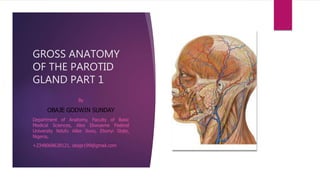
- 4. Gross anatomy of parotid gland Introduction Parotid gland is wrapped around the mandibular ramus and extends to a position anterior and inferior to the ear within the parotid space. ECA It has superficial and deep lobes. The ECA forms its two terminal branches within the parotid gland: maxillary and superficial temporal artery Definition Parotid gland is the largest of the salivary glands and secretes saliva via the parotid duct into the oral cavity to facilitate mastication and swallowing. It is located in the parotid space.
- 5. Gross anatomy of parotid gland cont’d Structures passing… The facial nerve and its branches pass through the parotid gland, as does the external carotid artery (ECA) and retromandibular vein. There is moderate fatty infiltration or fatty replacement of the parotid glands with age Lymph nodes The gland usually contains several intraparotid lymph nodes. These are typically situated in two locations within the gland. These lymph nodes are the first station of lymphatic drainage of the skin of the pinna and peri-auricular skin. Fibrous capsule The fibrous capsule surrounds the gland: formed by the split layers of the investing layer (deep cervical fascia). Posteriorly this fascia condenses forming the stylomandibular ligament. The inferior projection of the parotid is often referred to as the "tail" which overlies the angle of the mandible.
- 6. Gross anatomy of parotid gland cont’d Poles Superior pole: external acoustic meatus, temporomandibular joint Lower pole: behind the angle of the mandible, anterior to the sternocleidomastoid and posterior belly of the digastric Surfaces Lateral surface: subcutaneous tissue anterior surface: the ramus of the mandible with the masseter on its outer surface and medial pterygoid on its inner surface inferiorly (separated by the stylomandibular ligament) Anterior border Masseter, the parotid duct and 5 facial nerve branches. Sternocleidomastoid and posterior belly of the digastric), styloid process and its attached muscles (stylohyoid, styloglossus, stylop haryngeus) and two ligaments (stylomandibular, stylohyoid)
- 7. Gross anatomy of parotid gland cont’d Blood supply arterial: ECA and a specific branch of the artery, the transverse facial artery venous drainage: plexus of veins into the internal jugular vein Lymphatic drainage Intra-parotid nodes drain into the deep cervical chain. Innervation sensory: auriculotemporal nerve, greater auricular nerve parasympathetic: via auriculotemporal nerve sympathetic: via plexus surrounding external carotid artery from superior cervical ganglion.
- 8. Gross anatomy of parotid gland cont’d Cranial nerves passing Parotid gland Sensory innervation is supplied by the auriculotemporal nerve (gland) and the great auricular nerve (fascia). The parasympathetic innerv ation to the parotid gland has a complex path. It begins with the glossopharyngeal nerve (cranial nerve IX) Stones The most common cause of swollen sa livary glands, salivary stones are buildups of crystallized saliva deposits. Sometimes salivary stones can block the flow of saliva. When saliva can't exit through the ducts, it backs up into the gland, causing pain and swelling Drugs for parotiditis Staphylococcus aureus is the most common organism in community-acquired parotitis and first- line antibiotic therapy should include antistaphylococcal antibiotic (nafcillin, oxacillin, cefazolin)
- 9. Parotid duct The parotid duct or Stensen duct is a duct and the route that saliva takes from the major salivary gland, the parotid gland into the mouth The parotid duct, a long excretory duct, emerges from the front of each gland, superficial to the masseter muscle. The duct pierces the buccinator muscle, then opens into the mouth on the inner surface of the cheek, usually opposite the maxillary second molar
- 10. Parotid gland infection Risk factors • Age • Lifestyle • Children not immunized against mumps • Disease (HIV, Diabetes) Clinical anatomy
- 11. Sialolithiasis Refers to the formation of concrements (sialoliths) inside the ducts or parenchyma of salivary glands and most commonly occurs in the submandibular glands and their ducts
- 12. The SOAR (Summer Opportunities in Anatomy Research) program at the University of North Texas Health Science Center provides undergraduate students the opportunity to gain research, education, and outreach experience in the anatomical sciences. Summer Opportunities in Anatomy Research