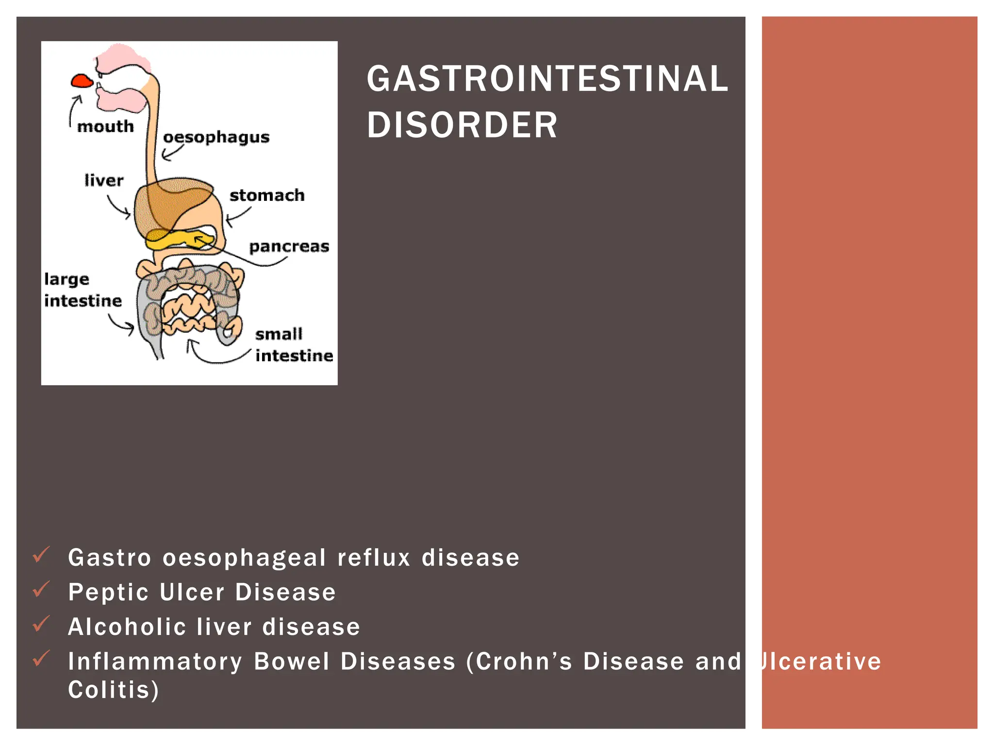
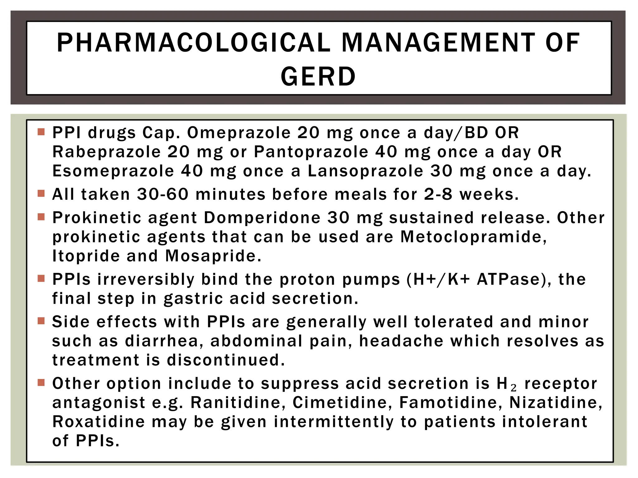
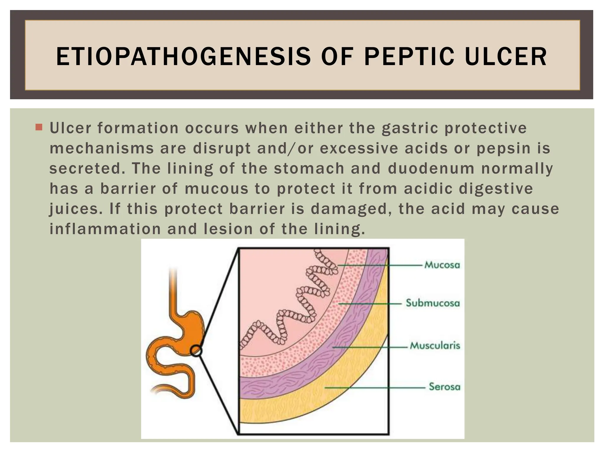
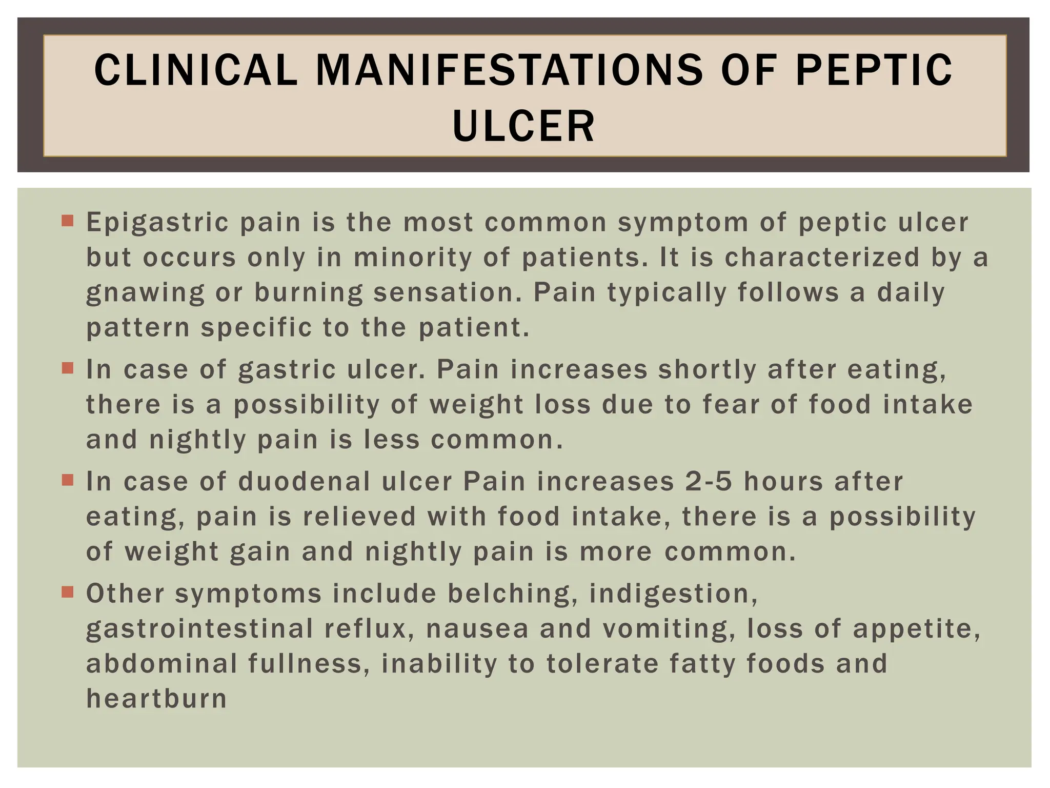
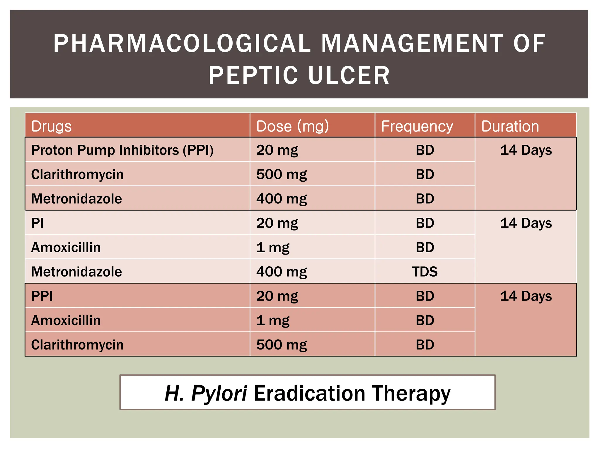
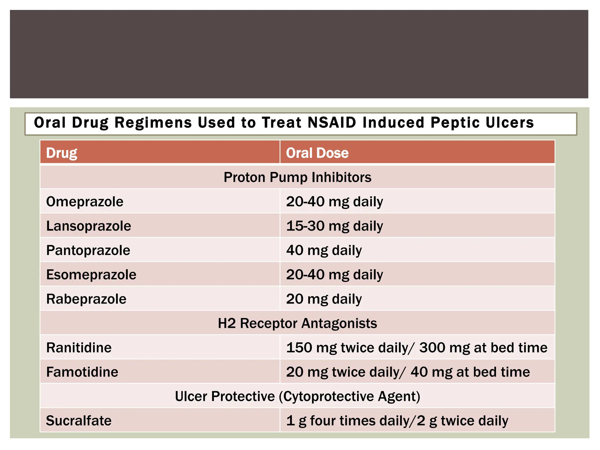
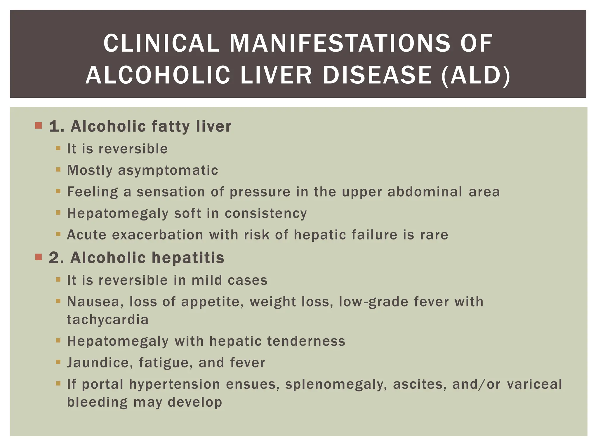
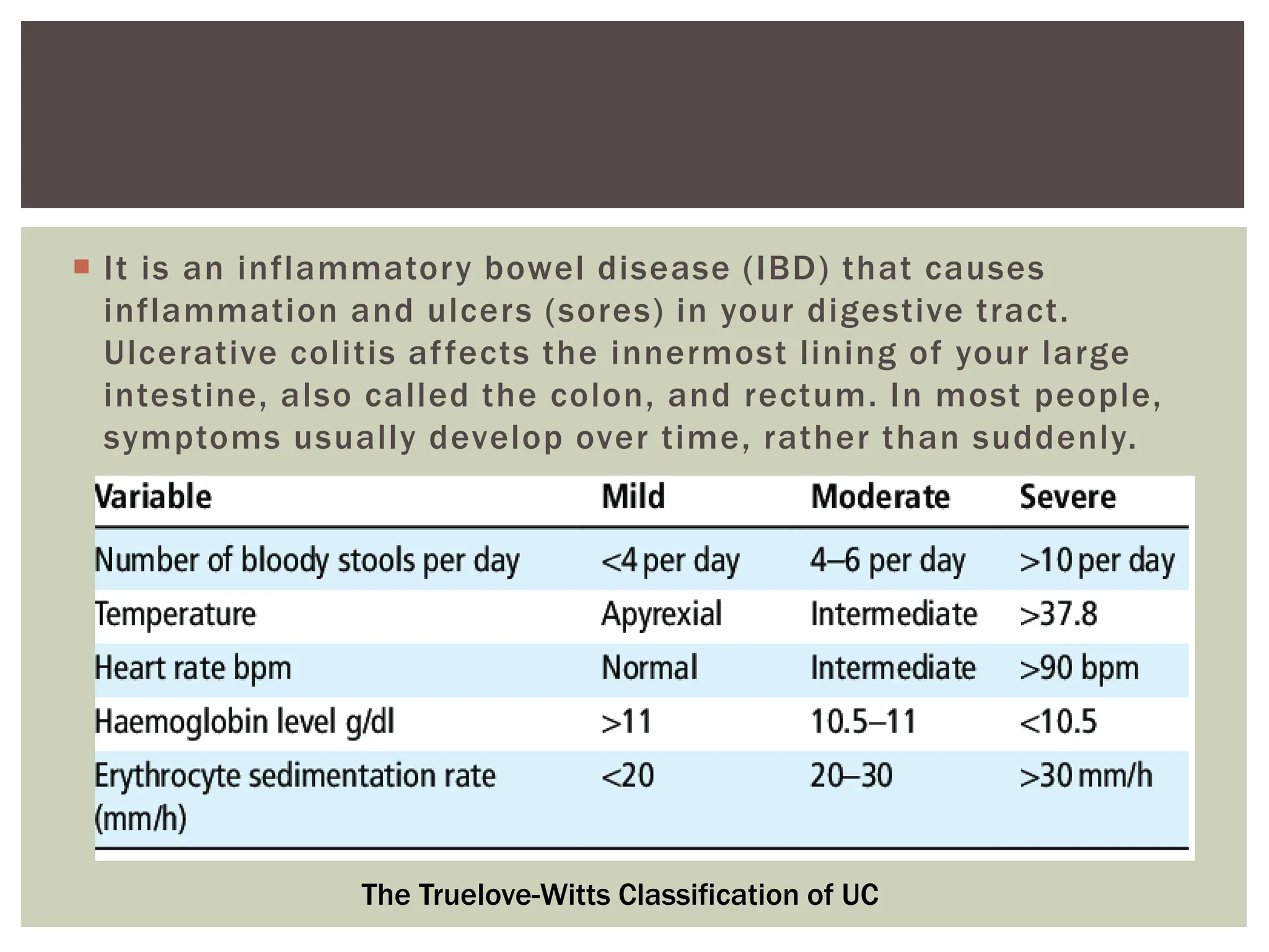
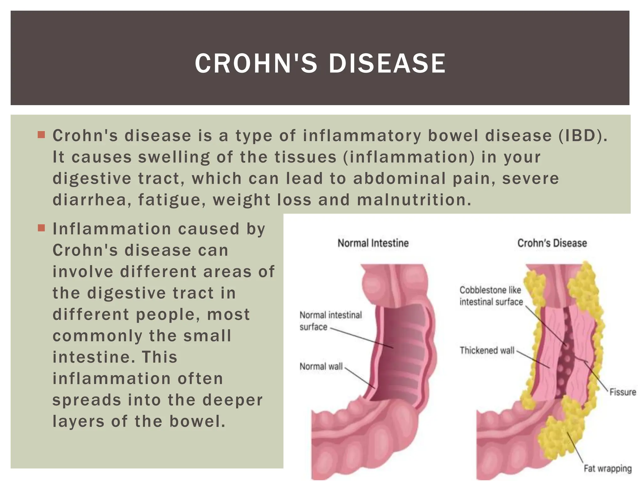
Mr. Sankappa
Definition
Gastro esophageal reflux disease is a chronic and relapsing condition in which prolonged reflux of hydrochloric acid, pepsin and bile salts in the esophagus, oral cavity and respiratory system occurs that leads to esophagitis
Excessive intake of junk foods, coffee, chocolate
Excessive intake of onion, tomato, and beverages
Heavy exercise
Alcoholic and smoking
Medications
Heartburn
Discomfort
Chest pain
Difficulty in respiration
Aspiration pneumonia
After endoscopy the lesions are graded for severity using the Savary Miller grading system;
Grade 1: single or multiple erosions on a single fold.
Grade 2: multiple erosions affecting multiple folds. Erosions may be confluent.
Grade 3: multiple circumferential or rounded erosions.
Grade 4: ulcer, stenosis or esophageal shortening.
Grade 5: Barrett's epithelium. Columnar metaplasia (cellular changes on the microscopic level) in the form of circular or non-circular (islands or tongues) extensions.
Pathophysiology
Management
Antacids: An antacid is a substance which neutralizes stomach acidity, used to relieve heartburn, indigestion or an upset stomach (ex: Rantac, Zantac)
H2receptor antagonist: H2 antagonists block histamine-induced gastric acid secretion from the parietal cells of the gastric mucosa. They include cimetidine, famotidine, nizatidine
Proton Pump Inhibitors: Proton pump inhibitors (PPIs) reduce the production of acid by blocking the enzyme in the wall of the stomach that produces acid (Omeprazole, Rabeprazole, pantoprazole)
Cholinergic drugs:Cholinergic drug, any of various drugs that inhibit, enhance, or mimic the action of the neurotransmitter (acetylcholine, carbachol, methacholine)
Cytoprotective drugs: is a process by which chemical compounds provide protection to cells against harmful agents (carbenoxolone, sucralfate, misoprostol)
Prokinetic drugs: prokinetic, is a type of drug which enhances gastrointestinal motility by increasing the frequency of contractions in the small intestine or making them stronger, but without disrupting their rhythm. (Benzamide, Cisapride, Domperidone).
Endoscopic intraluminal valvuloplasty
Gastric tissue is utilised to increase the integrity of LES By creating a valve like structure.
CHRONIC DYSPEPSIA
Seminar Prepared by :-
Ali Abdulazeem
Shilan Adnan Abdulrahman
Alaa Shamil
Guldan Hameed
Internal Medicine
College of Medicine - University of Kirkuk
inflammatory bowel disease is a diagnosis of exclusion and it has two form known as crohn's disease which can affect all GI tract from ''gum to bum'' with skip lesion and the formation of cobblestones. ulcerative colitis affect only the colon and also causes proctitis and toxic megacolon. both of the disease has extraGI symptoms like sclerosing cholangitis, uveitis, ankylosing spondylitis,conjunctivitis, liver cirrhosis, pyoderma gangrenosum, arthropathy and althralgia, etc .
Acid reflux occurs when the sphincter muscle at the lower end of your esophagus relaxes at the wrong time, allowing stomach acid to back up into your esophagus.
Gastro esophageal Reflux Disease (GERD) and its managementDr. Ankit Gaur
In this presentation I have tried to explain in brief about gastro esophageal Reflux Disease (GERD), its etiology, risk factors, diagnosis, and its management via pharmacotherapy.
Gastrointestinal Diseases
Group 5:
Leticia Bernal Leon
Daydig Rodriguez
Maria Rodriguez
Karina Silveira
Instructor:
Dr. Alain Llanes Rojas, DNP, APRN, FNP-BC
Miami Regional University
Diagnosis, Symptoms & Illness Management
MSN5600
Gastroesophageal Reflux
Gastroesophageal reflux that does not cause symptoms is known as physiologic reflux. In nonerosive reflux disease (NERD), individuals have symptoms of reflux disease but no visible or minimal esophageal mucosal injury
Gastroesophageal reflux disease (GERD) is the reflux of acid and pepsin or bile salts from the stomach to the esophagus that causes esophagitis. The severity of the esophagitis depends on the composition of the gastric contents and esophageal mucosa exposure time.
Definition & Classification
Gastroesophageal Reflux
Causes
GERD can be caused by abnormalities or alterations in
1. Lower esophageal sphincter function
2. Esophageal motility
3. Gastric motility or emptying
Esophageal function studies include the following:
Determination of the lower esophageal sphincter (LES) pressure (manometry)
Graphic recording of esophageal swallowing waves, or swallowing pattern (manometry)
Detection of reflux of gastric acid back into the esophagus (acid reflux)
Detection of the ability of the esophagus to clear acid (acid clearing)
An attempt to reproduce symptoms of heartburn (Bernstein test)
Gastroesophageal Reflux
Risk Factors
Obesity
Hiatal hernia
Use of drugs or chemicals that relax the LES (anticholinergics, nitrates, calcium channel blockers, nicotine)
Cigarette smoke.
Trigger Factors
Coughing
Vomiting
Straining at stool
Asthma
Chronic cough
Sinusitis.
Gastroesophageal Reflux
Common Symptoms
Heartburn that occurs 30 to 60 minutes after meals and when the patient bends over or lies down.
Regurgitation of sour or bitter gastric contents
Belching, and fullness of the stomach
Upper abdominal pain within 1 hour of eating.
Atypical Symptoms
chronic cough
asthma attacks
chronic laryngitis
sinusitis
discomfort during swallowing.
Noncardiac chest pain.
Dysphagia
Gastroesophageal Reflux
Clinical manifestations are related to mucosal injury from acid regurgitation and the frequency and duration of reflux events.
The symptoms worsen if the individual lies down or if intraabdominal pressure increases because of coughing, vomiting, or straining at stool.
Uncomplicated GERD that is responsive to first-line therapy does not require an endoscopy.
Patients who do not respond to therapy and those with suspected complications should undergo an endoscopic examination
Management & Evaluation
Differential diagnosis
Gastritis
Peptic ulcer
Gastric cancer
Cholelithiasis
Angina pectoris.
Gastroesophageal Reflux
Diagnosis of GERD is based on the history and clinical manifestations.
An upper endoscopy with biopsy is the standard diagnostic procedure for GERD. It confirms the diagnosis and documents the type and extent of tissue damage.
Esophageal endoscopy: shows hyperemia ...
Central Nervous System, Epilepsy, Parkinson, Alzheimer, Stroke and Migraine.Dr. Kiran Dhamak
Central Nervous System is one of the unit in Pharmacotherapeutics Subject which is for Second Year Diploma in Pharmacy. The unit covers diseases like Epilepsy, Parkinson, Alzheimer, Stroke and Migraine. The presentation includes the point as per diploma in pharmacy students may understand very easily. The syllabus is framed by Pharmacy Council of India which is implemented by MSBTE ER 2020-2021
Read| The latest issue of The Challenger is here! We are thrilled to announce that our school paper has qualified for the NATIONAL SCHOOLS PRESS CONFERENCE (NSPC) 2024. Thank you for your unwavering support and trust. Dive into the stories that made us stand out!
The French Revolution, which began in 1789, was a period of radical social and political upheaval in France. It marked the decline of absolute monarchies, the rise of secular and democratic republics, and the eventual rise of Napoleon Bonaparte. This revolutionary period is crucial in understanding the transition from feudalism to modernity in Europe.
For more information, visit-www.vavaclasses.com
We all have good and bad thoughts from time to time and situation to situation. We are bombarded daily with spiraling thoughts(both negative and positive) creating all-consuming feel , making us difficult to manage with associated suffering. Good thoughts are like our Mob Signal (Positive thought) amidst noise(negative thought) in the atmosphere. Negative thoughts like noise outweigh positive thoughts. These thoughts often create unwanted confusion, trouble, stress and frustration in our mind as well as chaos in our physical world. Negative thoughts are also known as “distorted thinking”.
Model Attribute Check Company Auto PropertyCeline George
In Odoo, the multi-company feature allows you to manage multiple companies within a single Odoo database instance. Each company can have its own configurations while still sharing common resources such as products, customers, and suppliers.
Instructions for Submissions thorugh G- Classroom.pptxJheel Barad
This presentation provides a briefing on how to upload submissions and documents in Google Classroom. It was prepared as part of an orientation for new Sainik School in-service teacher trainees. As a training officer, my goal is to ensure that you are comfortable and proficient with this essential tool for managing assignments and fostering student engagement.
How to Create Map Views in the Odoo 17 ERPCeline George
The map views are useful for providing a geographical representation of data. They allow users to visualize and analyze the data in a more intuitive manner.
Ethnobotany and Ethnopharmacology:
Ethnobotany in herbal drug evaluation,
Impact of Ethnobotany in traditional medicine,
New development in herbals,
Bio-prospecting tools for drug discovery,
Role of Ethnopharmacology in drug evaluation,
Reverse Pharmacology.
This is a presentation by Dada Robert in a Your Skill Boost masterclass organised by the Excellence Foundation for South Sudan (EFSS) on Saturday, the 25th and Sunday, the 26th of May 2024.
He discussed the concept of quality improvement, emphasizing its applicability to various aspects of life, including personal, project, and program improvements. He defined quality as doing the right thing at the right time in the right way to achieve the best possible results and discussed the concept of the "gap" between what we know and what we do, and how this gap represents the areas we need to improve. He explained the scientific approach to quality improvement, which involves systematic performance analysis, testing and learning, and implementing change ideas. He also highlighted the importance of client focus and a team approach to quality improvement.
Unit 8 - Information and Communication Technology (Paper I).pdfThiyagu K
This slides describes the basic concepts of ICT, basics of Email, Emerging Technology and Digital Initiatives in Education. This presentations aligns with the UGC Paper I syllabus.
2. The term gastrointestinal disorders refer to any condition or
disease that occurs within the gastrointestinal tract. For
example, gastro esophageal reflux disease (GERD), peptic
ulcer disease (PUD), alcoholic liver disease (ALD),
inflammatory bowel disease (IBD), etc.
Gastrointestinal tract includes the mouth, esophagus,
stomach, small intestine, large intestine, colon, rectum,
biliary tract, gallbladder, liver, and pancreas.
INTRODUCTION
3. Gastro esophageal reflux disease (GERD) is a chronic
condition in which gastric content flow back into the
esophagus, causing irritation to the esophageal mucosa with
or without tissue damage.
The GERD with tissue damage is known as esophagitis or
erosion GERD and without tissue damage is known as non-
erosive reflux disease (NERD)
GASTRO ESOPHAGEAL REFLUX DISEASE
(GERD)
4. Risk Factors Linked to GERD:
Age and male sex are associated with a higher incidence of
esophagitis.
Obesity - Obese people 2.5 times more likely to have GERD
than those with normal body mass index (BMI).
Alcohol consumption
Stress
Smoking
Coffee, chocolate, acidic and fatty foods (e.g. spicy foods,
citrus, carbonated drinks
Certain eating habits (e.g., heavy meals or lying down shortly
after eating)
ETIOPATHOGENESIS OF GERD
5. Hiatal hernia (It is a condition in which part of the stomach
pushes up through the diaphragm muscle).
Medication. A number of common drugs are associated with
GERD e.g. Anticholinergic, benzodiazepines, calcium channel
blockers, dopamine, nicotine nitrates, theophylline, estrogens,
progesterone, glucagon, some prostaglandin, NSAIDs,
bisphosphonates, antibiotics, etc.
Pregnancy
Gastrointestinal malformations and tumors
latrogenic (e.g. after gastrectomy)
Scleroderma
Infectious esophagitis
Asthma
6. The various mechanisms involved in the production of
gastroesophageal reflux are inappropriate, transient
relaxation of the lower esophageal sphincter (LES), pressure
abnormalities in the LES (due to hormonal and neural
mediators, food, drugs and patient lifestyle), poor esophageal
clearance, delayed gastric emptying time and hiatal hernia.
Untreated GERD may lead to Barrett esophagus, esophagitis,
ulceration, stricture, and rarely adenocarcinoma (An
esophageal stricture is an abnormal tightening of the
esophagus)
7. Clinical manifestations of GERD can be classified as esophageal and
extraesophageal manifestations of GERD.
(A) Esophageal (Typical) Manifestations
Retrosternal pain (Heart burn)
Chest pain
Epigastric pain
Regurgitation mostly after meals
Dysphagia (difficulty or discomfort in swallowing)
Odynophagia (Painful swallowing)
Nausea
Belching
Early satiety
Bloating
Water brash (Excessive salivation)
Halitosis (Bad breath)
CLINICAL MANIFESTATIONS OF GERD
8. (B) Extraesophageal (Atypical) Manifestations
Chronic cough
Asthma
Laryngitis
Bronchospasm
Recurrent pulmonary infections
Otitis media
Dental erosion
Discomfort in the nose
Trouble sleeping
9. Diagnosis and Investigation of GERD
A diagnosis of GERD based on the presence of typical symptoms
is correct in only 70% of patients.
Diagnostic studies, e.g, upper GI endoscopy, esophageal
manometry test, 24-hours pH probe test and nuclear medicine
gastric emptying study, may be indicated to confirm the
diagnosis or to rule out other causes of symptoms.
10. The goals of GERD treatment are to control symptoms, to heal
esophagitis, and prevent recurrent esophagitis or other
complications.
The treatment of GERD consists of lifestyle modification and
initiating acid suppression therapy, preferably with proton
pump inhibitors (PPIs).
Sometimes surgical treatment with corrective anti reflux
surgery is considered in select cases, e.g, patients who
develop complications despite receiving optimal drug therapy.
Patients with signs of GERD complications or other illness or
do not respond to drug therapy should be considered for
further diagnostic testing
MANAGEMENT OF GERD
11. Non-pharmacological management of GERD includes patient
education, lifestyle modification and surgical therapy.
All patients should be educated regarding factors that may
worsen the symptoms.
Lifestyle modification is considered to be the first line of
treatment of GERD.
Lifestyle modification includes
Avoid alcohol, chocolate, citrus juice, and tomato-based products.
Weight loss (if overweight)
Avoid peppermint, coffee, and possibly the onion family
Avoid smoking
Avoid eating at least 3 hours before bedtime, elevating the head of the
bed
Avoid skipping meals
Avoid drugs that lower LES pressure [e.g., calcium channel blockers, beta
alpha-adrenergic agonists, theophylline, nitrates, PDE-S inhibitors) or in
esophagus (e.g, Ferrous sulfate, NSAIDs, bisphosphonates, etc.)
NON-PHARMACOLOGICAL MANAGEMENT
OF GERD
12. PPI drugs Cap. Omeprazole 20 mg once a day/BD OR
Rabeprazole 20 mg or Pantoprazole 40 mg once a day OR
Esomeprazole 40 mg once a Lansoprazole 30 mg once a day.
All taken 30-60 minutes before meals for 2-8 weeks.
Prokinetic agent Domperidone 30 mg sustained release. Other
prokinetic agents that can be used are Metoclopramide,
Itopride and Mosapride.
PPIs irreversibly bind the proton pumps (H+/K+ ATPase), the
final step in gastric acid secretion.
Side effects with PPIs are generally well tolerated and minor
such as diarrhea, abdominal pain, headache which resolves as
treatment is discontinued.
Other option include to suppress acid secretion is H₂ receptor
antagonist e.g. Ranitidine, Cimetidine, Famotidine, Nizatidine,
Roxatidine may be given intermittently to patients intolerant
of PPIs.
PHARMACOLOGICAL MANAGEMENT OF
GERD
13. Antacids and alginates are effective in short-
term or intermittent symptom antacid-alginate
combination is recommended for episodic and
postprandial symptoms.
These preparations are usually taken after each
meal and at bed time Further evaluation may
be considered in patients non-responsive to
this therapy include an upper Gl endoscopy with
or without enhanced imaging and function
testing.
15. Peptic ulcer disease is the presence of ulceration (an erosion
or lesion) in the esophagus, stomach, and duodenum
secondary due to pepsin and gastric acid secretion.
The diameter of peptic ulcer is at least 0.5 cm and a depth
that reaches the muscularis mucosae.
Peptic ulcer is the main cause of upper gastrointestinal
haemorrhage.
INTRODUCTION
16. Types of peptic ulcer are
(A) According to site of ulcer
1. Gastric ulcer (Stomach ulcer): It is a peptic ulcer of the gastric mucosa.
2 Duodenal ulcer: It is a peptic ulcer of the duodenal mucosa
(B) According to cause
1. Helicobacter pylori associated peptic ulcers
2 NSAID induced peptic ulcers
17. Ulcer formation occurs when either the gastric protective
mechanisms are disrupt and/or excessive acids or pepsin is
secreted. The lining of the stomach and duodenum normally
has a barrier of mucous to protect it from acidic digestive
juices. If this protect barrier is damaged, the acid may cause
inflammation and lesion of the lining.
ETIOPATHOGENESIS OF PEPTIC ULCER
18. Peptic ulcer disease (PUD) may be due to any of the following
Helicobacter pylori infection (most common)
Prolonged nonselective NSAID use (possibly in combination
with glucocorticoids)
Medications such as steroids, SSRI, bisphosphonates,
anticoagulants, potassium chloride or chemotherapeutic
agents
Lifestyle factors (e.g, alcohol use, smoking, caffeine, spicy
food, etc.).
Psychological factors (e.g, anxiety, stress, post-traumatic
stress disorder (PTSD)
20. Epigastric pain is the most common symptom of peptic ulcer
but occurs only in minority of patients. It is characterized by a
gnawing or burning sensation. Pain typically follows a daily
pattern specific to the patient.
In case of gastric ulcer. Pain increases shortly after eating,
there is a possibility of weight loss due to fear of food intake
and nightly pain is less common.
In case of duodenal ulcer Pain increases 2-5 hours after
eating, pain is relieved with food intake, there is a possibility
of weight gain and nightly pain is more common.
Other symptoms include belching, indigestion,
gastrointestinal reflux, nausea and vomiting, loss of appetite,
abdominal fullness, inability to tolerate fatty foods and
heartburn
CLINICAL MANIFESTATIONS OF PEPTIC
ULCER
21. In advance cases upper abdominal pain, anorexia, cachexia.
Hematemesis, melena, or anemia may suggest bleeding.
Patient may show iron deficiency anaemia.
Up to 70% of patients with peptic ulcers do not experience
symptoms.
Diagnosis and Investigation of Peptic Ulcer
Stool Antigen Test
Polymerase Chain Reaction (PCR)
Urea breath test
Biopsy
CBC
Liver Function Test
GI Endoscopy
22. Drugs Dose (mg) Frequency Duration
Proton Pump Inhibitors (PPI) 20 mg BD 14 Days
Clarithromycin 500 mg BD
Metronidazole 400 mg BD
PI 20 mg BD 14 Days
Amoxicillin 1 mg BD
Metronidazole 400 mg TDS
PPI 20 mg BD 14 Days
Amoxicillin 1 mg BD
Clarithromycin 500 mg BD
PHARMACOLOGICAL MANAGEMENT OF
PEPTIC ULCER
H. Pylori Eradication Therapy
27. Alcoholic liver disease (ALD) refers to a range of progressive
liver damaging conditions caused by chronic and excessive
alcohol consumption.
Liver is the primary site for ethanol metabolism. Each time
liver filters alcohol, some of its cells die. The liver can
regenerate new cells, but chronic and excessive alcohol
consumption an reduce its ability to regenerate. This can
result in serious and permanent damage to the liver.
INTRODUCTION
28. Chronic and excessive alcohol consumption produces a wide
spectrum of hepatic lesions which are steatosis, hepatitis,
fibrosis/cirrhosis and hepatocellular carcinoma.
Steatosis is characterized by the deposition of fat in
hepatocytes Steatosis can progress to steatohepatitis, which
is a more severe, inflammatory type of liver injury.
ETIOPATHOGENESIS OF ALCOHOLIC LIVER
DISEASE (ALD)
Steatohepatitis can lead to the
development of fibrosis, during
which there is excessive deposition
of extracellular matrix proteins This
stage may progress to cirrhosis,
characterized by excessive liver
scarring, vascular alterations, and
eventual liver failure.
29. The three stages of ALD, which may or may not occur
sequentially, are as follows:
Stage I Alcoholic fatty liver: This is the stage of steatosis This
stage occurs after acute alcohol ingestion, is typically
asymptomatic and reversible with abstinence.
Stage II Alcoholic hepatitis: This is the stage of
steatohepatitis Alcoholic hepatitis can range in severity from
asymptomatic to liver failure and death.
Stage III Alcohol-related cirrhosis: This is the final stage of
ALD This stage involves replacement of the normal hepatic
parenchyma with extensive thick bands of fibrous tissue and
regenerative nodules, which results in the clinical
manifestations of portal hypertension and liver failure.
30. The mechanism of development of ALD involves hepatic
degradation of ethanol to acetyl-CoA by alcohol
dehydrogenase results in excess of NADH.
The excess of NADH drives the formation of glycerol 3-
phosphate (G3P) from dihydroxyacetone phosphate (DHAP
Increase in both G3P and fatty acids cause increased
triglyceride synthesis in the live accompanying inflammation
(steatohepatitis).
Chronic inflammation leads to hepatic fibrosis and sclerosis
leading to portal hypertension and eventually cirrhosis.
PATHOGENESIS OF ALD
31. Cofactors in the development of alcoholic liver disease (ALD)
are
Genetic factors: Rate of alcohol metabolism plays a role
Gender: Women are more susceptible compared to men
Diet and nutrition: Under nutrition and over nutrition
Cigarette smoking
Co-morbid conditions e.g. Non-alcoholic steatohepatitis (NASH), non-
alcoholic fatty liver disease (NAFLD) viral hepatitis and
hemochromatosis
32. 1. Alcoholic fatty liver
It is reversible
Mostly asymptomatic
Feeling a sensation of pressure in the upper abdominal area
Hepatomegaly soft in consistency
Acute exacerbation with risk of hepatic failure is rare
2. Alcoholic hepatitis
It is reversible in mild cases
Nausea, loss of appetite, weight loss, low-grade fever with
tachycardia
Hepatomegaly with hepatic tenderness
Jaundice, fatigue, and fever
If portal hypertension ensues, splenomegaly, ascites, and/or variceal
bleeding may develop
CLINICAL MANIFESTATIONS OF
ALCOHOLIC LIVER DISEASE (ALD)
33. 3. Alcohol-related cirrhosis
It is irreversible and final stage of ALD
Patients are often initially asymptomatic
Nausea, vomiting, fatigue, malaise, anorexia, and weight loss
Hepatomegaly
Splenomegaly
Pruritus
Jaundice
Ascites
Telangiectasia (These are dilated small blood vessels on the skin or
mucous membranes)
Caput medusa (It is the cluster of swollen veins in abdomen).
Palmar erythema (It is a skin condition that makes the palms of
hands turn red)
Petechiae and purpura
Hormonal disorders
Gynecomastia
Hypogonadism
Peripheral edema
34. Patient history of habitual alcohol intake of sufficient length and
intensity, transaminase levels, and imaging studies are crucial
for diagnosis of ALD.
Laboratory tests
Aspartate aminotransferase (AST) > Alanine aminotransferase
(ALT) (both ↑ AST and ↑ ALT)
↑ Gamma-glutamyl transpeptidase (GGT)
↑ Serum ferritin
↑ CDT(carbohydrate-deficient transferring)
↑ Bilirubin
↑ Prothrombin time
↑ Glutamate dehydrogenase (GLDH)
↑ Ammonia
↓ Serum albumin
↓Cholinesterase
DIAGNOSIS AND INVESTIGATION OF
ALCOHOLIC LIVER DISEASE (ALD)
35. Treatment options include medication, lifestyle changes, and
liver transplants.
There are still no FDA approved pharmacological or nutritional
therapies for treating patients with ALD.
Treatment of ALD requires complete cessation of alcohol use.
Also, patients should be encouraged to stop smoking.
Disulfiram
Naltrexone
Acamprosate
This drugs are approved abstinence and relapse prevention
medications to treat alcohol addiction.
MANAGEMENT OF ALCOHOLIC LIVER
DISEASE (ALD)
37. There are two forms of idiopathic inflammatory bowel disease
(IBD):
Ulcerative Colitis
Crohn's Disease
38. It is an inflammatory bowel disease (IBD) that causes
inflammation and ulcers (sores) in your digestive tract.
Ulcerative colitis affects the innermost lining of your large
intestine, also called the colon, and rectum. In most people,
symptoms usually develop over time, rather than suddenly.
The Truelove-Witts Classification of UC
40. Crohn's disease is a type of inflammatory bowel disease (IBD).
It causes swelling of the tissues (inflammation) in your
digestive tract, which can lead to abdominal pain, severe
diarrhea, fatigue, weight loss and malnutrition.
CROHN'S DISEASE
Inflammation caused by
Crohn's disease can
involve different areas of
the digestive tract in
different people, most
commonly the small
intestine. This
inflammation often
spreads into the deeper
layers of the bowel.
43. Diarrhea, often with blood or pus
Rectal bleeding — passing small amount of blood with stool
Abdominal pain and cramping
Rectal pain
Urgency to defecate
Inability to defecate despite urgency
Weight loss
Fatigue
Fever
In children, failure to grow
CLINICAL MANIFESTATION OF UC
44. Diarrhea
Fever
Fatigue
Abdominal pain and cramping
Blood in your stool
Mouth sores
Reduced appetite and weight loss
Pain or drainage near or around the anus due to inflammation
from a tunnel into the skin (fistula)
Inflammation of skin, eyes and joints
Inflammation of the liver or bile ducts
Kidney stones
Iron deficiency (anemia)
Delayed growth or sexual development, in children
CLINICAL MANIFESTATION OF CD
45. Rehydration
Supplementation of Nutritional Deficiencies
Pharmacotherapy
Supportive care:
Antidiarrheal Agents
Anticholinergic Drugs: Relieves Abdominal cramping
Mild Disease:
5-aminosalicylic acid Derivatives: Mesalamine, Sulfasalazine,
olsalazine
Can be administered orally, as suppositories or as enemas
If no improvement or 5-ASA agents are not tolerated
Topical Corticosteroids: Budesonide
Oral systemic Corticosteroids
Pharmacological Management of UC
46. Pharmacological Management of CD
Aminosalicylates: Sulfasalazine, Mesalazine, Olsalazine
Antimicrobials: Ciprofloxacin, Metronidazole
Corticosteroids: Prednisolone, Budesonide
Thiopurines: Azathioprine or Mercaptopurine
Calcineurin Antagonists: Cyclosporine