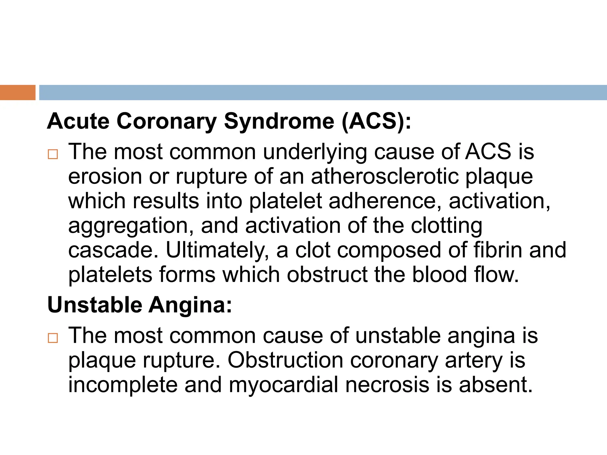
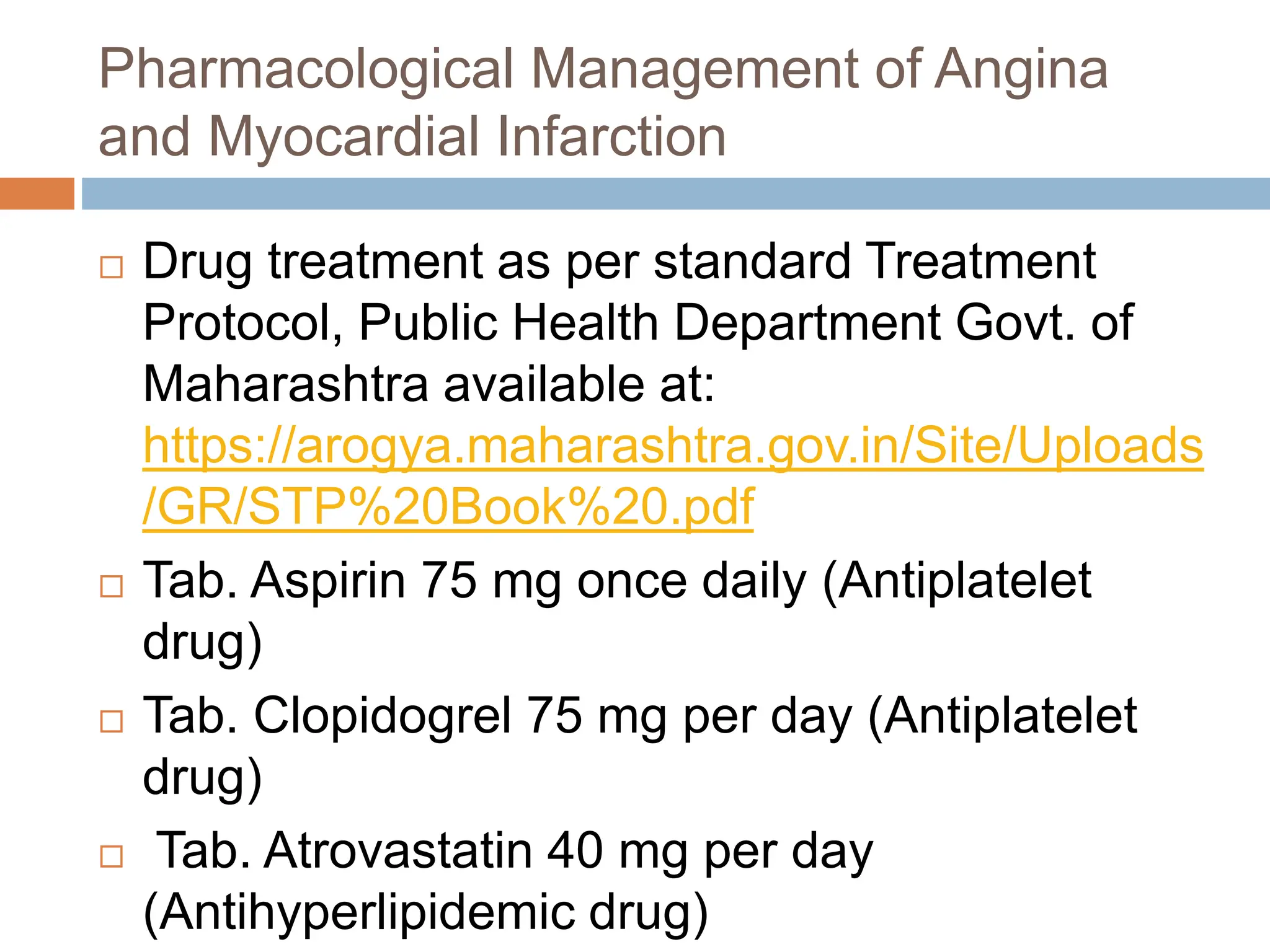
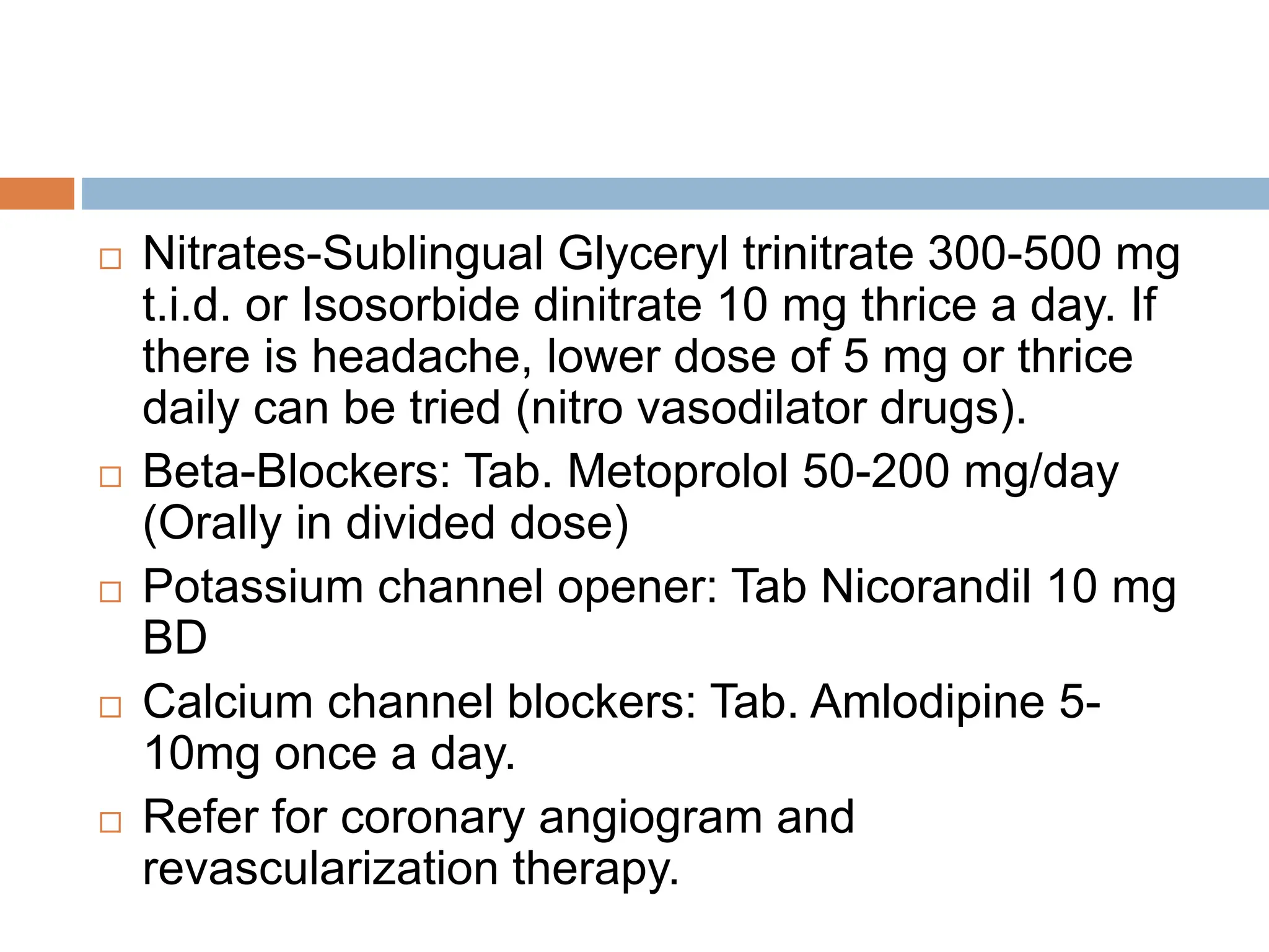
CARDIOVASCULAR
SYSTEM
By: Mr. Kiran B. Dhamak
 Hypertension
 Angina and Myocardial
Infarction
 Hyperlipidaemia
 Congestive Heart Failure
Hypertension
 Defination:
Hypertension is defined as systolic blood
pressure greater than 140 mmHg and/or
diastolic blood pressure than 90 mmHg.
Classification
 Hypertension can be
classified as:
 Primary or Essential
Hypertension: When
the cause is
unknown (90 to 95%
cases)
 Secondary
Hypertension:
Specific organ
dysfunction is
detected i.e. cause is
known (05 to 10%
Etiopathogenesis of
Hypertension
 (Etiopathogenesis means the cause and
development of a disease or abnormal
condition)
 The etiology of primary hypertension is not
clear.
 Secondary hypertension can be secondary to:
 Renal diseases e.g. chronic diffuse
glomerulonephritis, pyelonephritis, polycystic
kidneys, etc.
 Endocrine diseases e.g. Cushing's syndrome,
pheochromocytoma, primary hyperaldosteronism
 Hypertension is an asymptomatic condition If
untreated, it can lead to target organ damage
(TOD) such as coronary artery disease (CAD),
left ventricular hypertrophy (LVH) stroke
transient ischemic attacks (TIA), retinopathy,
peripheral vascular diseases including
dissecting aneurysm renal disease, etc.
Clinical Manifestations of Hypertension:
 Often asymptomatic (silent killer)
 Non specific symptoms are fatigue headache
epistasis, vomiting, giddiness, breathlessness and
palpitations
 Stroke, acute myocardial infarction due to vascular
disease
 Symptoms of underlying organ affected due to
underlying disease
 Bruits over carotid
 Spells of sweating tachycardia indicates
 Management of Hypertension
 The overall aim of the management of
hypertension is not only reduction of BP to
target levels but also to lower the
cardiovascular risk of the patient Target BP
150/90 mm Hg in elderly and 140/90 mm Hg in
all others (including diabetes mellitus, chronic
kidney disease)
 Non-Pharmacological Management of
Hypertension:
 Reduce dietary sodium intake to not more than 24 gm
sodium or 6 gm sodium chloride per day
 Adopt DASH eating plan, Diet rich in fruits, vegetables
with low fat dairy product with a reduced content of
saturated and total fat (DASH Dietary Approaches to
Stop Hypertension).
 Physical activity: Regular aerobic activity, brisk walking
for 30 min/day. Maintain weight Target BMI 18.5-22.9
kg/m²
 Reduce alcohol consumption
 Stop smoking
 Stress management
 Yoga
 Patients with Grade 1 hypertension may require only
Pharmacological Management
 ACE is Angiotensin converting enzyme (ACE)
inhibitors (e.g. Enalapril) or Angiotensin II
receptor blockers (ARBS) (e.g. Losartan) Use
ARBs if the patient is intolerance ACE
inhibitors.
 Bb is Beta Blockers (e.g. Atenolol)
 CCB is Calcium Channel Blockers (e.g.
Amlodipine)
 TD is Thiazide Diuretics (e.g.
Hydrochorthiazide)
 MRA Mineral Corticoid Receptor Antagonist
Grade 1 Hypertension:
 If not controlled after 3 months of lifestyle
modification or involves more than 3 risk factors
such as age men >55 years, women> 65 years,
smoking, obesity, including abdominal obesity,
dyslipidaemia, impaired fasting glucose family
history of early coronary artery disease, begin the
drug therapy as ACEI or CCB or TD.
 If response is not adequate within 2-4 weeks, add
second drug CCB+ACE or ACEI+TD or CCB+TD
 If response is not adequate within next 2-4 weeks,
add third drug-ACEI+CCB+TD
Grade 2 Hypertension:
 Begin the drug therapy as soon as grade 2
hypertension is diagnosed. Begin with ACEI or
CCB or TD.
 If response is not adequate within 2-4 weeks
add second drug as add on therapy CCB+ACE
or ACE+TD or CCB+TD
 If response is not adequate within next 2-4
weeks add third drug- ACEI+CCB+TD
Grade 3 Hypertension:
 Use two drugs ACEI+CCB or CCB+TD or
ACE+TD
 If response is not adequate within next 2-4
weeks, either increase the dose of drugs or
add third drug ACEI+CCB+TD.
All Grades of Hypertension with Associated
Clinical Conditions:
 Coronary artery disease: Bb+ACEI, CCB (CCB
is added only if it is required to ach the target
BP)
 Congestive heart failure: TD+ACEI+Bb, MRA
 Diabetes mellitus: ACEI or CCB TD
 Chronic kidney disease: ACEI or CCB or TD
(Patients with CKD may require loop diuretics
if the glomerular filtration rate is low)
ACE inhibitors should not be combined
with angiotensin receptor blockers.
Avoid prescribing a combination of beta-
blockers and diuretics as they can increase
the risk of diabetes mellitus in those at
risk
Contraindications: (action that is opposite)
 Amlodipine: Cardiogenic shock unstable angina,
significant aortic stenosis worsening angina or
increased risk of myocardial infarction may be
seen.
 Enalapril: Use of all ACE inhibitors is
contraindicated in pregnancy, Bilateral renal artery
stenosis History of hypersensitivity (including
angioedemal).
 Hydrochlorthiazide: Severe hypokalemia,
hyponatremia, hypercalcemia
Hypertension in Pregnancy:
 Tablet Methyl dopa: 500 mg-1000 mg/day in
three divided doses.
 Tablet Nifedipine Extended Release
Preparation: 30-60 mg OD/BD
 Other Drugs that can be given: Labetalol,
Hydralazine, Beta Blocker
Angina and Myocardial
Infarction
 Angina and Myocardial infarction (MI) are
ischemic heart diseases (IHD).
 Ischemia is a condition in which there is an
inadequate supply of blood and oxygen to a
portion of the myocardium.
Angina
 It is a pain syndrome due to myocardial
ischemia caused by critical obstruction in
coronary arteries due to atherosclerosis or
emboli or calcific aortic stenosis or due to
vasospasm.
 Angina
 Stable, exertional angina (Classical angina):
 Microvascular angina
 Variant/prinzmetal/vasospastic angina
 Acute Coronary syndrome (ACS)
 Unstable angina
 Acute myocardial infarction
 Non-ST-Segment Elevation MI (NSTEMI)
 ST-Segment Elevation MI (STEMI)
 Stable, extertional angina (Classical Angina): it
is usually provoked by physical exertion or
emotional stress when the oxygen demand of
myocardium is increased and is relieved by rest
and nitrates when the increased oygen demand is
withdrawn.
 Microvascular angina: it is more common in
females. Ischaemia is due to endothelial or
microvascular dysfunction and no coronary artery
blockage is there.
 Variant Angina: attacks are unpredictable and
almost always occur at rest or during sleep.
 Acute coronary syndromes (ACS) refer to
range of conditions associated with sudden
reduced blood flow to the heart producing
symptoms compatible with acute myocardial
ischemia which may be life threatening.
According to severity, acute coronary
syndromes (ACS) may be classified as:
 Unstable Angina (UA): It is more severe than
stable angina It occurs at rest (or with minimal
exertion), usually lasting for more than 10 minutes
It is progressive angina with prolonged or more
frequent attacks superimposed on chronic stable
angina (crescendo angina).
 Obstruction of the coronary artery is incomplete
and myocardial necrosis is absent. Biochemical
markers of ischaemia (raised troponin 1,
myoglobin CK-MB (creatine kinase myocardial
band)) do not appear in blood, and ST segment is
not elevated in ECG
 Acute Myocardial Infarction (AMI):
Myocardial infarction (MI) refers to death of
myocytes (necrosis) of a portion of the
myocardium due to sudden occlusion of a
branch of coronary artery (ischemia). It is
commonly known as heart attack.
 (i) Non-ST-Segment Elevation Myocardial
Infarction (NSTEMI): It is more severe than
unstable angina. Obstruction of the coronary
artery is incomplete but myocardial necrosis up to
some extent is there. Biochemical markers of
ischaemia (raised troponin 1 myoglobin, CK MB)
appear in blood but ST segment is not elevated in
ECG.
 (ii) ST-Segment Elevation Myocardial Infarction
(STEMI): It is the most serious type of heart
attack. Obstruction of the coronary artery is
complete and full thickness of ventricular wall and
larger area of myocardium is necrosed. ST
segment in FCG is elevated and biochemical
markers of ischaemia (raised troponin I
myoglobin, CK MB) in blood are prominent.
Etiopathogenesis of IHD
Angina:
 The cause of angina is less supply of oxygen
to the myocardium than the d Occurrence of
angina depends upon two factors (a) Coronary
blood flow, and (b) consumption by the
myocardium. The coronary blood flow may be
obstructed by atherosclerosis or vasospasm.
Acute Coronary Syndrome (ACS):
 The most common underlying cause of ACS is
erosion or rupture of an atherosclerotic plaque
which results into platelet adherence, activation,
aggregation, and activation of the clotting
cascade. Ultimately, a clot composed of fibrin and
platelets forms which obstruct the blood flow.
Unstable Angina:
 The most common cause of unstable angina is
plaque rupture. Obstruction coronary artery is
incomplete and myocardial necrosis is absent.
Myocardial Infarction (MI):
 The most common cause of MI is the formation
of occlusive thrombus (blood clot) at the site of
rupture of an atheromatous plaque in a coronary
artery.
 STEMI takes place when a coronary artery
thrombus forms rapidly.
 Unstable angina can progress to MI and sudden
death Possible STEMI complications are:
 Arrhythmias
 Rupture of papillary muscle
 Ventricular remodelling
 Acute heart failure
 Embolism leading to stroke
 Ventricular aneurysm
Clinical Manifestations of Ischemic Heart
Disease (IHD)
Angina:
 Chest pain: Retrosternal pain (pain behind the
sternum), heaviness or discomfort which may
radiate to the neck, shoulder, back or the arm.
Pain is usually precipitated by exertion or
stress and relieved by rest or nitrates.
 Associated symptoms include, sweating,
nausea and/or dizziness.
Unstable angina:
 New onset angina
 Angina occurring at rest or with minimal
exertion.
 Attack is prolonged, usually >20 minutes.
 Increasing or crescendo angina. Progressive
angina with prolonged or more frequent
attacks superimposed on chronic stable
angina).
 Investigation and diagnosis of IHD involves:
 Typical clinical features
 Determination of biochemical markers of ischaemia
(raised troponin 1, myoglobin creatinine kinase-
myocardial band (CK-MB)) in blood
 Complete blood count (CBC).
 Lipid profile.
 ECG
 Chest X-ray
 Coronary CT angiography
 Echocardiography
 MRI
 Stable angina is diagnosed when the chest pain
remains unchanged in severity. frequency and
duration over several weeks Usually, a treadmill
 Management of Angina and Myocardial
Infarction
Risk factors for Ischemic Heart Disease (IHD)
are
 Smoking
 Diabetes
 Dyslipidemia
 Hypertension
 Obesity
 History of coronary artery disease (CAD)
Non Pharmacological Management of and
Myocardial Infarction:
Stable Angina:
 Daily exercise
 Stop smoking
 Avoid alcohol
 Dietary modification: Low cholesterol, low fat diet with
high roughage
 Avoid any activity known to precipitate anginal attack
 Weight reduction in obese patients
Acute Coronary Syndrome (ACS):
 Complete bed rest
 Coronary angiography and revascularization therapy
should be advised
 Revascularization Therapy:
Revascularization is the restoration of
perfusion to a body organ that has suffered
ischemia. It relieves angina or ongoing
myocardial infarction and prevents death due
to myocardial infarction. Revascularization
procedures include:
 percutaneous coronary intervention (PCI) and
 coronary artery bypass graft (CABG)
Coronary Artery Bypass Graft
Pharmacological Management of Angina
and Myocardial Infarction
 Drug treatment as per standard Treatment
Protocol, Public Health Department Govt. of
Maharashtra available at:
https://arogya.maharashtra.gov.in/Site/Uploads
/GR/STP%20Book%20.pdf
 Tab. Aspirin 75 mg once daily (Antiplatelet
drug)
 Tab. Clopidogrel 75 mg per day (Antiplatelet
drug)
 Tab. Atrovastatin 40 mg per day
(Antihyperlipidemic drug)
 Nitrates-Sublingual Glyceryl trinitrate 300-500 mg
t.i.d. or Isosorbide dinitrate 10 mg thrice a day. If
there is headache, lower dose of 5 mg or thrice
daily can be tried (nitro vasodilator drugs).
 Beta-Blockers: Tab. Metoprolol 50-200 mg/day
(Orally in divided dose)
 Potassium channel opener: Tab Nicorandil 10 mg
BD
 Calcium channel blockers: Tab. Amlodipine 5-
10mg once a day.
 Refer for coronary angiogram and
revascularization therapy.
Hyperlipidaemia
 Hyperlipidaemia, also known as dyslipidemia
or high cholesterol, means you have too many
lipids (fats) in your blood.
 Your liver creates cholesterol to help you
digest food and make things like hormones.
But you also eat cholesterol in foods from the
meat and dairy aisles.
 Too much cholesterol (200 to 239 mg/dL is
borderline high and 240 mg/dL is high) is not
healthy because it can create roadblocks in
your artery highways where blood travels
around to your body.
 This damages the organs. Bad cholesterol
(LDL) is the most dangerous type because it
causes hardened cholesterol deposits (plaque)
to collect inside of your blood vessels. This
makes it harder for your blood to get through,
which puts you at risk for a stroke or heart
Clinical Manifestation
 Most people don't have symptoms when their
cholesterol is high.
 People who have a genetic problem with
cholesterol clearance that causes very high
cholesterol levels may get xanthomas (waxy,
fatty plaques on the skin) or corneal arcus
(cholesterol rings around the iris of the eye).
Etiopathogenesis
 Various hyperlipidaemia causes include :
 Smoking
 Drinking a lot of alcohol
 Eating foods that have a lots of saturated fats or
trans fats.
 Sitting too much instead of being active
 Being stressed
 Inheriting genes that make your cholesterol levels
unhealthy
 Being overweight.
Non-Pharmacological
Management
 Exercising
 Quitting smoking
 Sleeping at least seven hours each night
 Keeping your stress level under control
 Eating healthier foods
 Limiting how much alcohol you drink
 Losing a few pounds to reach a healthy
weight.
Pharmacological Management
Congestive Heart Failure
 Heart failure sometimes known as congestive
heart failure occurs when the heart muscle
doesn’t - pump blood as well as it should.
 When this happens, blood often backs up and
fluid can build up in the lungs, causing
shortness of breath.
 Certain heart conditions, such as narrowed
arteries in the heart (coronary artery disease)
or high blood pressure, gradually leave the
heart too weak or stiff to fill and pump blood
properly.
 Heart failure can be on-going (chronic), or it may
start suddenly (acute). Heart failure signs and
symptoms may include :
 Shortness of breath with activity or when lying
down.
 Fatigue and weakness.
 Swelling in the legs, ankles and feet.
 Rapid or irregular heartbeat.
 Reduced ability to exercise.
 Persistent cough or wheezing with white or pink
blood-tinged mucus.
 Swelling of the belly area (abdomen).
 Very rapid weight gain from fluid build-up.
 Nausea and lack of appetite.
Etiopathogenesis
 In heart failure, the main
pumping chambers of the heart
(the ventricles) may become
stiff and not fill properly
between beats. In some
people, the heart muscle may
become damaged and
weakened. The ventricles may
stretch to the point that the
heart can’t pump enough blood
through the body.
 Over time, the heart can no longer keep up
with the typical demands placed on it to pump
blood to the rest of the body.
 Heart failure can involve the left side (left
ventricle), right side (right ventricle) or both
sides of your- heart. Generally, heart failure
begins with the left side, specifically the left
ventricle your heart’s main pumping chamber.
Non-Pharmacological Management
 No Smoking
 Reduce Alcohol
 Eat Healthy
 Avoid Stress
 Proper Rest
 Maintain Weight
 Drink Fluids
 Physically Active
Pharmacological Management
 ACE Inhibitors: Enalapril, Captopril
 Angiotensin II Receptor Blocker: Candesartan
 β Blocker: Atenolol, metoprolol
 Diuretics: Furosemide
 Aldosterone antagonists : Spironolactone
Cardiovascular_System, Pharmacotherapeutics, Second Year D. Pharm

Cardiovascular_System, Pharmacotherapeutics, Second Year D. Pharm

  • 1.
    CARDIOVASCULAR SYSTEM By: Mr. KiranB. Dhamak  Hypertension  Angina and Myocardial Infarction  Hyperlipidaemia  Congestive Heart Failure
  • 2.
    Hypertension  Defination: Hypertension isdefined as systolic blood pressure greater than 140 mmHg and/or diastolic blood pressure than 90 mmHg.
  • 3.
    Classification  Hypertension canbe classified as:  Primary or Essential Hypertension: When the cause is unknown (90 to 95% cases)  Secondary Hypertension: Specific organ dysfunction is detected i.e. cause is known (05 to 10%
  • 4.
    Etiopathogenesis of Hypertension  (Etiopathogenesismeans the cause and development of a disease or abnormal condition)  The etiology of primary hypertension is not clear.  Secondary hypertension can be secondary to:  Renal diseases e.g. chronic diffuse glomerulonephritis, pyelonephritis, polycystic kidneys, etc.  Endocrine diseases e.g. Cushing's syndrome, pheochromocytoma, primary hyperaldosteronism
  • 5.
     Hypertension isan asymptomatic condition If untreated, it can lead to target organ damage (TOD) such as coronary artery disease (CAD), left ventricular hypertrophy (LVH) stroke transient ischemic attacks (TIA), retinopathy, peripheral vascular diseases including dissecting aneurysm renal disease, etc.
  • 6.
    Clinical Manifestations ofHypertension:  Often asymptomatic (silent killer)  Non specific symptoms are fatigue headache epistasis, vomiting, giddiness, breathlessness and palpitations  Stroke, acute myocardial infarction due to vascular disease  Symptoms of underlying organ affected due to underlying disease  Bruits over carotid  Spells of sweating tachycardia indicates
  • 7.
     Management ofHypertension  The overall aim of the management of hypertension is not only reduction of BP to target levels but also to lower the cardiovascular risk of the patient Target BP 150/90 mm Hg in elderly and 140/90 mm Hg in all others (including diabetes mellitus, chronic kidney disease)
  • 8.
     Non-Pharmacological Managementof Hypertension:  Reduce dietary sodium intake to not more than 24 gm sodium or 6 gm sodium chloride per day  Adopt DASH eating plan, Diet rich in fruits, vegetables with low fat dairy product with a reduced content of saturated and total fat (DASH Dietary Approaches to Stop Hypertension).  Physical activity: Regular aerobic activity, brisk walking for 30 min/day. Maintain weight Target BMI 18.5-22.9 kg/m²  Reduce alcohol consumption  Stop smoking  Stress management  Yoga  Patients with Grade 1 hypertension may require only
  • 9.
    Pharmacological Management  ACEis Angiotensin converting enzyme (ACE) inhibitors (e.g. Enalapril) or Angiotensin II receptor blockers (ARBS) (e.g. Losartan) Use ARBs if the patient is intolerance ACE inhibitors.  Bb is Beta Blockers (e.g. Atenolol)  CCB is Calcium Channel Blockers (e.g. Amlodipine)  TD is Thiazide Diuretics (e.g. Hydrochorthiazide)  MRA Mineral Corticoid Receptor Antagonist
  • 10.
    Grade 1 Hypertension: If not controlled after 3 months of lifestyle modification or involves more than 3 risk factors such as age men >55 years, women> 65 years, smoking, obesity, including abdominal obesity, dyslipidaemia, impaired fasting glucose family history of early coronary artery disease, begin the drug therapy as ACEI or CCB or TD.  If response is not adequate within 2-4 weeks, add second drug CCB+ACE or ACEI+TD or CCB+TD  If response is not adequate within next 2-4 weeks, add third drug-ACEI+CCB+TD
  • 11.
    Grade 2 Hypertension: Begin the drug therapy as soon as grade 2 hypertension is diagnosed. Begin with ACEI or CCB or TD.  If response is not adequate within 2-4 weeks add second drug as add on therapy CCB+ACE or ACE+TD or CCB+TD  If response is not adequate within next 2-4 weeks add third drug- ACEI+CCB+TD
  • 12.
    Grade 3 Hypertension: Use two drugs ACEI+CCB or CCB+TD or ACE+TD  If response is not adequate within next 2-4 weeks, either increase the dose of drugs or add third drug ACEI+CCB+TD.
  • 13.
    All Grades ofHypertension with Associated Clinical Conditions:  Coronary artery disease: Bb+ACEI, CCB (CCB is added only if it is required to ach the target BP)  Congestive heart failure: TD+ACEI+Bb, MRA  Diabetes mellitus: ACEI or CCB TD  Chronic kidney disease: ACEI or CCB or TD (Patients with CKD may require loop diuretics if the glomerular filtration rate is low)
  • 15.
    ACE inhibitors shouldnot be combined with angiotensin receptor blockers. Avoid prescribing a combination of beta- blockers and diuretics as they can increase the risk of diabetes mellitus in those at risk
  • 16.
    Contraindications: (action thatis opposite)  Amlodipine: Cardiogenic shock unstable angina, significant aortic stenosis worsening angina or increased risk of myocardial infarction may be seen.  Enalapril: Use of all ACE inhibitors is contraindicated in pregnancy, Bilateral renal artery stenosis History of hypersensitivity (including angioedemal).  Hydrochlorthiazide: Severe hypokalemia, hyponatremia, hypercalcemia
  • 17.
    Hypertension in Pregnancy: Tablet Methyl dopa: 500 mg-1000 mg/day in three divided doses.  Tablet Nifedipine Extended Release Preparation: 30-60 mg OD/BD  Other Drugs that can be given: Labetalol, Hydralazine, Beta Blocker
  • 18.
    Angina and Myocardial Infarction Angina and Myocardial infarction (MI) are ischemic heart diseases (IHD).  Ischemia is a condition in which there is an inadequate supply of blood and oxygen to a portion of the myocardium.
  • 19.
    Angina  It isa pain syndrome due to myocardial ischemia caused by critical obstruction in coronary arteries due to atherosclerosis or emboli or calcific aortic stenosis or due to vasospasm.
  • 20.
     Angina  Stable,exertional angina (Classical angina):  Microvascular angina  Variant/prinzmetal/vasospastic angina  Acute Coronary syndrome (ACS)  Unstable angina  Acute myocardial infarction  Non-ST-Segment Elevation MI (NSTEMI)  ST-Segment Elevation MI (STEMI)
  • 21.
     Stable, extertionalangina (Classical Angina): it is usually provoked by physical exertion or emotional stress when the oxygen demand of myocardium is increased and is relieved by rest and nitrates when the increased oygen demand is withdrawn.  Microvascular angina: it is more common in females. Ischaemia is due to endothelial or microvascular dysfunction and no coronary artery blockage is there.  Variant Angina: attacks are unpredictable and almost always occur at rest or during sleep.
  • 22.
     Acute coronarysyndromes (ACS) refer to range of conditions associated with sudden reduced blood flow to the heart producing symptoms compatible with acute myocardial ischemia which may be life threatening. According to severity, acute coronary syndromes (ACS) may be classified as:
  • 23.
     Unstable Angina(UA): It is more severe than stable angina It occurs at rest (or with minimal exertion), usually lasting for more than 10 minutes It is progressive angina with prolonged or more frequent attacks superimposed on chronic stable angina (crescendo angina).  Obstruction of the coronary artery is incomplete and myocardial necrosis is absent. Biochemical markers of ischaemia (raised troponin 1, myoglobin CK-MB (creatine kinase myocardial band)) do not appear in blood, and ST segment is not elevated in ECG
  • 24.
     Acute MyocardialInfarction (AMI): Myocardial infarction (MI) refers to death of myocytes (necrosis) of a portion of the myocardium due to sudden occlusion of a branch of coronary artery (ischemia). It is commonly known as heart attack.
  • 25.
     (i) Non-ST-SegmentElevation Myocardial Infarction (NSTEMI): It is more severe than unstable angina. Obstruction of the coronary artery is incomplete but myocardial necrosis up to some extent is there. Biochemical markers of ischaemia (raised troponin 1 myoglobin, CK MB) appear in blood but ST segment is not elevated in ECG.  (ii) ST-Segment Elevation Myocardial Infarction (STEMI): It is the most serious type of heart attack. Obstruction of the coronary artery is complete and full thickness of ventricular wall and larger area of myocardium is necrosed. ST segment in FCG is elevated and biochemical markers of ischaemia (raised troponin I myoglobin, CK MB) in blood are prominent.
  • 27.
    Etiopathogenesis of IHD Angina: The cause of angina is less supply of oxygen to the myocardium than the d Occurrence of angina depends upon two factors (a) Coronary blood flow, and (b) consumption by the myocardium. The coronary blood flow may be obstructed by atherosclerosis or vasospasm.
  • 28.
    Acute Coronary Syndrome(ACS):  The most common underlying cause of ACS is erosion or rupture of an atherosclerotic plaque which results into platelet adherence, activation, aggregation, and activation of the clotting cascade. Ultimately, a clot composed of fibrin and platelets forms which obstruct the blood flow. Unstable Angina:  The most common cause of unstable angina is plaque rupture. Obstruction coronary artery is incomplete and myocardial necrosis is absent.
  • 29.
    Myocardial Infarction (MI): The most common cause of MI is the formation of occlusive thrombus (blood clot) at the site of rupture of an atheromatous plaque in a coronary artery.  STEMI takes place when a coronary artery thrombus forms rapidly.  Unstable angina can progress to MI and sudden death Possible STEMI complications are:  Arrhythmias  Rupture of papillary muscle  Ventricular remodelling  Acute heart failure  Embolism leading to stroke  Ventricular aneurysm
  • 30.
    Clinical Manifestations ofIschemic Heart Disease (IHD) Angina:  Chest pain: Retrosternal pain (pain behind the sternum), heaviness or discomfort which may radiate to the neck, shoulder, back or the arm. Pain is usually precipitated by exertion or stress and relieved by rest or nitrates.  Associated symptoms include, sweating, nausea and/or dizziness.
  • 31.
    Unstable angina:  Newonset angina  Angina occurring at rest or with minimal exertion.  Attack is prolonged, usually >20 minutes.  Increasing or crescendo angina. Progressive angina with prolonged or more frequent attacks superimposed on chronic stable angina).
  • 32.
     Investigation anddiagnosis of IHD involves:  Typical clinical features  Determination of biochemical markers of ischaemia (raised troponin 1, myoglobin creatinine kinase- myocardial band (CK-MB)) in blood  Complete blood count (CBC).  Lipid profile.  ECG  Chest X-ray  Coronary CT angiography  Echocardiography  MRI  Stable angina is diagnosed when the chest pain remains unchanged in severity. frequency and duration over several weeks Usually, a treadmill
  • 33.
     Management ofAngina and Myocardial Infarction Risk factors for Ischemic Heart Disease (IHD) are  Smoking  Diabetes  Dyslipidemia  Hypertension  Obesity  History of coronary artery disease (CAD)
  • 34.
    Non Pharmacological Managementof and Myocardial Infarction: Stable Angina:  Daily exercise  Stop smoking  Avoid alcohol  Dietary modification: Low cholesterol, low fat diet with high roughage  Avoid any activity known to precipitate anginal attack  Weight reduction in obese patients Acute Coronary Syndrome (ACS):  Complete bed rest  Coronary angiography and revascularization therapy should be advised
  • 35.
     Revascularization Therapy: Revascularizationis the restoration of perfusion to a body organ that has suffered ischemia. It relieves angina or ongoing myocardial infarction and prevents death due to myocardial infarction. Revascularization procedures include:  percutaneous coronary intervention (PCI) and  coronary artery bypass graft (CABG)
  • 36.
  • 37.
    Pharmacological Management ofAngina and Myocardial Infarction  Drug treatment as per standard Treatment Protocol, Public Health Department Govt. of Maharashtra available at: https://arogya.maharashtra.gov.in/Site/Uploads /GR/STP%20Book%20.pdf  Tab. Aspirin 75 mg once daily (Antiplatelet drug)  Tab. Clopidogrel 75 mg per day (Antiplatelet drug)  Tab. Atrovastatin 40 mg per day (Antihyperlipidemic drug)
  • 38.
     Nitrates-Sublingual Glyceryltrinitrate 300-500 mg t.i.d. or Isosorbide dinitrate 10 mg thrice a day. If there is headache, lower dose of 5 mg or thrice daily can be tried (nitro vasodilator drugs).  Beta-Blockers: Tab. Metoprolol 50-200 mg/day (Orally in divided dose)  Potassium channel opener: Tab Nicorandil 10 mg BD  Calcium channel blockers: Tab. Amlodipine 5- 10mg once a day.  Refer for coronary angiogram and revascularization therapy.
  • 39.
  • 40.
     Hyperlipidaemia, alsoknown as dyslipidemia or high cholesterol, means you have too many lipids (fats) in your blood.  Your liver creates cholesterol to help you digest food and make things like hormones. But you also eat cholesterol in foods from the meat and dairy aisles.
  • 41.
     Too muchcholesterol (200 to 239 mg/dL is borderline high and 240 mg/dL is high) is not healthy because it can create roadblocks in your artery highways where blood travels around to your body.  This damages the organs. Bad cholesterol (LDL) is the most dangerous type because it causes hardened cholesterol deposits (plaque) to collect inside of your blood vessels. This makes it harder for your blood to get through, which puts you at risk for a stroke or heart
  • 42.
    Clinical Manifestation  Mostpeople don't have symptoms when their cholesterol is high.  People who have a genetic problem with cholesterol clearance that causes very high cholesterol levels may get xanthomas (waxy, fatty plaques on the skin) or corneal arcus (cholesterol rings around the iris of the eye).
  • 43.
    Etiopathogenesis  Various hyperlipidaemiacauses include :  Smoking  Drinking a lot of alcohol  Eating foods that have a lots of saturated fats or trans fats.  Sitting too much instead of being active  Being stressed  Inheriting genes that make your cholesterol levels unhealthy  Being overweight.
  • 44.
    Non-Pharmacological Management  Exercising  Quittingsmoking  Sleeping at least seven hours each night  Keeping your stress level under control  Eating healthier foods  Limiting how much alcohol you drink  Losing a few pounds to reach a healthy weight.
  • 45.
  • 46.
  • 47.
     Heart failuresometimes known as congestive heart failure occurs when the heart muscle doesn’t - pump blood as well as it should.  When this happens, blood often backs up and fluid can build up in the lungs, causing shortness of breath.  Certain heart conditions, such as narrowed arteries in the heart (coronary artery disease) or high blood pressure, gradually leave the heart too weak or stiff to fill and pump blood properly.
  • 48.
     Heart failurecan be on-going (chronic), or it may start suddenly (acute). Heart failure signs and symptoms may include :  Shortness of breath with activity or when lying down.  Fatigue and weakness.  Swelling in the legs, ankles and feet.  Rapid or irregular heartbeat.  Reduced ability to exercise.  Persistent cough or wheezing with white or pink blood-tinged mucus.  Swelling of the belly area (abdomen).  Very rapid weight gain from fluid build-up.  Nausea and lack of appetite.
  • 49.
    Etiopathogenesis  In heartfailure, the main pumping chambers of the heart (the ventricles) may become stiff and not fill properly between beats. In some people, the heart muscle may become damaged and weakened. The ventricles may stretch to the point that the heart can’t pump enough blood through the body.
  • 50.
     Over time,the heart can no longer keep up with the typical demands placed on it to pump blood to the rest of the body.  Heart failure can involve the left side (left ventricle), right side (right ventricle) or both sides of your- heart. Generally, heart failure begins with the left side, specifically the left ventricle your heart’s main pumping chamber.
  • 51.
    Non-Pharmacological Management  NoSmoking  Reduce Alcohol  Eat Healthy  Avoid Stress  Proper Rest  Maintain Weight  Drink Fluids  Physically Active
  • 52.
    Pharmacological Management  ACEInhibitors: Enalapril, Captopril  Angiotensin II Receptor Blocker: Candesartan  β Blocker: Atenolol, metoprolol  Diuretics: Furosemide  Aldosterone antagonists : Spironolactone