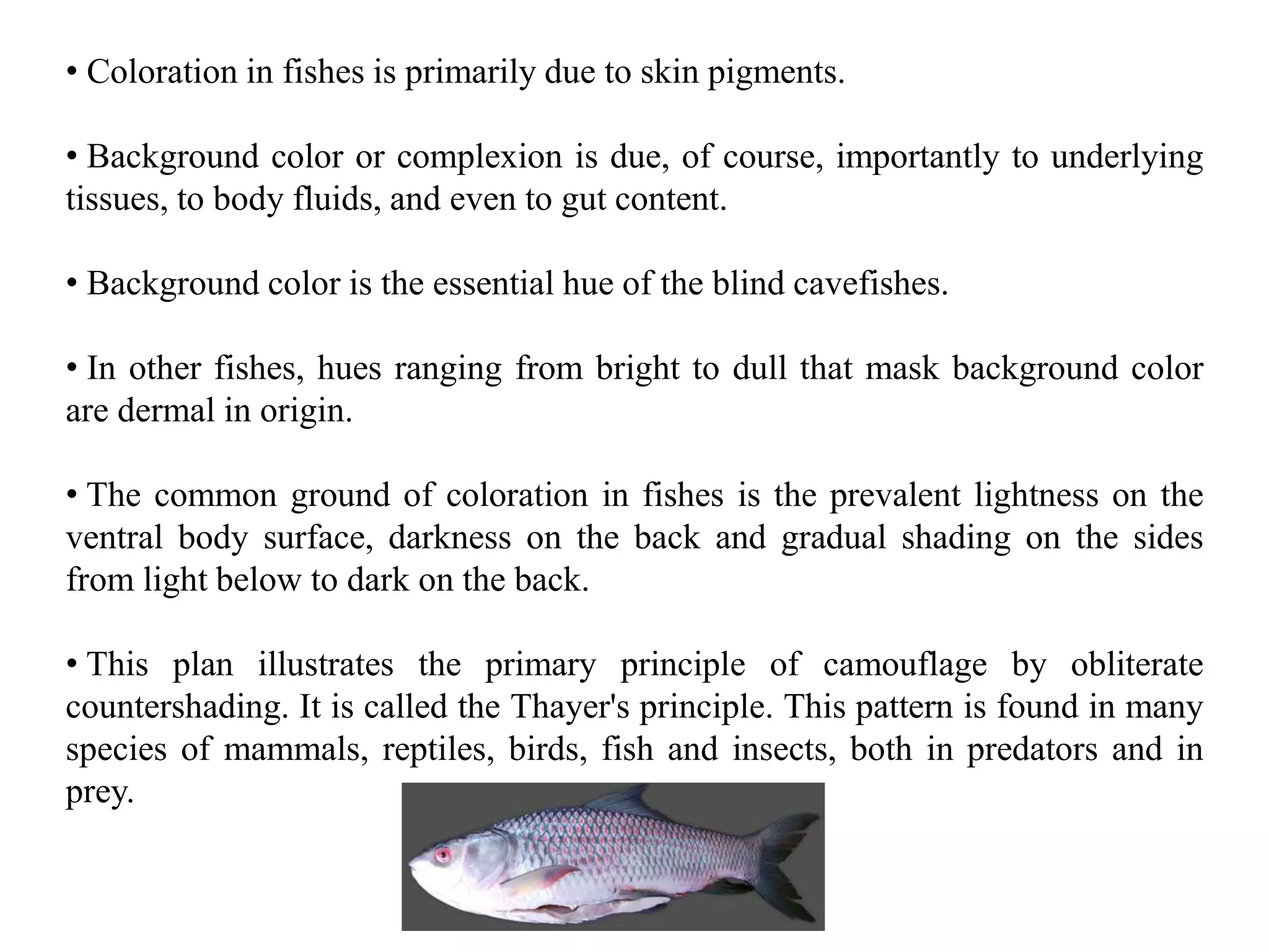
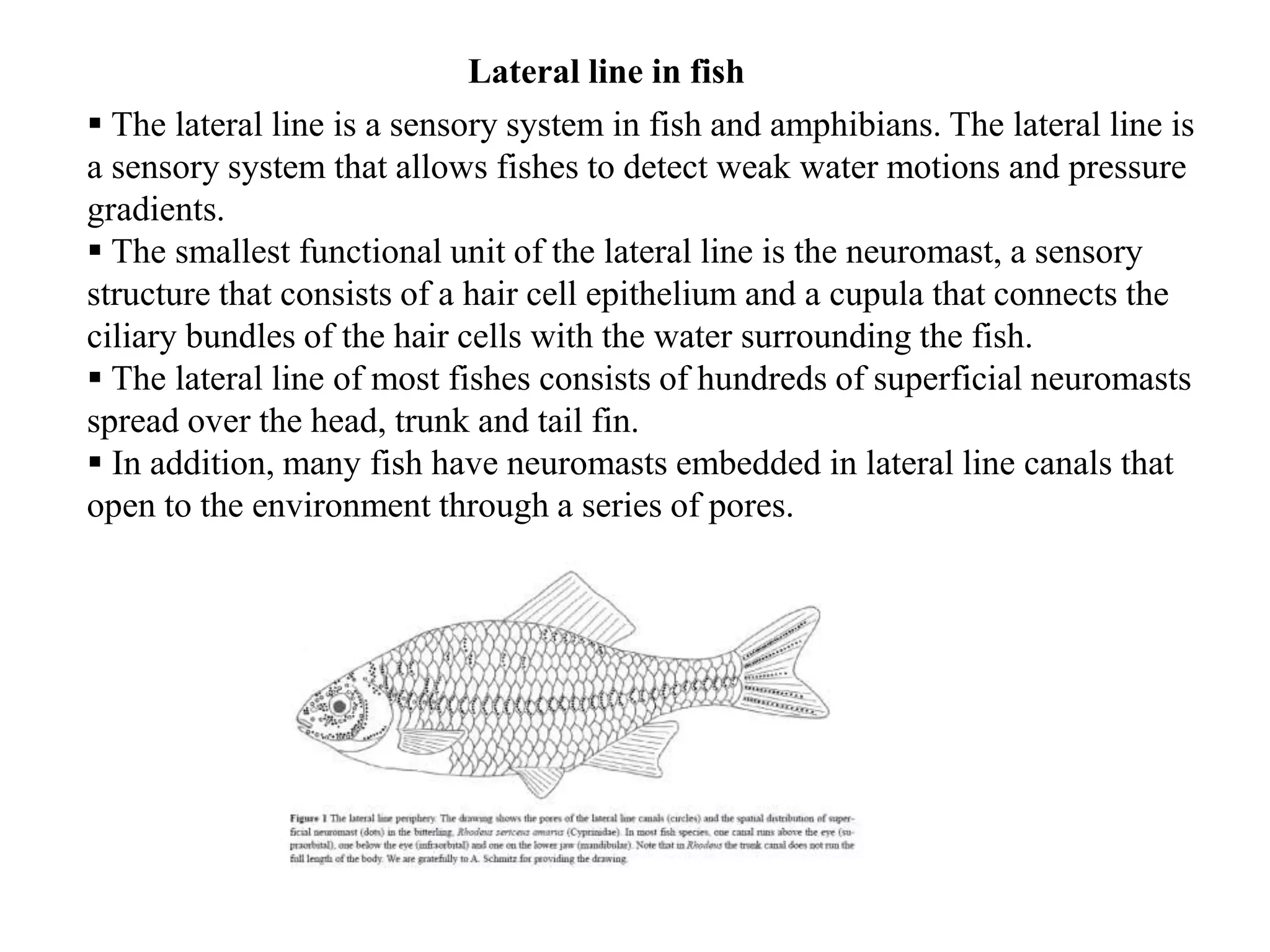
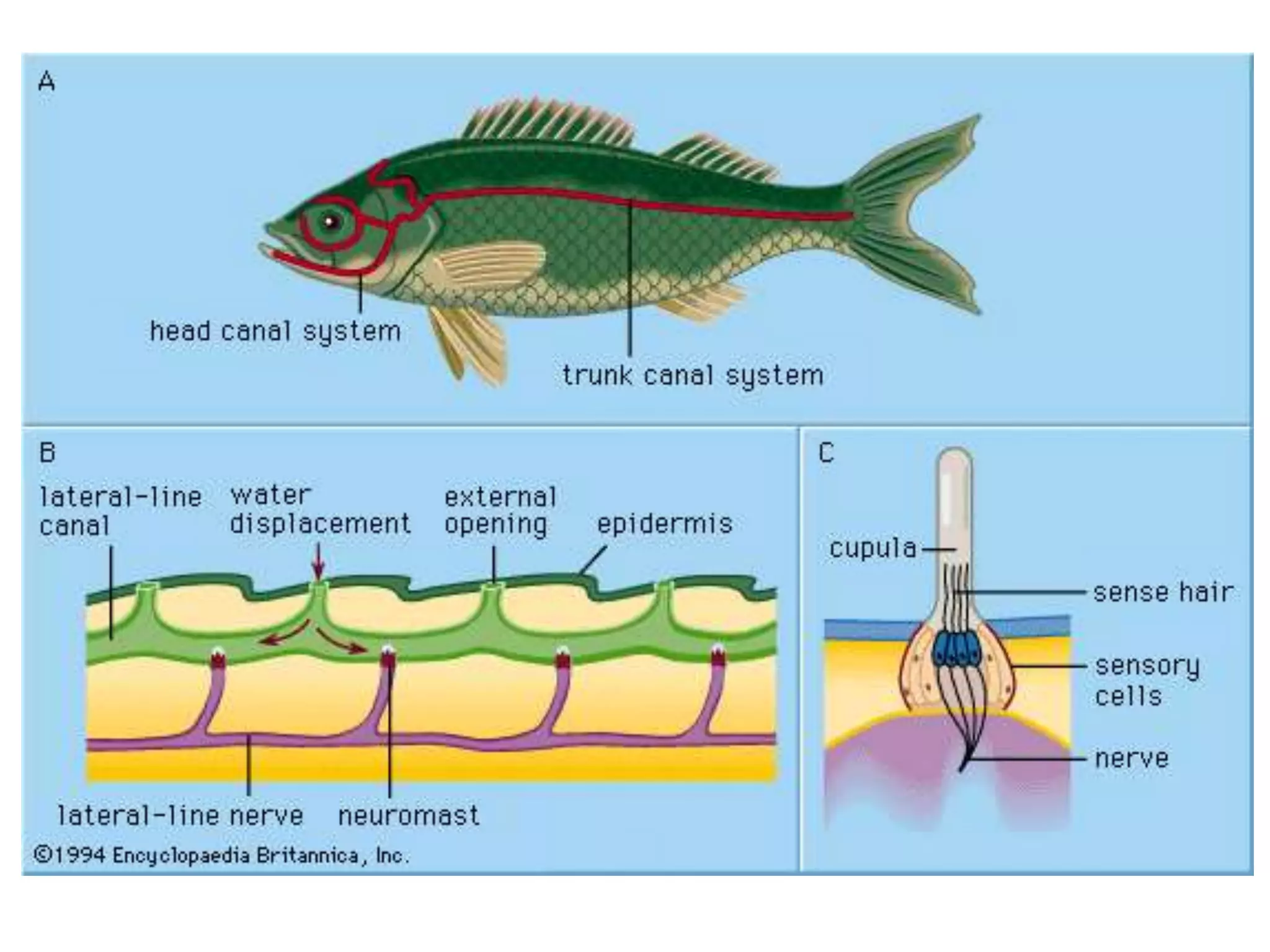
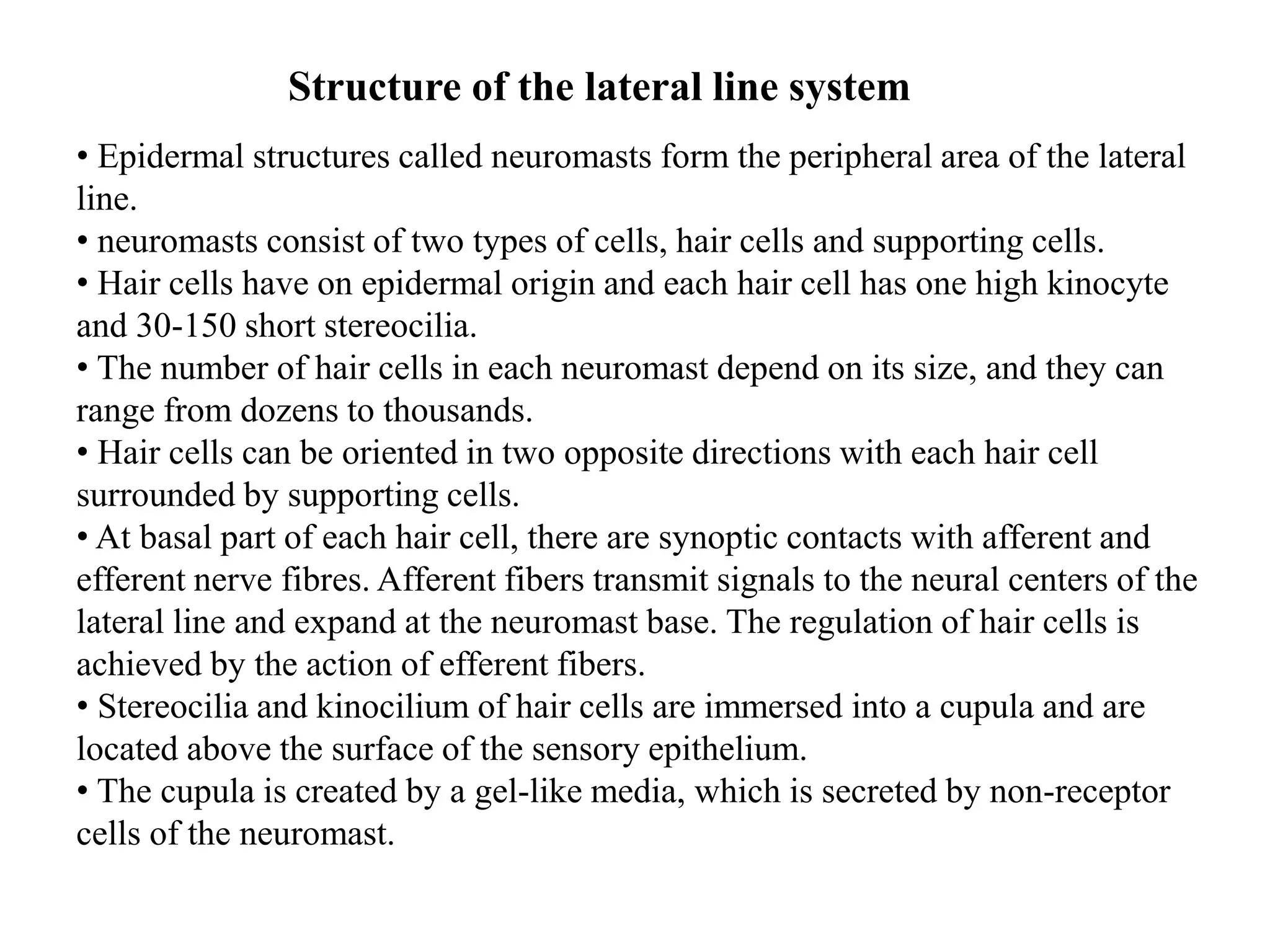
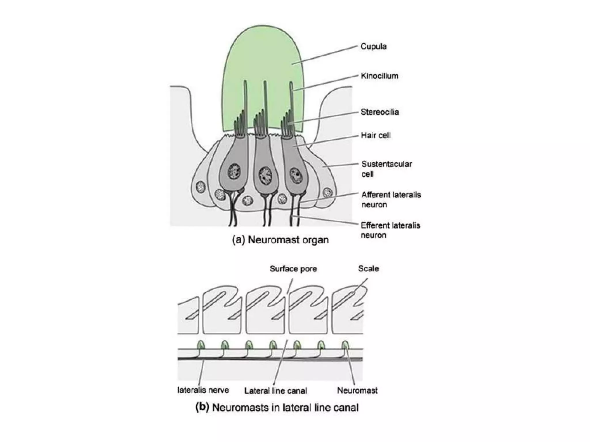
OSCamp Kubernetes 2024 | A Tester's Guide to CI_CD as an Automated Quality Co...
Gas Bladder Structure and Functions in 40 Characters
1. Gas bladder
The gas bladder, less precisely designated as the swim bladder or the air bladder is
characteristics of true fishes and reaches its fullest development as a hydrostatic
organ among spiny rayed fishes.
The gas bladder functions also
as an accessory breathing organ
As a sound producer
As a resonator in sound perception.
In certain deep sea mouth fish relatives the bladder may also serve as a fat-storage
organ of possible buoyant function.
In its weight regulating (hydrostatic) function, gas secretion is accomplished
through a special structure in the bladder wall, the gas secreting complex with its
rete mirabile or wondernet of circulatory vessels and a gas gland.
2. Structure and location
Swim bladders can be paired or unpaired gas-filled sacs.
The pneumatic duct usually connects to the oesophagus ventrally in dipnoans
and chondrostean while dorsally in garfishes, the bowfins, and modern bony
fish.
Sometimes this pneumatic duct is linked with the pharynx or stomach.
Swim bladders are present above the coelomic lining of the peritoneal cavity,
below the dorsal aorta and vertebral column, and lies close to the kidney.
The walls are made up of elastic tissues and smooth muscles.
3. On the basis of the connection of the pneumatic duct with the gut, fish are
divided into two groups:
Physostomous fishes:
These types of fishes have open pneumatic duct i.e., linked with the gut. E.g.
dipnoans, chondrostean, holostean and few teleosts.
Physoclistous fishes:
These types of fishes have closed pneumatic duct i.e., it is not linked or any
connection with gut. e.g., cat fish, carp, eels, herring and salmon.
Gases in swim bladder:
Different fishes contain different gases in their swim bladder. Some fishes have
almost 99% pure nitrogen, while some fishes have 87% oxygen. The swim
bladder of all fishes contains at least traces of four gases i.e., nitrogen, oxygen,
argon, and carbon dioxide.
4.
5. Basic Structure of Swim-Bladder:
The swim-bladder in fishes varies greatly in structure, size and shape:
1. It is essentially a trough sac-like structure with an overlying capillary
network.
2. Beneath the capillary system the wall of the anterior part of swim-bladder
consists of the following layers outside to inside (Fig. 2.38A1).
(a) Tunica externa made up of dense collagenous fibrous material.
(b) Sub-mucosa, consisting of loose connective tissue.
(c) Muscularis mucosa, consisting of a thick layer of smooth muscle fibres.
(d) Lamina propria, formed of thin-layer of connective tissue and
(e) Innermost layer of epithelial cells.
3. In the posterior chamber of swim bladder, outside the layer of muscularis
mucosa there is a glandular layer. This layer is richly supplied with blood
capillaries from rete mirabile (Fig. 2.38A2).
4. The swim bladder opens into the oesophagus by a ductus pneumaticus, which is
short and wide in lower teleosts (Chondrostie and Holostei), while in others it is
longer and narrower. The gas secreted by the swim- bladder is mostly oxygen.
Nitrogen and little quantity of carbon dioxide are also present.
6.
7. Functions of Swim bladder
Hydrostatic organ:
It is primarily a hydrostatic organ and helps to keep the weight of the body
equal to the volume of the water the fish displaces.
It also serves to equilibrate the body in relation to the surrounding medium by
increasing or decreasing the volume of gas content.
In the physostomous fishes the expulsion of the gas from the swim-bladder
occurs through the ductus pneumaticus, but in the physoclistous fishes-where the
ductus pneumaticus is absent -the superfluous gas is removed by diffusion.
Adjustable float:
The swim-bladder also acts as an adjustable float to enable the fishes to swim
at any depth with the least effort.
When a fish likes to sink, the specific gravity of the body is increased.
When it ascends the swim-bladder is distended and the specific gravity is
diminished.
By such adjustment, a fish can maintain an equilibrium at any level.
8. Maintain proper centre of gravity:
The swim bladder helps to maintain the proper centre of gravity by shifting
the contained gas from one part of it to the other and thus facilitates in
exhibiting a variety of movement.
Respiration:
The respiratory function of the swim-bladder is quite significant.
In many fishes living in water in which oxygen content is considerably low,
the oxygen produced in the bladder may serve as a source of oxygen.
In few fishes, specially in the dipnoans, the swim-bladder becomes modified
into the ‘lung’. The ‘lung’ is capable of taking atmospheric air.
Resonator:
The swim bladder is regarded to act as a resonator. It intensifies the
vibrations of sound and transmits these to the ear through the Weberian
ossicles.
9. Production of sound:
The swim bladder helps in the production of sound.
Many fishes, Doras, Platystoma, Malapterurus, Trigla can produce grunting
or hissing or drumming sound.
The circulation of the contained air inside the swim bladder causes the
vibration of the incomplete septa.
The sound is produced as the consequence of vibration of the incomplete
septa present on the inner wall of the swim bladder.
The vibrations are caused by the movement of the contained air of the swim
bladder.
Sound may also be produced by the compression of the extrinsic and intrinsic
musculatures of the swim-bladder.
Polypterus, Protopterus and Lepidosiren can produce sound by compression
and forceful expulsion of the contained gas in the swim bladder. In Cynoscion
male, the musculus sonorificus probably helps in compression.
10. Coloration in Fishes
• Majority of fishes are vividly and brightly coloured. Colouration is one of the
most common phenomena found among the fishes.
• The enormous range of colours and patterns that produced in fishes are
generally related to their habits.
• Normally fishes are darker on the dorsal and lighter on sides or ventral side.
This gives them protection from above and below.
• However, some fishes have uniform colouration as found in the gold fish,
Carassius, which has brilliant colour all over the body.
• The bottom dwellers are often strongly and intricately coloured above and pale
below.
•Variation in colour may be seen in a single fish. The trunk fish (Ostracion) has
green body, orange tail and yellow belly with blue bands on the body.
11. • Coloration in fishes is primarily due to skin pigments.
• Background color or complexion is due, of course, importantly to underlying
tissues, to body fluids, and even to gut content.
• Background color is the essential hue of the blind cavefishes.
• In other fishes, hues ranging from bright to dull that mask background color
are dermal in origin.
• The common ground of coloration in fishes is the prevalent lightness on the
ventral body surface, darkness on the back and gradual shading on the sides
from light below to dark on the back.
• This plan illustrates the primary principle of camouflage by obliterate
countershading. It is called the Thayer's principle. This pattern is found in many
species of mammals, reptiles, birds, fish and insects, both in predators and in
prey.
12. •Beyond this general plan, there are many extraordinary features of color dress in
fishes.
•One of these is color uniformity due either to lack or excess of pigment in
albino or in melanistic fishes.
•Lack of pigment and resultant transparency characterizes the pelagic, free
swimming young of many kind of fishes.
•Another is almost uniform coloration by some one hue or another. Gold color in
goldfish (Carassius) or the magnificent orange shown by the garibaldi
(Hypsypops).
• Uniform coloration would include the overall blackness or melanism that
characterizes many deep sea fishes.
13. Source of colour
Coloration in fishes due to schemachromes (colors due to physical
configuration) and biochromes or true pigment (Fox, 1953).
schemachromes
White : skeleton, gas bladder, scales and testes
Blue and violet : Iris
Iridescent: scales, eyes and intestinal membranes, integument.
Biochromes
Carotenoids: yellow, red and other hues
Chromolipoids: yellow to brown
Indigoids: blue, red and green
Melanins: Mostly black or brown
Porphyrins and bile pigments: red, yellow, green, blue and brown
Flavins: yellow, greenish fluorescence
Purins: White or silvery
Pterins: White, yellow, red and orange
14. Presence of biochromes
• Carotenoids, melanins, flavins and purins appear in fish skin.
• The liver, eggs, and eyes have carotenoids
• Melanins occur in the endoderm and skin
• Muscles and blood have porphyrins
• The skeleton and bile have bile pigment
• Flavins are widespread in blood, muscle, spleen, gills, heart, kidneys, eggs, liver &
eyes.
• Purines are in the scales and eyes.
• Pterins are in the eyes, blood, liver, kidneys and stomach.
15. Special cells for color/pigmentation
• Two kinds of cells: Chromatophores and Iridocytes.
Chromatophores impart true color
Located in the dermis of skin, either outside or beneath the scales.
Also found in the peritonium and deeply around the brain and spinal cord.
The basic Chromatophores are:
Erythrophores: Red and orange
Xanthophores: yellow
Melanophores: Black
Leucophores: White
Iridocytes
• Called mirror cells because they contain reflecting materials that mirror colors
outside the fish.
• Both leucophores and irdophores contain purines, primarily guanine where
leucophores contain small crystals that can move back and forth in cytoplasm.
• Having purines of large crystals incapable of movement and usually stacked in
layers.
16. Significance of coloration
For communication with either the members of same species (Intraspecific) or
with different species (interspecific)
Intraspecific signal: Serve social and sexual purposes.
For recognition, threat or warning
For sexual purposes
Recognition of their parents e.g. Hemichromis which are bright during period of
caring of young ones
Interspecific signal:
For warning or intimidating potential predators.
For decoying or masking purposes directed toward either prey or predators.
17. Function of colorations
Cott (1940) grouped principal functions of coloration under three heading:
i) Concealment
• General color resemblance
• Variable color resemblance
• Obliterative shading
• Disruptive coloration
• Coincident disruptive coloration
ii) Disguise
iii) Advertisement
18. Concealment: General color resemblance
Important in resemblance between fish and its background- e.g. Many coral-
reef fishes are highly bright in color as the coral heads.
Some fishes living over light shaded bottom are light colored but over dark
bottoms the same species are dark.
19. Variable color resemblance
• Ability of fish to change color gradually or rapidly to match its background.
•Occurs in life history stages. e.g. In Rainbow trout (stream resident phase) is
multicolored including dark spots (young) and rosy sides in adult.
•While the same individuals in sea are blue above and white below.
•Variations also occurs according to in season, day or night.
•E.g. trout occupying bright or partly shaded in summer while in winter they are
evenly dark.
20. Obliterate shading
Countershading or Thayer’s law is a method of camouflage in which an
animal’s coloration is darker on the upper side and lighter on the underside of
the body.
Disruptive coloration
Also known as disruptive camouflage or disruptive patterning.
Surface of fish is covered by irregular patched of contrasted colorand
tones that draw attention away from the original shape which they have.
Prevention or delay as long as recognition of sight.
21. Disguise
Simply tends to reduce the resemblance of the fish to itself.
Deflective and directive marks are important here
Deflective marks are those which deflect the attack of an enemy from more or
less vital part of the body to some other part. E.g. Dark spot on the tail of the
bowfin.
Directive marks divert the attention of prey from the most dangerous part of
the predator parts. E.g., In stargazers feeding habit.
22. Advertisement
Coloration for advertise or reveal
E.g., in Darters American streams and also members of the prech family are
the most bright
Significance for sexual recognition
E.g. of Sticklebacks suggest the value of color in sexual recognition
23. Use in classification
• Used often as character to separate taxonomic units.
•Exact patterns of chromatophores are often in genetic control, use in
classification.
24.
25. Fishes have only four biochromes, varied color patterns are obtained by
mixing of these existing pigments in variable proportions.
Melanophores, Erythophores and Xanthophores are the light absorbing
chromatophores.
Iridiophores are called as the light reflecting chromatophores.
Chromatophores
26. The lateral line system consists of 100 or more sensory organ (neuromast) that are
typically arranged in lines on or just under the skin of the head and body.
It is absent from all reptiles, birds, and mammals, even those that are aquatic
(such as turtle, dolphins and whales)
In the head, the lateral line canal is separated into three canals, one posses forward
and below the eye and the other downward and below the jaw.
These canals have numerous pores and together with the lateral line canal, make
the lateral line system
It is made up of mechanoreceptors called neuromasts which are sensitive to water
movement (Diaz et al. 2003).
The lateral line system has an important role in the detection of stationary objects,
navigation, prey detection, capture and in swimming in schools (Gelman et al.
2007).
The receptor organ of the lateral line system is the neuromast. There are two types
of neuromasts, canal neuromasts which are located in the intradermal canals, and
the superficial neuromasts which are located in the intraepidermal canals.
Canal neuromasts are able to detect water flow acceleration, while superficial or
free neuromasts can detect velocity (Gelman et al. 2007).
27. Lateral line in fish
The lateral line is a sensory system in fish and amphibians. The lateral line is
a sensory system that allows fishes to detect weak water motions and pressure
gradients.
The smallest functional unit of the lateral line is the neuromast, a sensory
structure that consists of a hair cell epithelium and a cupula that connects the
ciliary bundles of the hair cells with the water surrounding the fish.
The lateral line of most fishes consists of hundreds of superficial neuromasts
spread over the head, trunk and tail fin.
In addition, many fish have neuromasts embedded in lateral line canals that
open to the environment through a series of pores.
28.
29. Structure of the lateral line system
• Epidermal structures called neuromasts form the peripheral area of the lateral
line.
• neuromasts consist of two types of cells, hair cells and supporting cells.
• Hair cells have on epidermal origin and each hair cell has one high kinocyte
and 30-150 short stereocilia.
• The number of hair cells in each neuromast depend on its size, and they can
range from dozens to thousands.
• Hair cells can be oriented in two opposite directions with each hair cell
surrounded by supporting cells.
• At basal part of each hair cell, there are synoptic contacts with afferent and
efferent nerve fibres. Afferent fibers transmit signals to the neural centers of the
lateral line and expand at the neuromast base. The regulation of hair cells is
achieved by the action of efferent fibers.
• Stereocilia and kinocilium of hair cells are immersed into a cupula and are
located above the surface of the sensory epithelium.
• The cupula is created by a gel-like media, which is secreted by non-receptor
cells of the neuromast.
30.
31. • Stereocilia and kinocilium of hair cells are immersed into a cupula and are located
above the surface of the sensory epithelium.
• The cupula is created by a gel-like media, which is secreted by non-receptor cells
of the neuromast.
•There are two types of neuromasts, superficial or free neuromasts
and canal neuromasts.
• Superficial neuromasts are located at the surface of the body and are affected by
the environment. Superficial neuromasts are categorized into primary or
paedomorphic neuromasts and secondary or neomorphic neuromasts.
• Canal neuromasts are primary neuromasts. These are found inside epidermal or
bony canals and are located on the head or body of the fish (Coombs et al. 1992).
32. Fig. A schematic transferred process for detecting underwater sound of the hair cell