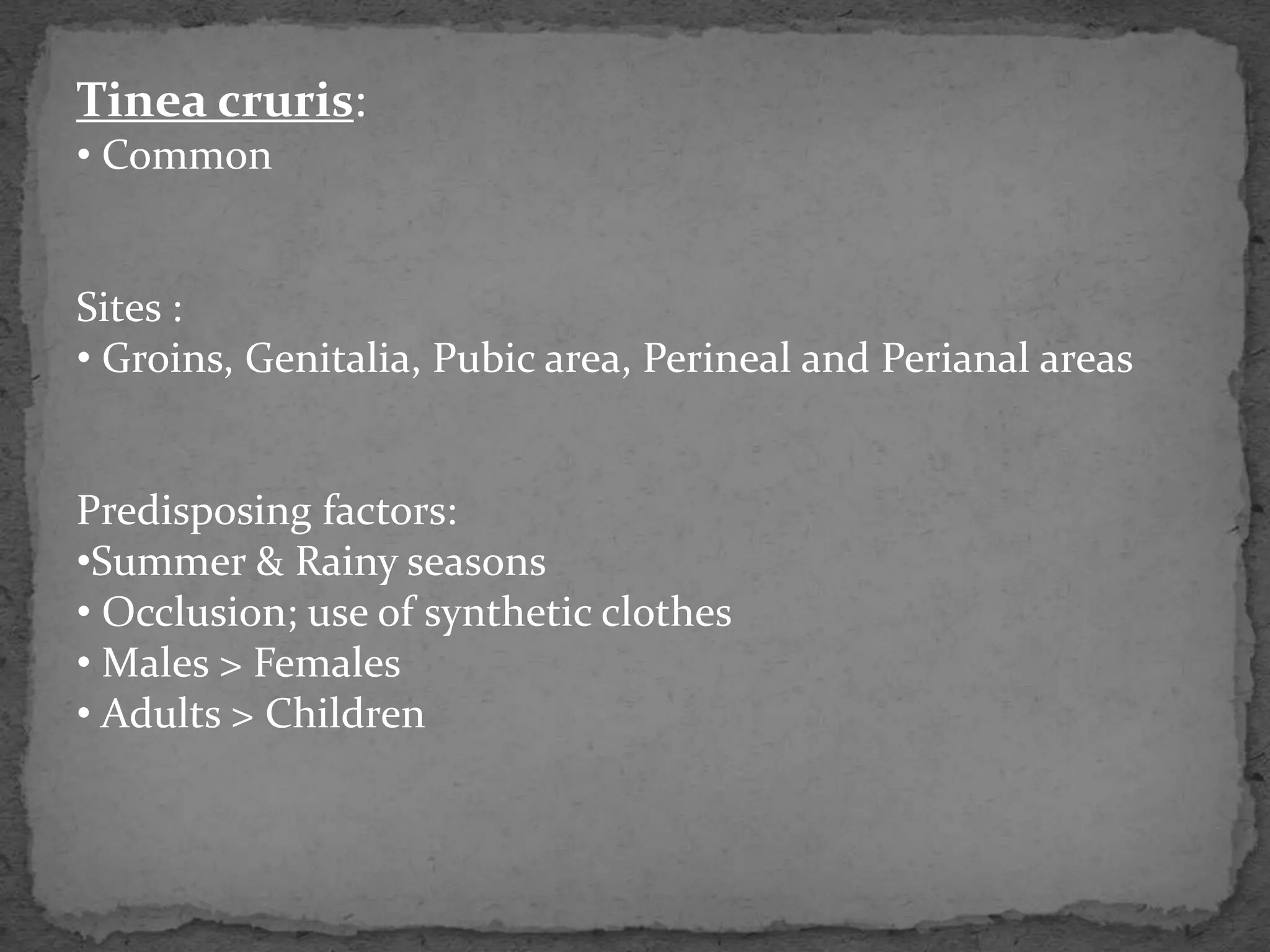
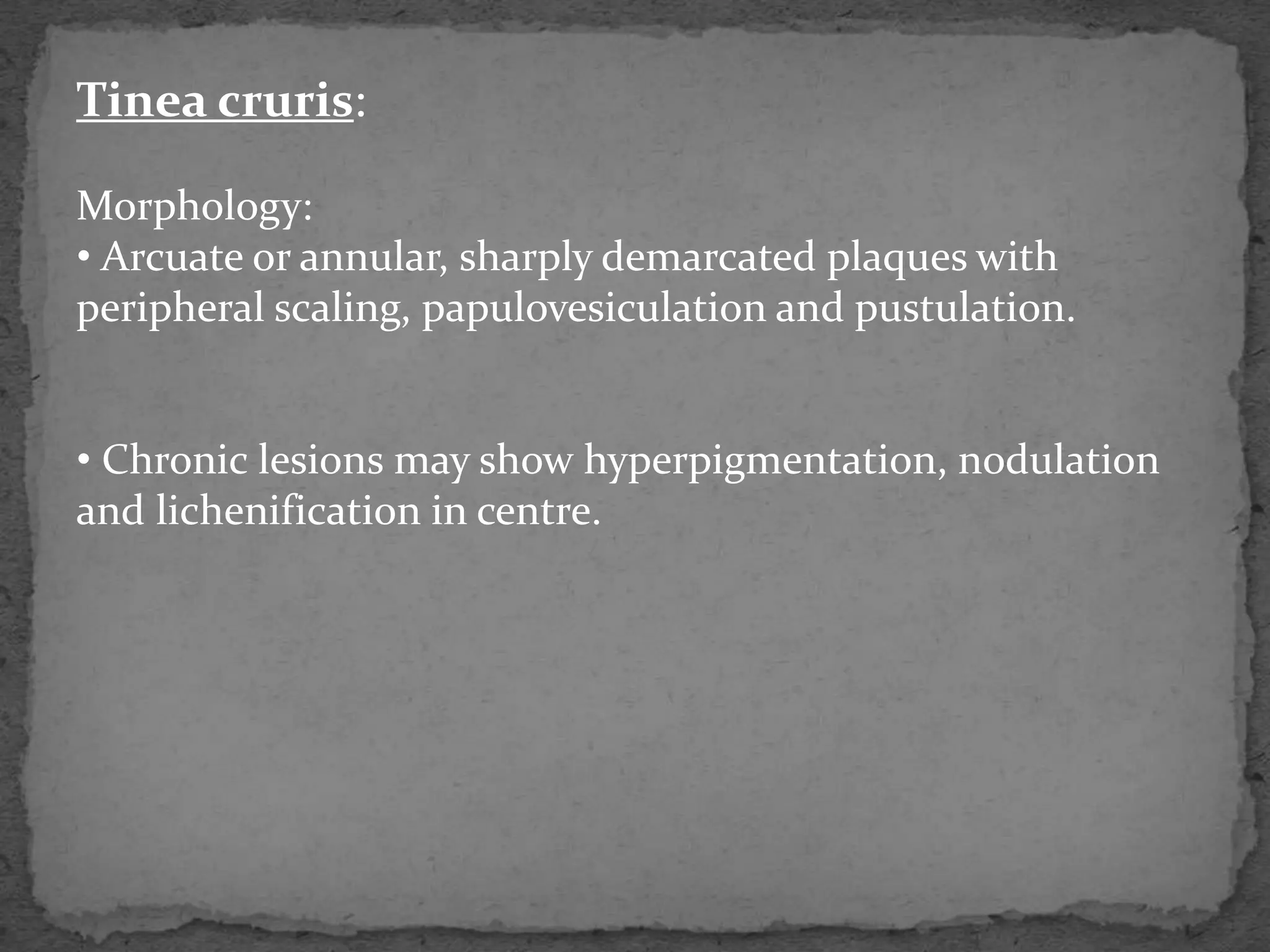
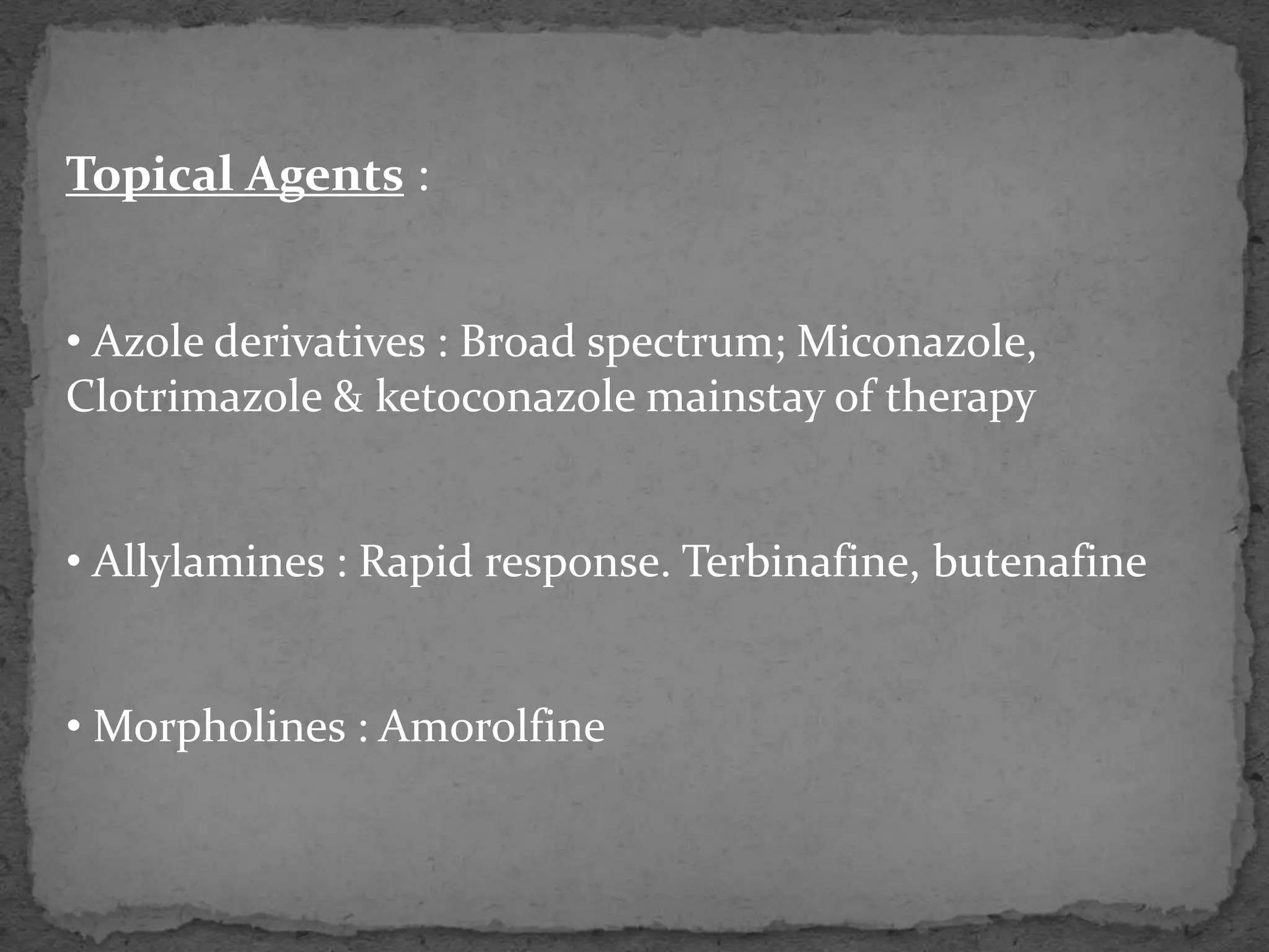
Dermatology
Fungal infections of skin:
1. Deep fungal infections
2. Superficial fungal infections
Deep Fungal Infections:
1. Mycetoma
2. Sporotrichosis
3. Chromoblastomycosis
4. Sub cutaneous zygomycosis
Superficial Fungal infections:
1. Dermatophyte infection
2. Pityriasis versicolor
3. Candidiasis
Dermatophyte Infection:
Cause
1. Trichophyton - skin, hair and nails
2. Microsporum - skin and hair
3. Epidermophyton - skin and nails
• Dermatophytes invade only onto Stratum corneum and
the inflammation caused is the outcome of delayed
hypersensitivity or the metabolic products of fungus.
• Severity of infection: Zoophilic fungi > Anthropophilic
fungi
Tinea pedis (Athelete’s foot):
• M/c type
• Organisms :
a. Trichophyton rubrum (m/c and most stubborn)
b. Trichophyton mentagrophytes var. interdigitale
c. Epidermophyton floccosum
• Clinical pattern:
a. Soggy interdigital scaling, particularly in 4th & 5th
interspace (all three organisms)
b. Diffuse dry scaling of soles (usually T. rubrum)
c. Recurrent episodes of vesication (T. mentagrophytes var.
interdigitale or E. floccosum)
Tinea unguium:
Nail shows following changes :
• Yellow-brown discoloration & thickening of nail plate that
crumbles easily and so appears tunnelled.
• Subungual hyperkeratosis
• Onycholysis
Patterns:
 Distal/lateral Subungual variety (m/c)
Superficial white variety
 Proximal subungual variety.
Tinea manuum:
• Often associated with Tinea pedis
• Lesions manifest as unilateral, well-defined plaques or as
diffuse erythema of the palms with accumulation of fine
scales in the creases.
Tinea cruris:
• Common
Sites :
• Groins, Genitalia, Pubic area, Perineal and Perianal areas
Predisposing factors:
•Summer & Rainy seasons
• Occlusion; use of synthetic clothes
• Males > Females
• Adults > Children
Tinea cruris:
Morphology:
• Arcuate or annular, sharply demarcated plaques with
peripheral scaling, papulovesiculation and pustulation.
• Chronic lesions may show hyperpigmentation, nodulation
and lichenification in centre.
Tinea corporis:
Sites : Glabrous skin, except palms, soles and groins
Variants:
• Tinea incognito ( modified by steroid therapy)
• Tinea faciae ( Cheeks of children)
• Tinea barbae ( Inflammatory swelling with alopecia, in
beard region)
Diagnostic features of T. corporis/cruris:
•Itchy dermatosis
• Annular/ arcuate lesions
• Peripheral papulovesiculation & scaling with a relatively
clear centre
Tinea capitis:
• Usually a disease of children
• Anthropophilic fungi – bald and scaly areas, with minimal
inflammation and hairs broken off 3-4mm from scalp
• Zoophilic fungi – induce more intense inflammation with
boggy swelling, pustulations and lymphadenopathy.
Patterns:
• Noninflammatory tinea capitis
• Inflammatory tinea capitis
• Favus (T. schoenleinii/ cicatricial alopecia)
Diagnostic features of T. capitis:
• Patient usually children
• Non-inflammatory/inflammatory patch of alopecia
• Easy, painless pluckability of hair
Complications of Tinea:
• Dermatophytide reaction
• Cicatricial alopecia
Investigations :
• Microscopic Examination
• Culture (SDA )
• Wood’s light
Treatment :
Factors determining treatment modalities :
• Site of lesions
• Extent of lesions
• Chronicity
• Patient compliance
General Measures :
• Keeping area dry
• Avoid use of synthetic clothes
• In recurrent infection, prophylactic use of antifungal
talc.
Systemic Therapy recommended in :
• Extensive dermatophytic infections
• Tinea unguium
• Tinea capitis
Topical Agents :
• Azole derivatives : Broad spectrum; Miconazole,
Clotrimazole & ketoconazole mainstay of therapy
• Allylamines : Rapid response. Terbinafine, butenafine
• Morpholines : Amorolfine
Systemic Therapy :
• Terbinafine
• Griseofulvin
• Itraconazole
Terbinafine :
• Fungicidal
• Indications – Dermatophytic infections [ DOC for
extensive tinea infection and tinea unguium; ineffective
in pityriasis versicolor and candidal infection.]
• Dose – 250mg OD. With development of resistance,
500mg OD being used.
•S/Es – GI side effects, Alteration in tastes and skin
rashes
Griseofulvin :
• Fungistatic
• Indications – resurgent use, with development of
resistance to terbinafine. [DOC in T. capitis; ineffective
in pityriasis versicolor and candidal infection.]
• Dose – 10mg/kg daily of ultramicronized formulation,
after fatty meal
• S/Es – may cause persistant headdache, GI side effects
and skin eruptions. (Common cause of photosensitive
reaction)
• Avoid in – Pregnants and in pt. with liver failure,
porphyria and systemic lupus.
Itraconazole :
• Fungistatic
• Indications – broad-spectrum antifungal agent,
effective in dermatophytic infections, pityriasis versicolor
and candidal infection. (Generally used in
onychomycosis)
• Dose – 200-400 mg, to be taken with meals
•S/Es – GI side effects and hepatotoxicity.
• Avoid in – Children, Pregnant and lactating women.
References :
• Illustrated Synopsis of Dermatology and
Sexually Transmitted Diseases. Khanna Neena
• Textbook of Clinical Dermatology. Sehgal
Virendra N.
Thank You !

Fungal Infections.pptx

  • 1.
  • 2.
    Fungal infections ofskin: 1. Deep fungal infections 2. Superficial fungal infections
  • 3.
    Deep Fungal Infections: 1.Mycetoma 2. Sporotrichosis 3. Chromoblastomycosis 4. Sub cutaneous zygomycosis
  • 4.
    Superficial Fungal infections: 1.Dermatophyte infection 2. Pityriasis versicolor 3. Candidiasis
  • 5.
    Dermatophyte Infection: Cause 1. Trichophyton- skin, hair and nails 2. Microsporum - skin and hair 3. Epidermophyton - skin and nails • Dermatophytes invade only onto Stratum corneum and the inflammation caused is the outcome of delayed hypersensitivity or the metabolic products of fungus. • Severity of infection: Zoophilic fungi > Anthropophilic fungi
  • 6.
    Tinea pedis (Athelete’sfoot): • M/c type • Organisms : a. Trichophyton rubrum (m/c and most stubborn) b. Trichophyton mentagrophytes var. interdigitale c. Epidermophyton floccosum • Clinical pattern: a. Soggy interdigital scaling, particularly in 4th & 5th interspace (all three organisms) b. Diffuse dry scaling of soles (usually T. rubrum) c. Recurrent episodes of vesication (T. mentagrophytes var. interdigitale or E. floccosum)
  • 8.
    Tinea unguium: Nail showsfollowing changes : • Yellow-brown discoloration & thickening of nail plate that crumbles easily and so appears tunnelled. • Subungual hyperkeratosis • Onycholysis Patterns:  Distal/lateral Subungual variety (m/c) Superficial white variety  Proximal subungual variety.
  • 10.
    Tinea manuum: • Oftenassociated with Tinea pedis • Lesions manifest as unilateral, well-defined plaques or as diffuse erythema of the palms with accumulation of fine scales in the creases.
  • 12.
    Tinea cruris: • Common Sites: • Groins, Genitalia, Pubic area, Perineal and Perianal areas Predisposing factors: •Summer & Rainy seasons • Occlusion; use of synthetic clothes • Males > Females • Adults > Children
  • 13.
    Tinea cruris: Morphology: • Arcuateor annular, sharply demarcated plaques with peripheral scaling, papulovesiculation and pustulation. • Chronic lesions may show hyperpigmentation, nodulation and lichenification in centre.
  • 15.
    Tinea corporis: Sites :Glabrous skin, except palms, soles and groins Variants: • Tinea incognito ( modified by steroid therapy) • Tinea faciae ( Cheeks of children) • Tinea barbae ( Inflammatory swelling with alopecia, in beard region)
  • 17.
    Diagnostic features ofT. corporis/cruris: •Itchy dermatosis • Annular/ arcuate lesions • Peripheral papulovesiculation & scaling with a relatively clear centre
  • 18.
    Tinea capitis: • Usuallya disease of children • Anthropophilic fungi – bald and scaly areas, with minimal inflammation and hairs broken off 3-4mm from scalp • Zoophilic fungi – induce more intense inflammation with boggy swelling, pustulations and lymphadenopathy. Patterns: • Noninflammatory tinea capitis • Inflammatory tinea capitis • Favus (T. schoenleinii/ cicatricial alopecia)
  • 20.
    Diagnostic features ofT. capitis: • Patient usually children • Non-inflammatory/inflammatory patch of alopecia • Easy, painless pluckability of hair
  • 21.
    Complications of Tinea: •Dermatophytide reaction • Cicatricial alopecia
  • 22.
    Investigations : • MicroscopicExamination • Culture (SDA ) • Wood’s light
  • 23.
  • 24.
    Factors determining treatmentmodalities : • Site of lesions • Extent of lesions • Chronicity • Patient compliance
  • 25.
    General Measures : •Keeping area dry • Avoid use of synthetic clothes • In recurrent infection, prophylactic use of antifungal talc.
  • 26.
    Systemic Therapy recommendedin : • Extensive dermatophytic infections • Tinea unguium • Tinea capitis
  • 27.
    Topical Agents : •Azole derivatives : Broad spectrum; Miconazole, Clotrimazole & ketoconazole mainstay of therapy • Allylamines : Rapid response. Terbinafine, butenafine • Morpholines : Amorolfine
  • 28.
    Systemic Therapy : •Terbinafine • Griseofulvin • Itraconazole
  • 29.
    Terbinafine : • Fungicidal •Indications – Dermatophytic infections [ DOC for extensive tinea infection and tinea unguium; ineffective in pityriasis versicolor and candidal infection.] • Dose – 250mg OD. With development of resistance, 500mg OD being used. •S/Es – GI side effects, Alteration in tastes and skin rashes
  • 30.
    Griseofulvin : • Fungistatic •Indications – resurgent use, with development of resistance to terbinafine. [DOC in T. capitis; ineffective in pityriasis versicolor and candidal infection.] • Dose – 10mg/kg daily of ultramicronized formulation, after fatty meal • S/Es – may cause persistant headdache, GI side effects and skin eruptions. (Common cause of photosensitive reaction) • Avoid in – Pregnants and in pt. with liver failure, porphyria and systemic lupus.
  • 31.
    Itraconazole : • Fungistatic •Indications – broad-spectrum antifungal agent, effective in dermatophytic infections, pityriasis versicolor and candidal infection. (Generally used in onychomycosis) • Dose – 200-400 mg, to be taken with meals •S/Es – GI side effects and hepatotoxicity. • Avoid in – Children, Pregnant and lactating women.
  • 32.
    References : • IllustratedSynopsis of Dermatology and Sexually Transmitted Diseases. Khanna Neena • Textbook of Clinical Dermatology. Sehgal Virendra N.
  • 33.