
More Related Content
What's hot
What's hot (20)
Similar to FUNGAL INFECTIONS.pptx
Similar to FUNGAL INFECTIONS.pptx (20)
Recently uploaded
Recently uploaded (20)
FUNGAL INFECTIONS.pptx
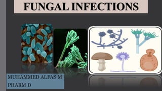
- 1. FUNGAL INFECTIONS MUHAMMED ALFAS M PHARM D
- 2. DEFENITION : • Fungi are ubiquitous microorganisms that differ from bacteria in their cellular structure, and this makes them naturally resistant to antibacterial agents .Fungi are broadly divided into yeasts and moulds. Yeasts are typically round or oval shaped microscopically, grow flat round colonies on culture plates and reproduce by forming buds from their cells. Moulds (e.g. Aspergillus , Mucor) appear as a collection or mass (mycelium) of individual tubular structures called hyphae that grow by branching and longitudinal extension. • There are hundreds of species of fungi found in the environment , but some are mainly act as human pathogen . • Fungal spore are spread by air, water and direct contact with infected source. Humans usually become infected by inhalation of airborne spores or by inoculation into traumatized skin and mucous membrane. • This increase can be attributed, in part, to the growing numbers of immunocompromised hosts as a result of organ transplants, cancer chemotherapy, and the acquired immunodeficiency syndrome (AIDS) epidemic.
- 5. • fungi are a cause of superficial infections of the skin and mucous membranes. In some susceptible hosts whose immune system is heavily compromised, deep-seated infections involving organs like lungs and brain can manifest as ‘difficult to cure’ infections, for example, pulmonary aspergillosis or cryptococcal meningitis. 1. Superficial fungal infection . 2. Invasive fungal infection .
- 6. SUPERFICIAL FUNGAL INFECTIONS • Superficial fungal infections are benign infections of the skin, scalp and nails caused by Candida albicans or dermatophytes. • It grow in dark and moist areas and invade various parts of the body. These infections are easily treatable in immunocompetent individuals. • Superficial mycoses are among the most common infections in the world and the second most common vaginal infections in North America. Mucocutaneous candidiasis can occur in three forms— oropharyngeal, esophageal, and vulvovaginal disease—with oropharyngeal and vulvovaginal disease being the most common. Over the past 15 to 20 years, the occurrence rates of some fungal infections have increased dramatically. The prevalence of fungal skin infections varies throughout different parts of the world, from the most common causes of skin infections in the tropics to relatively rare disorders in the United States.
- 7. VULVOVAGINAL CANDIDIASIS FUNGUS • Vulvovaginal candidiasis (VVC) refers to infections in individuals with or without symptoms who have positive vaginal cultures for Candida species. Depending on episodic frequency, VVC can be classified as either sporadic or recurrent. This classification is essential to understand the pathophysiology, as well as the pharmacotherapy, of VVC. Furthermore, VVC may be defined as uncomplicated, which refers to sporadic infections that are susceptible to all forms of antifungal therapy regardless of the duration of treatment, or complicated, in which consideration of factors affecting the host, microorganism, and pharmacotherapy all have an essential role in successful treatment. Complicated VVC includes recurrent VVC, severe disease, non– Candida albicans candidiasis, and host factors, including diabetes mellitus, immunosuppression, and pregnancy.
- 10. PATHOPHYSIOLOGY • C. albicans is the major pathogen responsible for VVC, accounting for 80% to 92% of symptomatic episodes. • Candida species can act as commensal members of the vaginal flora. Asymptomatic colonization with Candida species has been found in 10% to 20% of women of reproductive age. Candida organisms are dimorphic; blastospores are responsible for colonization (transmission and spread), whereas germinated Candida forms are associated with tissue invasion and symptomatic infections.To colonize the vagina, Candida species must be able to attach to the mucosa. The attachment process is complex. Not only are candidal surface structures important for attachment, but appropriate receptors for attachment must be present in the epithelial tissue. Not all women have the same range of receptors, which may explain variation in colonization. Changes in the host’s vaginal environment or response are necessary to induce a symptomatic infection. Unfortunately, in most cases of symptomatic VVC, no precipitating factor can be identified
- 11. TREATMENTS
- 12. • Complicated VVC : It occurs in patients who are immunocompromised or have uncontrolled diabetes mellitus.These individuals need a more aggressive treatment plan.Current recommendations are to lengthen therapy to 10 to 14 days regardless of the route of administration. Therapeutic options include those listed in un complicated treatment plan . however, regimens should be continued for 10 to 14 days. A study of oral fluconazole therapy in women with complicated VVC demonstrated that cure rates increased from 67% with single-dose therapy to 80% when the 150 mg dose of fluconazole was repeated 72 hours after the initial dose. • Recurrent Vulvovaginal Candidiasis :Recurrent vulvovaginal candidiasis (RVVC) is defined as having more than four episodes of VVC within a 12- month period. A proper diagnosis should be obtained to rule out other infections or nonmycotic contact dermatitis. RVVC is best treated in two stages: an initial intensive stage followed by prolonged antifungal therapy to achieve mycologic remission. The Infectious Diseases Society of America recommends 10 to 14 days of induction therapy with a topical or oral azole, followed by 150 mg of fluconazole once weekly for 6 months for recurring Candida VVC.
- 13. ANTIFUNGAL-RESISTANT VULVOVAGINAL CANDIDIASIS • Resistance to azole antimycotics should be considered in individuals who have persistently positive yeast cultures and fail to respond to therapy despite adherence to prescribed regimens.These infections can be treated with boric acid or 5-flucytosine • Boric acid is administered as a 600 mg intravaginal capsule daily for 14 days of induction therapy, followed by a maintenance regimen of one capsule intravaginally twice weekly. • Boric acid is administered as a 600 mg intravaginal capsule daily for 14 days of induction therapy, followed by a maintenance regimen of one capsule intravaginally twice weekly
- 14. OROPHARYNGEALAND ESOPHAGEAL CANDIDIASIS • Oropharyngeal candidiasis (OPC), or thrush, is a common and localized infection of the oral mucosa caused by the yeast Candida. C. albicans, a common oral commensal organism, is the most frequent infecting species. OPC is also referred to as candidiasis (or the more correct but less commonly used term candidosis). The infection may extend into the esophagus, causing esophageal candidiasis. • Candida carriage increases under immunocompromised conditions and also among hospitalized patients . Even in the era of highly active antiretroviral therapy (HAART) up to 80% of human immunodeficiency virus (HIV)- infected persons may demonstrate oral yeast colonization. The organism is capable of transition to a pathogen causing symptomatic mucosal infections in association with predisposing host factors
- 15. • C. albicans is the predominant colonizing Candida species (70%-80%), but any of the non-C. albicans species such as C. glabrata and C. tropicalis which may account for 5% to 8%, respectively, can be colonizers. RISK FACTORS FOR THE DEVELOPMENT OF OROPHARYNGEAL OR ESOPHAGEAL CANDIDIASIS
- 19. Pathophysiology • The pathogenesis of OPC is most clearly elucidated in the setting of HIV infection. There appear to be several levels of immune defense against the development of OPC in HIV-infected persons, and they involve both systemic and local immunity. The primary line of host defense against C. albicans is cell-mediated immunity (CMI) at the mucosal surfaces, which is mediated by CD4 T cells. • When the number of CD4 T cells drops too low, recruitment of these cells to the oral cavity is impaired. The CD4 T-cell count is the hallmark predictor for development of OPC. similar immunosuppression, such as lymphoma and bone marrow transplant. • When the primary line of defense fails, the secondary host defenses become crucial. These include the CD8 T cells, salivary cytokines, and other innate immune cells, such as the neutrophils, macrophages, and epithelial cells (with anti-Candida activity). • The problem with the CD8 T cells is caused more by a dysfunction of the microenvironment, specifically, reduction in the E-cadherin adhesion molecule that promotes migration of the cells through mucosal tissues
- 20. CLINICAL PRESENTATION OF OROPHARYNGEAL AND ESOPHAGEAL CANDIDIASIS OROPHARYNGEAL CANDIDIASIS ESOPHAGEAL CANDIDIASIS Symptoms : Symptoms are diverse and range from none to a sore, painful mouth, burning tongue, metallic taste, and dysphagia and odynophagia with involvement of the hypopharynx Symptoms :Typically, the symptoms are dysphagia, odynophagia, and retrosternal chest pain but can be asymptomatic in some patients; although rare, epigastric pain can be the dominant symptom Signs :Signs are variable and can include diffuse erythema and white patches on the surfaces of the buccal mucosa, throat, tongue, or gums; constitutional signs are absent Signs : Constitutional signs, including fever, occasionally occur; physical findings can range from a few to numerous white or beige plaques of variable size Plaques can be hyperemic or edematous, with ulceration in more severe cases Most advanced cases can occur with increased mucosal friability and narrowing of lumen Uncommon complications include perforation and aortic–esophageal fistula formation
- 21. OROPHARYNGEAL CANDIDIASIS ESOPHAGEAL CANDIDIASIS Laboratory tests : Scraping of an active lesion for microscopic examination can help confirm the diagnosis (presence of pseudohyphae and budding yeast) but is usually not necessary Cultures are not necessary because isolation of Candida species does not distinguish between colonization and true infection; cultures can be taken in patients responding poorly to therapy to determine the infecting species and to predict likely drug resistance Laboratory tests : The best test is upper GI endoscopy (more useful than barium swallow); helps exclude other causes of esophagitis (eg, viral, aphthous ulcers); diagnosis is confirmed by the histologic presence of Candida species in biopsy lesions taken during endoscopy Cultures to look for drug-resistant Candida species are warranted in patients who require endoscopy
- 22. THERAPEUTIC OPTIONS FOR MUCOSAL CANDIDIASIS
- 24. MYCOTIC INFECTIONS OF THE SKIN, HAIR, AND NAILS • Superficial cutaneous mycoses affect up to 20% to 25% of the global population.65 The usual pathogens are the dermatophytes classified by genera: Trichophyton, Epidermophyton, and Microsporum. • Dermatophytes have the ability to penetrate keratinous structures of the body and therefore infections are limited to hair, nails and skin. These infections affect both male and female genders and all races. • Risk factors for the development of an infection include prolonged exposure to sweat or soaking in water, maceration, intertriginous folds, sharing personal belongings such as combs, close living quarters • Diagnosis usually is based on patient history, as well as the physical examination. Diagnostic tests include direct microscopic examination of a specimen after the addition of KOH or fungal cultures.
- 25. TINEA PEDIS TINEA MANUUM Tinea pedis, also known as athlete's foot or foot ringworm is an infection of the feet affecting soles, interdigital clefts of toes, and nails with a dermatophyte fungus. Tinea pedis is characterized by erythema, scaling, maceration, and/or bulla formation. In most cases of epidermal dermatophytosis, the infection occurs initially on the feet, and, in time, spreads to sites such as the inguinal area (tinea cruris), trunk (tinea corporis), hands (tinea manuum). Tinea manuum is a superficial fungal infection of one or infrequently both hands, and can involve the feet (tinea pedis). The infection presents with dry and hyperkeratotic palmar surface of the hand. The fingernails, when involved, may present with vesicles and scaling. Contact dermatitis, eczema, psoriasis and callus formation should be in the differential diagnosis Tinea is also called ringworm, and manuum refers to it being on the hands.
- 26. TINEA CRURIS TINEA CORPORIS Tinea cruris is an infection of the proximal thighs and buttocks. It is referred to as “jock itch” and is more common in males. Tinea cruris and tinea pedis often occur concurrently. High humidity and warm temperatures along with wet or tight- fitting clothes contribute to the development of tinea cruris. The scrotum and penis often are spared from infection. The lesions are red, scaling with raised borders. Itching and burning are the most common patient complaint. Tinea corporis, also known as ringworm, is an infection of the glabrous skin of the trunk, extremities, or face. Lesions of tinea corporis may be singular or multiple and appear as round, scaly lesions with central clearing and a raised border with sharp margination. The border may exhibit pustules.
- 27. TINEA CAPITIS TINEA BARBAE Tinea capitis is a mycotic infection involving the scalp, hair follicles, and adjacent skin that primarily affects children Approximately, 90% to 95% of tinea capitis cases are due to Trichophyton tonsurans. Inanimate objects such as hats, brushes, or pillowcases are often the source of transmission particularly in the setting of poor hygiene. Viable organisms can be recovered from shed hairs for up to a year. The lesions are characterized by irregular, frequently well- demarcated areas of alopecia with scaling. The alopecia is a result of infected hairs breaking off a few millimeters from the scalp; sometimes called “black dot alopecia. Tinea barbae affects the hairs and follicles of beards and mustaches of adult men and hirsute women. The differential diagnosis included bacterial folliculitis, contact dermatitis, perioral dermatitis, pseudofolliculitis barbae and herpes simplex. One clue to the diagnosis of tinea barbae is that hair removal with shaving is painless. Treatment is similar to that for tinea capitis
- 28. PITYRIASIS VERSICOLOR ONYCHOMYCOSIS (TINEA UNGUIUM) Hyper- and hypopigmented scaly patches characterize pityriasis versicolor, which is also known as tinea versicolor. It is caused by yeasts of the Malassezia genus which with the exception of Malassezia pachydermatis, are all lipophilic. The seborrheic areas (scalp, face, back and front of the trunk) of the human body. This is not considered a contagious infection given the source is normal flora. Lesions are described as well-demarcated and scaling thin plaques with various degrees of pigmentation. Most patients are asymptomatic or may complain of mild pruritis. Many are concerned about the cosmetic appearance and possible contagion. Onychomycosis is a fungal infection of the nail apparatus and is the most common single cause of nail dystrophy, affecting up to 8% of the general population and accounting for up to 50% of all nail problems. Onychomycosis more commonly affects the toenails. This can be because of the slower growth of toenails (three times slower than fingernails), making it easier for fungi to establish infection. Onychomycosis has a significant impact on quality of life, both functional and psychosocial. In addition, the affected nails can disrupt the integrity of the surrounding skin, potentially increasing the risk of secondary bacterial infections.
- 29. TREATMENT OF MYCOSES OF THE SKIN, HAIR, AND NAILS
- 30. Invasive Fungal Infections Invasive fungal infection refers to rare cases in which the fungus spreads throughout the body via the blood stream and invades other organ systems. Once established, invasive fungal infections are extremely difficult to cure and, as a result, the associated death rate is extremely high. General Patterns of Susceptibility and Interpretive Breakpoints of Candida Species
- 31. General Patterns of In-Vitro Susceptibility of Non-Candida Fungal Pathogens PATHOGENESIS AND EPIDEMIOLOGY • Systemic mycoses caused by primary or pathogenic fungi include histoplasmosis, coccidioidomycosis, cryptococcosis, blastomycosis, paracoccidioidomycosis, and sporotrichosis. • In contrast, mycoses caused by opportunistic fungi such as C. albicans, Aspergillus species, Trichosporon, Torulopsis (Candida) glabrata, Fusarium, Alternaria, and Mucor generally are found only in the immunocompromised host.
- 32. DIAGNOSIS • evaluation of clinical symptoms : serologic tests, and histopathologic examination and culture of clinical specimens. TREATMENT Invasive Mycoses classified broadly as prophylaxis, early empirical therapy, empirical therapy, and secondary prophylaxis or suppression.. Prophylactic therapy with topical, oral, or intravenous antifungal agents. Prevention or treatment of infections caused by Candida and Aspergillus species in patients taking cytotoxic chemotherapy : Antifungal therapy is given Early empirical therapy systemic antifungal agents at the onset of fever and neutropenia Empirical therapy with systemic antifungal agents is administered to granulocytopenic patients with persistent or recurrent fever Secondary prophylaxis (or suppressive therapy) is the administration of systemic antifungal agents (generally prior to and throughout the period of granulocytopenia) to prevent relapse of a documented invasive fungal infection.
- 33. PROPHYLAXIS OF FUNGAL INFECTION IN THE HIV- INFECTED PATIENT • Fluconazole : Cryptococcosis and local Candida infections, including esophagitis, in HIV-infected patients. HISTOPLASMOSIS • It is caused by inhalation of dust-borne microconidia of the dimorphic fungus H. capsulatum. • Exist two dimorphic varieties : H. capsulatum, the small- celled (2–5 microns) form (var. capsulatum) the largecelled (8–15 microns) form (var. duboisii) PATHOPHYSIOLOGY
- 34. CLINICAL MANIFESTATIONS AND THERAPY OF HISTOPLASMOSIS
- 35. Histoplasmosis in HIV-Infected Patients • Adult patients with AIDS demonstrate an acute form of disseminated disease that resembles the syndrome seen in infants and children. • Progressive disseminated histoplasmosis (PDH) can occur as the direct result of initial infection or because of the reactivation of dormant foci. • PDH is characterized by fever (75% of patients), weight loss, chills, night sweats, enlargement of the spleen, liver, or lymph nodes, and anaemia.
- 36. DIAGNOSIS • Direct examination or by histologic study of blood smears or tissues should raise strong suspicion of infection with H. capsulatum because colonization does not occur as with Aspergillus or Candida infection. • Suspected disseminated or chronic cavitary histoplasmosis two to three blood, sputum, and bone marrow cultures and stains should be obtained using the lysis centrifugation technique • Radioimmunoassay (RIA), which measures immunoglobulin M (IgM) and immunoglobulin G (IgG) antibodies against a histoplasmin extract BLASTOMYCOSIS • It is a systemic fungal infection caused by Blastomyces dermatitidis, a dimorphic fungus that infects primarily the lungs. • Can present with a variety of pulmonary and extrapulmonary clinical manifestations. Pulmonary disease can be acute or chronic and can mimic infection with tuberculosis, pyogenic bacteria, other fungi, or malignancy • Approximately 40% of patients with blastomycosis present with skin, bone and joint, or genitourinary tract involvement without any evidence of pulmonary disease • Pulmonary infection probably occurs by inhalation of conidia, which convert to the yeast form in the lung. CLINICAL PRESENTATION • fever, shaking chills, and productive, purulent cough, with or without hemoptysis, in immunocompetent individuals. Sporadic pulmonary blastomycosis : low-grade fever, night sweats, weight loss, and productive cough that resembles tuberculosis rather than bacterial pneumonia
- 37. PATHOPHYSIOLOGY Chronic pulmonary blastomycosis: fever, malaise, weight loss, night sweats, chest pain, and productive cough. LABORATORY AND DIAGNOSTIC TESTS Direct microscopic visualization: of the large, multinucleated yeast with single, broad-based buds in sputum or other respiratory specimens following digestion of cells and debris with 10% potassium hydroxide. Histopathologic examination of tissue biopsies and culture of secretions also should be used to identify B. dermatitidis
- 38. TREATMENT
- 39. COCCIDIOIDOMYCOSIS • It is caused by infection with Coccidioides immitis • Coccidioides immitis grows in the soil as a mold, and mycelia proliferate during the rainy season. During the dry season, resistant arthroconidia form and become airborne when the soil is disturbed. CLINICAL PRESENTATION • feeling of tiredness, loss of smell and taste, fever, cough, headaches, rash, muscle pain, and joint pain • Chronic fibrocavitary disease: cough (sometimes productive of mucus), fevers, night sweats, and weight loss. • Valley fever: erythema nodosum and erythema multiforme of the upper trunk and extremities in association with diffuse joint aches or fever • Disseminated disease: skin, lymph nodes, bone, and meninges, although the spleen, liver, kidney, and adrenal gland also can be involved. • CNS infection: headache, weakness, changes in mental status (lethargy and confusion), neck stiffness, low-grade fever, weight loss, and occasionally, hydrocephalus. DIAGNOSIS • microscopic detection : cells in body fluids, exudates, sputum and biopsy tissue • PCR • Serologic analysis : fungal antigen or host IgM or IgG antibody produced against the fungus Imaging • Chest X-rays : demonstrate lung opacification, pleural effusions, or enlargement of lymph nodes associated with the lungs • CT scans
- 40. TREATMENT SPECIFIC AGENTS • chronic pulmonary or disseminated infections: Azole antifungals, primarily fluconazole and itraconazole • Respiratory failure because of infection with Coccidioides species, those with rapidly progressive coccidioidal infections, or women during pregnancy: Amphotericin B • Specific antifungals: intravenous amphotericin B (0.5 to 1.5 mg/kg per day), ketoconazole (400 mg/day orally), intravenous or oral fluconazole (usually 400 to 800 mg/day, although dosages as high as 1200 mg/day have been used without complications), and itraconazole (200 to 300 mg orally twice daily or three times daily, as either capsules or solution). • Amphotericin B: rapidly progressive disease PRIMARY RESPIRATORY INFECTION • Commonly prescribed therapies: oral azole antifungals at their recommended doses for courses of therapy ranging from 3 to 6 months • Diffuse pneumonia with bilateral reticulonodular or miliary infiltrates: amphotericin B, Consolidation therapy with oral azoles INFECTIONS OF THE PULMONARY CAVITY • Symptomatic patients can benefit from oral azole therapy.
- 41. • disease located outside the lungs : 400 mg/ of an oral azole • Amphotericin B is an alternative therapy ,Patients with worsening lesions or with disease in the vertebral column. EXTRAPULMONARY (DISSEMINATED) DISEASE Nonmeningeal Disease Meningeal Disease • Fluconazole 400 mg/day : coccidioidal meningitis. • Itraconazole 400 to 600 mg/day : comparably effective • The intrathecal dose: amphotericin B ranges from 0.01 to 1.5 mg daily to weekly CRYPTOCOCCOSIS It is a noncontagious, systemic mycotic infection caused by the ubiquitous encapsulated soil yeast Cryptococcus neoformans, which is found in soil, particularly in pigeon droppings, although disease occurs throughout the world, even in areas where pigeons are absent. Infection is acquired by inhalation of the organism. CLINICAL PRESENTATION • cough, rales, and shortness of breath • AIDS patients, the symptoms of cryptococcal meningitis are nonspecific ,fever and headache are common • Headache, fever, nausea, vomiting, mental status changes, and neck stiffness generally are observed • Less common symptoms include visual disturbances (photophobia and blurred vision), papilledema, seizures, and aphasia. Laboratory Tests CSF opening pressure generally is elevated. CSF pleocytosis (usually lymphocytes), leukocytosis, a decreased glucose concentration, and an elevated CSF protein concentration. There is also a positive cryptococcal antigen (detected by LA).
- 42. TREATMENT
- 44. CANDIDA INFECTIONS • Candida species are yeasts that exist primarily as small (4–6 microns), unicellular, thin-walled, ovoid cells that reproduce by budding. • On agar medium, they form smooth, white, creamy colonies resembling staphylococci. Although there are more than 150 species of Candida, eight species—C. albicans, C. tropicalis, Candida parapsilosis, C. krusei, Candida stellatoidea, C. guilliermondii, C. lusitaniae, and C. glabrata— are regarded as clinically important pathogens in human disease. • 13 Yeast forms, hyphae, and pseudohyphae can be found in clinical specimens 1. HEMATOGENOUS CANDIDIASIS Hematogenous candida endophthalmitis, which has a characteristic finding of single or multiple fluffy white cotton ball-like chorioretinal lesions often extending into vitreous, is the most fulminant manifestation of systemic candidiasis and may result in blindness
- 45. CLINICAL PRESENTATION • Dissemination of C. Albicans can result in infection in single or multiple organs, particularly the kidney, brain, myocardium, skin, eye, bone, and joints. • multiple micro- and macroabscesses are formed. 1. Patients present with the acute onset of fever, tachycardia, tachypnea, and occasionally, chills or hypotension.The clinical presentation generally is indistinguishable from that seen with sepsis of bacterial origin.. 2. Patients develop intermittent fevers and are ill only when febrile. 3. Patient manifests progressive deterioration of their condition with or without fever. 4. Hepatosplenic candidiasis often is manifested only as fever while the patient remains neutropenic (1000WBCs/mm3) • Candida protein antigens, serum antibodies to Candida, and antibodies to cell wall components such as mannan • cultures of the skin, mouth, sputum, feces, or urine • imaging studies can detect the presence of abscess or microabscesses in the liver and spleen • peptide nucleic acid (PNA) • fluorescence in situ hybridization (FISH) Laboratory Tests
- 46. TREATMENT
- 48. CANDIDURIA Within the urinary tract, most common lesions are either Candida cystitis or hematogenously disseminated renal abscesses. Candida cystitis often follows catheterization or therapy with broad-spectrum antimicrobial agents DIAGNOSIS : of Candida cystitis can be problematic because of the frequent presence of Candida pseudohyphae and yeast cells in urine specimens secondary to urethral colonization. TREATMENT • Initial therapy : should focus on removal of urinary catheters whenever possible • Fluconazole 200 mg/day for 14 days • Bladder irrigation with amphotericin B (50 mg in 500 mL sterile water instilled twice daily into the bladder via a three-way catheter) ASPERGILLOSIS Aspergillosis is an infection caused by a type of mold (fungus). The illnesses resulting from aspergillosis infection usually affect the respiratory system, but their signs and severity vary greatly. The mold that triggers the illnesses, aspergillus, is everywhere — indoors and outdoors. CLINICAL MANIFESTATIONS Cough , Wheezing, Coughing up blood , Shortness of breath , Fever, Stuffy nose, Runny nose, Headache, Chest pain, Skin lesions
- 50. Allergic Bronchopulmonary Aspergillosis • BPA, which is almost always caused by A. fumigatus, is characterized by severe asthma with wheezing, fever, malaise, weight loss, chest pain, and a cough productive of blood-streaked sputum. • Recurrent episodes of severe asthma, the disease usually progresses to fibrosis and bronchiectasis with granuloma formation. • When Aspergillus conidia become trapped in the viscous mucus of asthmatic patients, BPA develops. • An immunoglobulin E (IgE)-mediated (type I) immune reaction results in bronchospasm, eosinophilia, and immediate skin reactivity. • administration of parenteral corticosteroids clears lung infiltrates • Itraconazole 200 mg twice daily for 16 weeks
- 51. Aspergilloma • Aspergillus infections of the sinuses most commonly occur as saprophytic colonization (aspergillomas or “fungus balls”) of previously abnormal sinus tissue • Infection usually is localized in the maxillary sinus and rarely is associated with local invasion of adjacent bone or brain tissue • Sinus aspergillosis also can present as allergic sinusitis with nasal drainage of brownish mucous plugs • Therapy with corticosteroids and surgery • Combination of antifungal and surgical therapy generally is required • Diagnosis of aspergilloma : chest radiographs, on which aspergillomas appear as a solid rounded mass, sometimes mobile, of water density within a spherical or ovoid cavity and separated from the wall of the cavity by an airspace of variable size and shape. • Chest pain, dyspnea, and sputum production • Hemoptysis : because of ulceration of the epithelial lining of the cavity with formation of granulation tissue.
- 52. Invasive Aspergillosis • Aspergillus conidia causes ivasive aspergillosis • Phagocytes (neutrophils, monocytes, and macrophages) rather than antibodies or lymphocytes constitute the primary host defense system against invasive disease with aspergillosis • Corticosteroids • Macrophages prevent germination of conidia and also eradicate conidia, providing the first line of defense against invasive disease. • Neutrophils halt hyphal growth and dissemination and kill mycelia, constituting a second line of defense • Therapy with amphotericin B 0.5 mg/kg per day or itraconazole 200 to 600 mg/day. CLINICAL PRESENTATION • Acute pulmonary embolus: pleuritic chest pain, fever, hemoptysis, and friction rubs. • The CNS, liver, spleen, heart, GI tract, pericardium, and other body sites are involved in a substantial minority of cases. Diagnosis • Demonstration of Aspergillus by repeated culture and microscopic examination of tissue • Galactomannan levels : Double-sandwich enzyme immunosorbent assay (EIA) • BG test : Detection of BG in serum uses a chromogenic variant of the limulus amoebocyte lysate assay. • CT abnormalities are best documented in neutropenic marrow transplant recipients
- 53. TREATMENT
- 54. Patient Counseling: • Clean the oral cavity prior to administering the topical antifungal agent. Daily fluoride rinses can help reduce the risk of caries when using an agent containing sucrose or dextrose. • Use the topical antifungal agent after meals, as saliva flow and mouth movements can reduce the contact time • Troches should be slowly dissolved in the mouth, not chewed or swallowed whole, over 15-20 minutes, and the saliva swallowed. • Suspension should be swished around the mouth in the oral cavity to cover all areas for aslong as possible, ideally at least 1 minute, then gargled and swallowed. • Remove dentures while medication is being applied to the oral tissues. • Use a suspension or buccal mucoadhesive tablet instead of a troche if xerostomia is present; if a troche is preferred, the patient should rinse or drink water prior to dosing. For xerostomia, suggest nonpharmacologic measures for symptomatic relief (eg, ice chips, sugarless gum or hard candy, citrus beverages). • Dentures should be removed and disinfected overnight using an antiseptic solution (eg, chlorhexidine 0.12%- 0.2%). Disinfect oral tissues in addition to dental prosthesis. • Complete treatment course even though symptomatic improvement can occur in 48-72 hours. • Maintain good oral hygiene. Brush teeth daily (twice daily) and floss, rinse mouth, or brush teeth after eating sweets. • Stop smoking; avoid alcohol.
- 55. • Shower regularly and dry yourself completely before dressing • Avoid wearing tight garments such as jeans, leggings and jeggings. Wear loose fitting cotton garments • Don’t share your bed linen, towel and clothes. • Dry the clothes inside out. Wear well dried inner garments after about 3-4 days of washing if ironing is not possible. • Remove waistband, wristband etc. • In case of tinea cruris or jock itch, (fungal infections in the groin area), wear “boxer shorts” instead of the tight fitting ones(Frenchie) that hug the groin and cut into it. • Wear non-occlusive (open-loose) footwear like sandals etc. if possible. • Sensitize people about the morbidities associated with obesity and encourage them to lose weight. • Regular removal of hair on genitalia. • Keeping scalp clean and do not share combs, hairbrushes, hats or helmets. • Vacuuming is considered the best method if possible. Wet moping may be ideal in our country. • Washable surfaces should be cleaned thoroughly with detergent soap and hot water. • This should be repeated at least once daily for 4-6 weeks until all affected persons have eliminated fungal infection. • Please consult your dermatologist for any fungal infection and take your medicine as advised by your dermatologist.