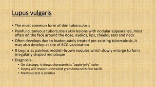
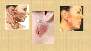
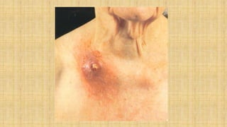
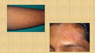
CUTANEOUS
TUBERCULOSIS
Introduction
• Cutaneous tuberculosis occurs rarely, despite a high and increasing
prevalence of tuberculosis worldwide. Mycobacterium tuberculosis,
Mycobacterrium bovis, and the Bacille Calmette-Guérin vaccine can cause
tuberculosis involving the skin
• Cutaneous tuberculosis can be acquired exogenously or endogenously and
present as a multitude of differing clinical morphologies
• Cutaneous TB- 1.5% of extrapulmonary TB
• Diagnosis of these lesions can be difficult, as they resemble many other
dermatological conditions that are often primarily considered.
• Commonest form
 in adults: Lupus Vulgaris
 in childhood: Scrofuloderma and Lichen scrofulosorum
• Skin manifestations present as a result of hematogenous spread or
direct extension from a latent or active foci of infection
• Cutaneous tuberculosis (CTB) is frequently elusive as it mimics a wide
differential diagnosis and also evades microbiological confirmation
despite recent advances in sophisticated techniques
• Although rare, given its worldwide prevalence, it is important to
recognize the many clinical variants of CTB to prevent missed or
delayed diagnoses
Route of infection Clinical type histology course
Inoculation tuberculosis
(exogenous source)
Tuberculosis chancre
Tuberculosis verrucosa cutis
Lupus vulgaris (occasionally)
Non specific
TB specific
TB specific
Localized
Localized
Localized
Secondary tuberculosis
( endogenous source)
Contiguous spread
Auto-inoculation
Scrofuloderma
Orificial tuberculosis
TB specific
TB specific
Localized
Progressive
Haematogenous tuberculosis Acute military tuberculosis
Lupus vulgaris
Tuberculous gumma
TB specific
TB specific
TB specific
Generalized
Localized
Localized
Eruptive tuberculosis (Tuberculids)
Micropapular
Papular
Nodular
Lichen scrofulosorum
Papular or papulonecrotic tuberculid
Erythma induratum of Bazin
Variable
variable
variable
Localized
Scattered Crops
Generalized
Lupus vulgaris
• The most common form of skin tuberculosis
• Painful cutaneous tuberculosis skin lesions with nodular appearance, most
often on the face around the nose, eyelids, lips, cheeks, ears and neck
• Often develops due to inadequately treated pre-existing tuberculosis, it
may also develop at site of BCG vaccination
• It begins as painless reddish-brown nodules which slowly enlarge to form
irregularly shaped red plaque
• Diagnosis-
• On diascopy, it shows characteristic "apple-jelly" color
• Biopsy will reveal tuberculoid granuloma with few bacilli
• Mantoux test is positive
Verrucosa cutis (prosector’s wart)
• Rash of small, red papular nodules in the skin that may appear 2–4
weeks after inoculation by Mycobacterium tuberculosis in a previously
infected and immunocompetent individual.
• Entry point usually is the site of a trauma, wound or puncture in the
skin (eg- during an autopsy), the most frequent site for the wart are
the hands
• The diagnosis is confirmed by a skin biopsy and a positive culture
for acid-fast bacilli
• A PPD test may also result positive
Scrofuloderma
• Condition caused by tuberculous involvement of the skin by direct
extension, usually from underlying tuberculous lymphadenitis.
• An asymptomatic reddish swelling which breaks down to form
sinuses, fistulate or tuberculous ulcers
• Characteristic caseous material discharges from lesions
• Most common sites – neck, chest
Miliary tuberculosis
• It is a rare haematogenous dissemination of tuberculosis
• Usually affects
– Young children
– Immunosuppressed patients
– Concurrent HIV infection
– Following viral infections
• Patient develops
– Crops of minute bluish papules, vesicles, pustules
– Erythematous nodules
– Haemorrhagic lesions
Tuberculid
• Hypersensitivity reaction to M. tuberculosis or its products in patient
with significant immunity
• Following criteria must be fulfilled to designate a condition as
tuberculid:
– Skin lesion must show tuberculoid histopathology
– Mycobacterium tuberculosis must not be demonstrated in the lesion
– Tuberculin test must be strongly positive
– Treatment of underlying TB focus must lead to resolution of skin lesion
Lichen scrofulosorum
• Second most common pattern of cutaneous TB in children
• Rare tuberculid that presents as a lichenoid eruption of
minute papules in children and adolescents with tuberculosis
• The lesions are usually asymptomatic, closely grouped, skin-colored
to reddish-brown papules, often perifollicular
• Mainly found on the abdomen, chest, back, and proximal parts of the
limbs
Diagnosis
• The diagnosis is usually on skin biopsy. Typical tubercles are caseating
epithelioid granulomas that contain acid-fast bacilli. These are
detected by tissue staining, culture and polymerase chain reaction
(PCR).
• Tuberculin skin test
• QFT-G
• X-ray
• Sputum culture
Treatment
• CTB treatment is the same as that for systemic TB and consists of
long, multidrug therapy
• The chemotherapeutic treatment of TB is divided into two phases:
• an intensive or bactericidal phase, designed to rapidly reduce the total body
burden of Mycobacterium tuberculosis
• a continuation or sterilizing phase
• The most commonly used drugs are isoniazid, rifampin, pyrazinamide,
and either ethambutol or streptomycin

Cutaneous tuberculosis

  • 1.
  • 2.
    Introduction • Cutaneous tuberculosisoccurs rarely, despite a high and increasing prevalence of tuberculosis worldwide. Mycobacterium tuberculosis, Mycobacterrium bovis, and the Bacille Calmette-Guérin vaccine can cause tuberculosis involving the skin • Cutaneous tuberculosis can be acquired exogenously or endogenously and present as a multitude of differing clinical morphologies • Cutaneous TB- 1.5% of extrapulmonary TB • Diagnosis of these lesions can be difficult, as they resemble many other dermatological conditions that are often primarily considered. • Commonest form  in adults: Lupus Vulgaris  in childhood: Scrofuloderma and Lichen scrofulosorum
  • 3.
    • Skin manifestationspresent as a result of hematogenous spread or direct extension from a latent or active foci of infection • Cutaneous tuberculosis (CTB) is frequently elusive as it mimics a wide differential diagnosis and also evades microbiological confirmation despite recent advances in sophisticated techniques • Although rare, given its worldwide prevalence, it is important to recognize the many clinical variants of CTB to prevent missed or delayed diagnoses
  • 4.
    Route of infectionClinical type histology course Inoculation tuberculosis (exogenous source) Tuberculosis chancre Tuberculosis verrucosa cutis Lupus vulgaris (occasionally) Non specific TB specific TB specific Localized Localized Localized Secondary tuberculosis ( endogenous source) Contiguous spread Auto-inoculation Scrofuloderma Orificial tuberculosis TB specific TB specific Localized Progressive Haematogenous tuberculosis Acute military tuberculosis Lupus vulgaris Tuberculous gumma TB specific TB specific TB specific Generalized Localized Localized Eruptive tuberculosis (Tuberculids) Micropapular Papular Nodular Lichen scrofulosorum Papular or papulonecrotic tuberculid Erythma induratum of Bazin Variable variable variable Localized Scattered Crops Generalized
  • 5.
    Lupus vulgaris • Themost common form of skin tuberculosis • Painful cutaneous tuberculosis skin lesions with nodular appearance, most often on the face around the nose, eyelids, lips, cheeks, ears and neck • Often develops due to inadequately treated pre-existing tuberculosis, it may also develop at site of BCG vaccination • It begins as painless reddish-brown nodules which slowly enlarge to form irregularly shaped red plaque • Diagnosis- • On diascopy, it shows characteristic "apple-jelly" color • Biopsy will reveal tuberculoid granuloma with few bacilli • Mantoux test is positive
  • 7.
    Verrucosa cutis (prosector’swart) • Rash of small, red papular nodules in the skin that may appear 2–4 weeks after inoculation by Mycobacterium tuberculosis in a previously infected and immunocompetent individual. • Entry point usually is the site of a trauma, wound or puncture in the skin (eg- during an autopsy), the most frequent site for the wart are the hands • The diagnosis is confirmed by a skin biopsy and a positive culture for acid-fast bacilli • A PPD test may also result positive
  • 9.
    Scrofuloderma • Condition causedby tuberculous involvement of the skin by direct extension, usually from underlying tuberculous lymphadenitis. • An asymptomatic reddish swelling which breaks down to form sinuses, fistulate or tuberculous ulcers • Characteristic caseous material discharges from lesions • Most common sites – neck, chest
  • 11.
    Miliary tuberculosis • Itis a rare haematogenous dissemination of tuberculosis • Usually affects – Young children – Immunosuppressed patients – Concurrent HIV infection – Following viral infections • Patient develops – Crops of minute bluish papules, vesicles, pustules – Erythematous nodules – Haemorrhagic lesions
  • 13.
    Tuberculid • Hypersensitivity reactionto M. tuberculosis or its products in patient with significant immunity • Following criteria must be fulfilled to designate a condition as tuberculid: – Skin lesion must show tuberculoid histopathology – Mycobacterium tuberculosis must not be demonstrated in the lesion – Tuberculin test must be strongly positive – Treatment of underlying TB focus must lead to resolution of skin lesion
  • 15.
    Lichen scrofulosorum • Secondmost common pattern of cutaneous TB in children • Rare tuberculid that presents as a lichenoid eruption of minute papules in children and adolescents with tuberculosis • The lesions are usually asymptomatic, closely grouped, skin-colored to reddish-brown papules, often perifollicular • Mainly found on the abdomen, chest, back, and proximal parts of the limbs
  • 17.
    Diagnosis • The diagnosisis usually on skin biopsy. Typical tubercles are caseating epithelioid granulomas that contain acid-fast bacilli. These are detected by tissue staining, culture and polymerase chain reaction (PCR). • Tuberculin skin test • QFT-G • X-ray • Sputum culture
  • 18.
    Treatment • CTB treatmentis the same as that for systemic TB and consists of long, multidrug therapy • The chemotherapeutic treatment of TB is divided into two phases: • an intensive or bactericidal phase, designed to rapidly reduce the total body burden of Mycobacterium tuberculosis • a continuation or sterilizing phase • The most commonly used drugs are isoniazid, rifampin, pyrazinamide, and either ethambutol or streptomycin