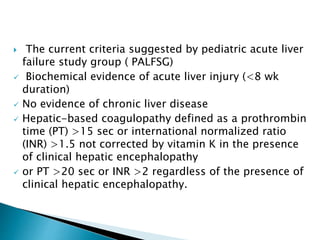
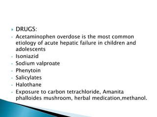
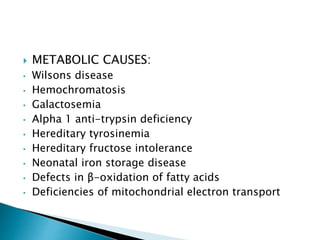
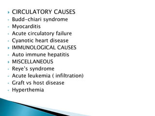
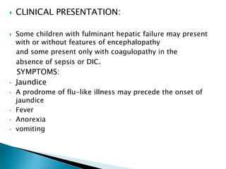
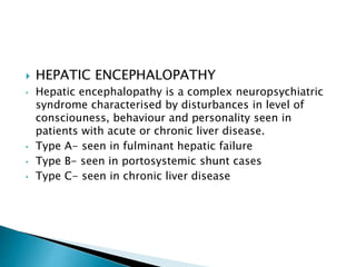
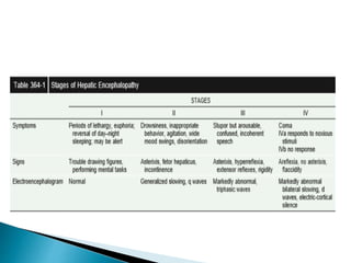
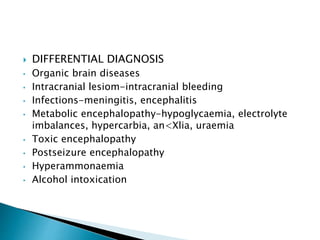
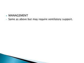
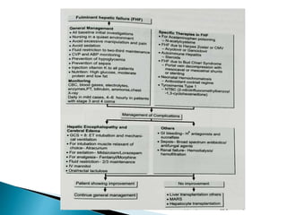
 Fulminant hepatic failure is a clinical syndrome of
coagulopathy and encephalopathy resulting from
massive necrosis of hepatocytes or from severe
functional impairment of hepatocytes.
 The disease evolves over a period of 8wks from the
onset of precipitating illness.
 Synthetic, excretory, and detoxifying functions of the
liver are all severely impaired.
 The current criteria suggested by pediatric acute liver
failure study group ( PALFSG)
 Biochemical evidence of acute liver injury (<8 wk
duration)
 No evidence of chronic liver disease
 Hepatic-based coagulopathy defined as a prothrombin
time (PT) >15 sec or international normalized ratio
(INR) >1.5 not corrected by vitamin K in the presence
of clinical hepatic encephalopathy
 or PT >20 sec or INR >2 regardless of the presence of
clinical hepatic encephalopathy.
 Previously asymptomatic children with chronic liver
disease like wilsons disesase, galactosemia and
other metabolic conditions can also present with
fulminant hepatic failure for the first time.
 ETIOLOGY:
 IDIOPATHIC: in 40-50% cases
 INFECTIVE
 VIRAL CAUSES
• Hepatitis a,b,c,d & e
• high risk of fulminant hepatic failure occurs in combined
infections with the hepatitis B virus (HBV) and hepatitis D.
• EBV
• Adenovirus
• Enterovirus
• Cmv
• Parvovirus B19
• Herpes simplex
• Varicella zoster
 BACTERIAL
• Enteric fever
• Weil’s disease
• Septicemia
 PROTOZOAL
• Facliparum malaria
 DRUGS:
• Acetaminophen overdose is the most common
etiology of acute hepatic failure in children and
adolescents
• Isoniazid
• Sodium valproate
• Phenytoin
• Salicylates
• Halothane
• Exposure to carbon tetrachloride, Amanita
phalloides mushroom, herbal medication,methanol.
 METABOLIC CAUSES:
• Wilsons disease
• Hemochromatosis
• Galactosemia
• Alpha 1 anti-trypsin deficiency
• Hereditary tyrosinemia
• Hereditary fructose intolerance
• Neonatal iron storage disease
• Defects in β-oxidation of fatty acids
• Deficiencies of mitochondrial electron transport
 CIRCULATORY CAUSES
• Budd-chiari syndrome
• Myocarditis
• Acute circulatory failure
• Cyanotic heart disease
 IMMUNOLOGICAL CAUSES
• Auto immune hepatitis
 MISCELLANEOUS
• Reye’s syndrome
• Acute leukemia ( infiltration)
• Graft vs host disease
• Hyperthemia
 CLINICAL PRESENTATION:
 Some children with fulminant hepatic failure may present
with or without features of encephalopathy
and some present only with coagulopathy in the
absence of sepsis or DIC.
SYMPTOMS:
• Jaundice
• A prodrome of flu-like illness may precede the onset of
jaundice
• Fever
• Anorexia
• vomiting
• Abdominal pain
• Foetor hepaticus
• Confusion
• Restlessness
• Neuropsychiatric changes
• Irritability
• poor feeding and disturbance in sleep pattern in infants
• Bleeding manifestations- hematemisis, hematochezia,
melena.
• Altered sensorium
• coma
 EVALUATION OF CHILD
• H/O fever
• H/O onset of jaundice
• Constitutional symptoms like vomitings
• H/O bleeding
• H/O decreased urine output
• H/O altered sensorium
• H/O i.v drug abuse
• H/O herbal medication
• Similar illness in family or community
• H/O mushroom eating
 Past history of ATT, anticonvulsant therapy
 Developemental history :
• Failure to thrive
• Developemental delay
• Neuromuscular weakness
 Family history of consanguinity , early infant death, liver
disease.
 Per abdominal examination: liver size, tenderness,
ascites
 CNS examination: level of consciousness, other causes
of altered sensorium
 INVESTIGATIONS
 Investigations for ascertaining liver cell injury
• Serum bilirubin-both direct and indirect are increased
• Hepatic enzymes do not correlate with the severity of
fulminant hepatic failure. ALT and AST are normal or
increased or even decreased. The enzymes are
decreased if the liver cell necrosis is severe.
• Liver biopsy – autoimmune hepatitis, metastatic liver
disease, lymphomas.
 Investigations for aetiological factors
• CBP- WBC increased in infections
• Viral markers-hepatitis B (HBV-DNA, antibody to HBV,
HBsAg, IgM core antigen), hepatitis A (IgM anti-HAV),
hepatitis C (anti-HCV, HCV-RNA) and hepatitis D (anti-
HDV antibody)
• Blood culture and sensitivity
• Serum paracetamol levels, other drug levels in
poisonings
• Autoantibodies-LKM, ANA and SMA
• Urine/serum screening test for metabolic disorders
• Blood ammonia is elevated in Reye's syndrome
• Seum ceruloplasmin levels
• Blood urea levels decreased in urea cycle disorders
 Investigations for complications:
• ABG- metabolic acidosis and respiratory alkalosis
• blood sugar levels
• Serum lactate levels
• RFT: Blood urea and Serum creatinine levels
• Serum electrolytes- hyponautremia , hyperkalemia.
• Prothrambin time elevated it doesnot respond to vit k
administration.
• Imaging: ultrasound abdomen, CT/MRI brain for
cerebral edema and to ruleout other causes of altered
sensorium.
 Management:
 supportive
• Children with fulminant hepatic failure should be
admitted in icu with continuous monitoring of vital
functions. Minimal handling of the child.
• Maintain a normal fluid balance. Avoid fluid overload.
• Correction hypoglycaemia.
• Maintain normal urine output.
• Administration of calcium, phosphorus, magnesium,
factor concentrate, magnesium and platelets.
• Coagulopathy can be treated with parenteral vitamin K,
fresh frozen plasma, cryoprecipitate.
• Prophylactic use of broad spectrum antobiotics,
antifungals
• Maintain a platelet count of more than 50,000.
• Gut sterilisation-using antibiotics such as
ampicillin/neomycin.
• Lactulose inhibits colonic organisms, decrease the
intestinal transit time.
• Fat soluble vitamin supplementation.
• Diet with decreased protein, high calories, high
carbohydrates and moderate fat should be given.
• Prophylactic use of proton pump inhibitors because of
the high risk of gastrointestinal bleeding.
• In complicated cases endotracheal intubation may be
required to prevent aspiration, to reduce cerebral
edema by hyperventilation, and to facilitate pulmonary
toilet.
• Mechanical ventilation and supplemental oxygen are
often necessary in advanced coma.
• Sedatives should be avoided unless needed in the
intubated patient because these agents can aggravate
or precipitate encephalopathy.
• Patients should be monitored closely for sepsis,
pneumonia, peritonitis, and urinary tract infections.
• Gastrointestinal hemorrhage, infection, constipation,
sedatives, electrolyte imbalance, and hypovolemia can
precipitate encephalopathy and should be identified and
corrected.
• liver dialysis with an albumin-containing dialysate, and
biologic liver support devices that involve perfusion of
the patient’s blood through a cartridge containing liver
cell lines or porcine hepatocytes can remove some
toxins, improve serum biochemical abnormalities.
• Orthotopic/auxillary orthotopic liver transplantation can
be lifesaving in patients who reach advanced stages of
hepatic coma.
 Specific
• Paracetamol poisoning-N-acetylcysteine
• Hepatitis B -antiviral agents lamivudine,entecavir
• Herpes simplex- acyclovir
• Galactosaemia, fructosaemia, hereditary tyrosinaemia
type I-dietary elimination.
• Autoimmune hepatitis-steroids, azathioprine
• BZD antagonist like flumazenil in BZD over dosage
• Enteric encephalopathy- ceftriaxone
• Wilson’s disease- D-pencillamine,zinc
• Hemochromatosis- desferoxamine, deferiprone
• Leptospirosis- pencillins, doxycycline
• Malaria- anti malarials
• Amanita posoning- pencillins
• L-ornithine and L-aspartate preparation (LOLA)
removes ammonia by conversion to urea through the
urea cycle.
 COMPLICATIONS:
• Infections
• Hepatic encephalopathy
• Cerebral edema
• Hepatorenal syndrome
• Hepatopulmonary syndrome
• Coagulopathy –GI bleeding
• Hypotension
• Electrolyte and acid base imbalance
 POOR PROGNOSTIC FACTORS:
• Age < 1 yr
• Depending on etiological factor
• Stage 4 encephalopathy
• INR > 4
• Plasma ammonia concentration > 200 micro mol/ L
• Sepsis
• Severe bleeding
 HEPATIC ENCEPHALOPATHY
• Hepatic encephalopathy is a complex neuropsychiatric
syndrome characterised by disturbances in level of
consciouness, behaviour and personality seen in
patients with acute or chronic liver disease.
• Type A- seen in fulminant hepatic failure
• Type B- seen in portosystemic shunt cases
• Type C- seen in chronic liver disease
 PATHOGENESIS:
• Accumulation of neurotoxins in brain
• Increased activity of the inhibitory GABA
neurotransmission system
• Imbalance between aromatic and branched amino acids
• Action of cytokines and bacterial lipopolysaccharide on
astrocytes
 DIFFERENTIAL DIAGNOSIS
• Organic brain diseases
• Intracranial lesiom-intracranial bleeding
• Infections-meningitis, encephalitis
• Metabolic encephalopathy-hypoglycaemia, electrolyte
imbalances, hypercarbia, an<Xlia, uraemia
• Toxic encephalopathy
• Postseizure encephalopathy
• Hyperammonaemia
• Alcohol intoxication
 MANAGEMENT
• Same as above but may require ventilatory support.
Fulminant hepatic failure

Fulminant hepatic failure

  • 2.
     Fulminant hepaticfailure is a clinical syndrome of coagulopathy and encephalopathy resulting from massive necrosis of hepatocytes or from severe functional impairment of hepatocytes.  The disease evolves over a period of 8wks from the onset of precipitating illness.  Synthetic, excretory, and detoxifying functions of the liver are all severely impaired.
  • 3.
     The currentcriteria suggested by pediatric acute liver failure study group ( PALFSG)  Biochemical evidence of acute liver injury (<8 wk duration)  No evidence of chronic liver disease  Hepatic-based coagulopathy defined as a prothrombin time (PT) >15 sec or international normalized ratio (INR) >1.5 not corrected by vitamin K in the presence of clinical hepatic encephalopathy  or PT >20 sec or INR >2 regardless of the presence of clinical hepatic encephalopathy.
  • 4.
     Previously asymptomaticchildren with chronic liver disease like wilsons disesase, galactosemia and other metabolic conditions can also present with fulminant hepatic failure for the first time.
  • 5.
     ETIOLOGY:  IDIOPATHIC:in 40-50% cases  INFECTIVE  VIRAL CAUSES • Hepatitis a,b,c,d & e • high risk of fulminant hepatic failure occurs in combined infections with the hepatitis B virus (HBV) and hepatitis D. • EBV • Adenovirus • Enterovirus • Cmv • Parvovirus B19 • Herpes simplex • Varicella zoster
  • 6.
     BACTERIAL • Entericfever • Weil’s disease • Septicemia  PROTOZOAL • Facliparum malaria
  • 7.
     DRUGS: • Acetaminophenoverdose is the most common etiology of acute hepatic failure in children and adolescents • Isoniazid • Sodium valproate • Phenytoin • Salicylates • Halothane • Exposure to carbon tetrachloride, Amanita phalloides mushroom, herbal medication,methanol.
  • 8.
     METABOLIC CAUSES: •Wilsons disease • Hemochromatosis • Galactosemia • Alpha 1 anti-trypsin deficiency • Hereditary tyrosinemia • Hereditary fructose intolerance • Neonatal iron storage disease • Defects in β-oxidation of fatty acids • Deficiencies of mitochondrial electron transport
  • 9.
     CIRCULATORY CAUSES •Budd-chiari syndrome • Myocarditis • Acute circulatory failure • Cyanotic heart disease  IMMUNOLOGICAL CAUSES • Auto immune hepatitis  MISCELLANEOUS • Reye’s syndrome • Acute leukemia ( infiltration) • Graft vs host disease • Hyperthemia
  • 10.
     CLINICAL PRESENTATION: Some children with fulminant hepatic failure may present with or without features of encephalopathy and some present only with coagulopathy in the absence of sepsis or DIC. SYMPTOMS: • Jaundice • A prodrome of flu-like illness may precede the onset of jaundice • Fever • Anorexia • vomiting
  • 11.
    • Abdominal pain •Foetor hepaticus • Confusion • Restlessness • Neuropsychiatric changes • Irritability • poor feeding and disturbance in sleep pattern in infants • Bleeding manifestations- hematemisis, hematochezia, melena. • Altered sensorium • coma
  • 12.
     EVALUATION OFCHILD • H/O fever • H/O onset of jaundice • Constitutional symptoms like vomitings • H/O bleeding • H/O decreased urine output • H/O altered sensorium • H/O i.v drug abuse
  • 13.
    • H/O herbalmedication • Similar illness in family or community • H/O mushroom eating  Past history of ATT, anticonvulsant therapy  Developemental history : • Failure to thrive • Developemental delay • Neuromuscular weakness
  • 14.
     Family historyof consanguinity , early infant death, liver disease.  Per abdominal examination: liver size, tenderness, ascites  CNS examination: level of consciousness, other causes of altered sensorium
  • 15.
     INVESTIGATIONS  Investigationsfor ascertaining liver cell injury • Serum bilirubin-both direct and indirect are increased • Hepatic enzymes do not correlate with the severity of fulminant hepatic failure. ALT and AST are normal or increased or even decreased. The enzymes are decreased if the liver cell necrosis is severe. • Liver biopsy – autoimmune hepatitis, metastatic liver disease, lymphomas.
  • 16.
     Investigations foraetiological factors • CBP- WBC increased in infections • Viral markers-hepatitis B (HBV-DNA, antibody to HBV, HBsAg, IgM core antigen), hepatitis A (IgM anti-HAV), hepatitis C (anti-HCV, HCV-RNA) and hepatitis D (anti- HDV antibody) • Blood culture and sensitivity
  • 17.
    • Serum paracetamollevels, other drug levels in poisonings • Autoantibodies-LKM, ANA and SMA • Urine/serum screening test for metabolic disorders • Blood ammonia is elevated in Reye's syndrome • Seum ceruloplasmin levels • Blood urea levels decreased in urea cycle disorders
  • 18.
     Investigations forcomplications: • ABG- metabolic acidosis and respiratory alkalosis • blood sugar levels • Serum lactate levels • RFT: Blood urea and Serum creatinine levels • Serum electrolytes- hyponautremia , hyperkalemia. • Prothrambin time elevated it doesnot respond to vit k administration. • Imaging: ultrasound abdomen, CT/MRI brain for cerebral edema and to ruleout other causes of altered sensorium.
  • 19.
     Management:  supportive •Children with fulminant hepatic failure should be admitted in icu with continuous monitoring of vital functions. Minimal handling of the child. • Maintain a normal fluid balance. Avoid fluid overload. • Correction hypoglycaemia. • Maintain normal urine output.
  • 20.
    • Administration ofcalcium, phosphorus, magnesium, factor concentrate, magnesium and platelets. • Coagulopathy can be treated with parenteral vitamin K, fresh frozen plasma, cryoprecipitate. • Prophylactic use of broad spectrum antobiotics, antifungals
  • 21.
    • Maintain aplatelet count of more than 50,000. • Gut sterilisation-using antibiotics such as ampicillin/neomycin. • Lactulose inhibits colonic organisms, decrease the intestinal transit time. • Fat soluble vitamin supplementation. • Diet with decreased protein, high calories, high carbohydrates and moderate fat should be given.
  • 22.
    • Prophylactic useof proton pump inhibitors because of the high risk of gastrointestinal bleeding. • In complicated cases endotracheal intubation may be required to prevent aspiration, to reduce cerebral edema by hyperventilation, and to facilitate pulmonary toilet. • Mechanical ventilation and supplemental oxygen are often necessary in advanced coma.
  • 23.
    • Sedatives shouldbe avoided unless needed in the intubated patient because these agents can aggravate or precipitate encephalopathy. • Patients should be monitored closely for sepsis, pneumonia, peritonitis, and urinary tract infections. • Gastrointestinal hemorrhage, infection, constipation, sedatives, electrolyte imbalance, and hypovolemia can precipitate encephalopathy and should be identified and corrected.
  • 24.
    • liver dialysiswith an albumin-containing dialysate, and biologic liver support devices that involve perfusion of the patient’s blood through a cartridge containing liver cell lines or porcine hepatocytes can remove some toxins, improve serum biochemical abnormalities. • Orthotopic/auxillary orthotopic liver transplantation can be lifesaving in patients who reach advanced stages of hepatic coma.
  • 25.
     Specific • Paracetamolpoisoning-N-acetylcysteine • Hepatitis B -antiviral agents lamivudine,entecavir • Herpes simplex- acyclovir • Galactosaemia, fructosaemia, hereditary tyrosinaemia type I-dietary elimination. • Autoimmune hepatitis-steroids, azathioprine
  • 26.
    • BZD antagonistlike flumazenil in BZD over dosage • Enteric encephalopathy- ceftriaxone • Wilson’s disease- D-pencillamine,zinc • Hemochromatosis- desferoxamine, deferiprone • Leptospirosis- pencillins, doxycycline • Malaria- anti malarials • Amanita posoning- pencillins • L-ornithine and L-aspartate preparation (LOLA) removes ammonia by conversion to urea through the urea cycle.
  • 27.
     COMPLICATIONS: • Infections •Hepatic encephalopathy • Cerebral edema • Hepatorenal syndrome • Hepatopulmonary syndrome • Coagulopathy –GI bleeding • Hypotension • Electrolyte and acid base imbalance
  • 28.
     POOR PROGNOSTICFACTORS: • Age < 1 yr • Depending on etiological factor • Stage 4 encephalopathy • INR > 4 • Plasma ammonia concentration > 200 micro mol/ L • Sepsis • Severe bleeding
  • 29.
     HEPATIC ENCEPHALOPATHY •Hepatic encephalopathy is a complex neuropsychiatric syndrome characterised by disturbances in level of consciouness, behaviour and personality seen in patients with acute or chronic liver disease. • Type A- seen in fulminant hepatic failure • Type B- seen in portosystemic shunt cases • Type C- seen in chronic liver disease
  • 30.
     PATHOGENESIS: • Accumulationof neurotoxins in brain • Increased activity of the inhibitory GABA neurotransmission system • Imbalance between aromatic and branched amino acids • Action of cytokines and bacterial lipopolysaccharide on astrocytes
  • 32.
     DIFFERENTIAL DIAGNOSIS •Organic brain diseases • Intracranial lesiom-intracranial bleeding • Infections-meningitis, encephalitis • Metabolic encephalopathy-hypoglycaemia, electrolyte imbalances, hypercarbia, an<Xlia, uraemia • Toxic encephalopathy • Postseizure encephalopathy • Hyperammonaemia • Alcohol intoxication
  • 33.
     MANAGEMENT • Sameas above but may require ventilatory support.