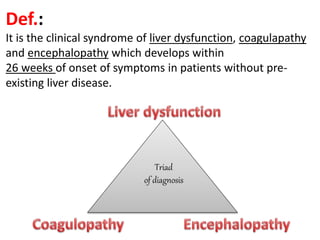
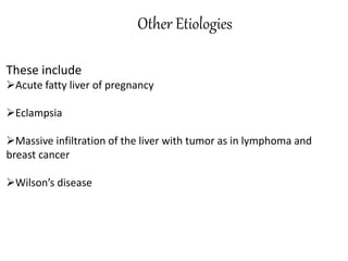
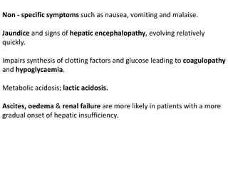
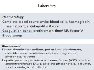
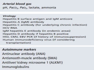
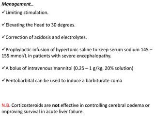
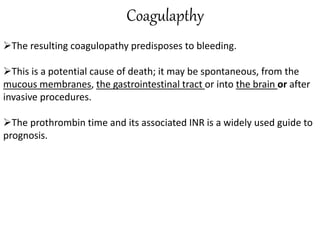
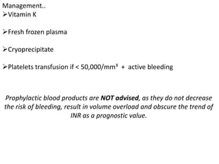
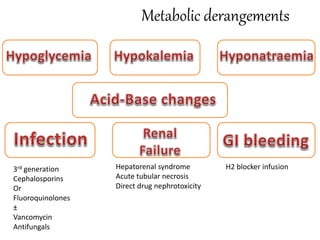
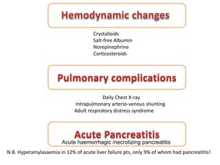
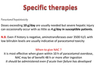
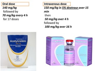
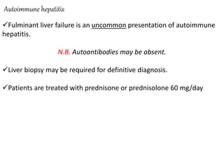
Fulminant Hepatitis
(Acute liver cell failure)
By,
Kerollos Motwade
Demonstrator of Internal Medicine,
Faculty of Medicine - Assiut University.
Def.:
It is the clinical syndrome of liver dysfunction, coagulapathy
and encephalopathy which develops within
26 weeks of onset of symptoms in patients without pre-
existing liver disease.
Triad
of diagnosis
Disease duration of greater than 28 weeks before the onset of
encephalopathy is categorized as chronic liver disease.
Etiology
Viral Hepatitis
-HAV
-HBV
It may occur due to
Acute Hepatitis B
Reactivation of viral replication in inactive carriers either
spontaneously or after chemotherapy or immunosuppressive therapy.
Delta superinfection
-HEV
The susceptible groups: *Chronic liver disease patients
*Pregnant women.
±HCV
-Herpes simplex 1 and 2
-Varicella zoster virus
Paracetamol toxicity
Paracetamol is a dose-related toxin.
Paracetamol is usually undetectable at the time of presentation, and
etiology often has to be based on clinical presentation, history and
typical laboratory results.
POD characterized by extreme elevations of serum aminotransferase
(usually >10,000 IU/L) and relatively low bilirubin levels.
Metabolic acidosis, elevated serum lactate, hypoglycemia and acute
kidney injury (AKI) can occur in early stages of clinical evolution.
Idiosyncratic drug
reaction
Cases are subacute with moderately elevated aminotransferases and
high bilirubin levels.
The most frequent drugs are anti-tuberculosis medications, NSAIDs,
anaesthetic agents and anti-seizure medications.
Herbal remedies, especially those containing green tea extract, have
been implicated in acute liver failure.
Vascular causes
These include
Low cardiac output states “ Shock Liver”= “Hypoxic hepatitis”
N.B. The absence of a documented episode of hypotension or hypoxia
does not exclude this condition.
Acute Budd-Chiari syndrome
Other Etiologies
These include
Acute fatty liver of pregnancy
Eclampsia
Massive infiltration of the liver with tumor as in lymphoma and
breast cancer
Wilson’s disease
Clinical Picture
Non - specific symptoms such as nausea, vomiting and malaise.
Jaundice and signs of hepatic encephalopathy, evolving relatively
quickly.
Impairs synthesis of clotting factors and glucose leading to coagulopathy
and hypoglycaemia.
Metabolic acidosis; lactic acidosis.
Ascites, oedema & renal failure are more likely in patients with a more
gradual onset of hepatic insufficiency.
Investigations
Laboratory
Abdominal imaging
&
liver biopsies
Abdominal U/S
…to assess for vasculature patency and mass lesions.
CT scanning
…it will show a reduction in liver size
Liver biopsy
…if diagnosis can’t be reached
with non-invasive techniques
Brain CT
Non - contrast computed tomography of the brain may help rule out
other pathology, such as haemorrhage.
N.B. The yield of such studies is low and may not justify the risk of
moving a critically ill patient
Complications & Management
Treatment has focused on the management of complications except for
a few etiology specific therapies.
Hepatic Encephalopathy
Lactulose ; it has shown no benefit
*It may increase aspiration risk
*It may cause bowel distension
Rifaximine ; there is not enough evidence to recommend non -
absorbable antibiotics in acute liver failure
l- ornithine l- aspartate ; it treats encephalopathy by increasing
muscle metabolism of ammonia but a recent randomized controlled trial
showed no benefit in acute liver failure
These may complicate transplanation
Progression of encephalopathy is often
triggered by infection and
empiric antibiotics
should be administered to these patients
Cerebral oedema
&
Intracranial hypertension
Acute liver failure is uniquely associated with cerebral oedema, which
can lead to an increase in intracerebral pressure.
This is uncommon in patients with stage 1 or 2 encephalopathy. But
develops in the majority with stage 4.
Clinically , increased intracranial pressure is suggested by :
Systolic hypertension (sustained or intermittent)
Increased muscle tone and myoclonus
Extension and hyperpronation of the arms and extension of the legs
(decerebrate posturing).
Dysconjugate eye movements
Brainstem herniation
Limiting stimulation.
Elevating the head to 30 degrees.
Correction of acidosis and electrolytes.
Prophylactic infusion of hypertonic saline to keep serum sodium 145 –
155 mmol/L in patients with severe encephalopathy.
A bolus of intravenous mannitol (0.25 – 1 g/kg, 20% solution)
Pentobarbital can be used to induce a barbiturate coma
N.B. Corticosteroids are not effective in controlling cerebral oedema or
improving survival in acute liver failure.
Management…
Coagulapthy
The resulting coagulopathy predisposes to bleeding.
This is a potential cause of death; it may be spontaneous, from the
mucous membranes, the gastrointestinal tract or into the brain or after
invasive procedures.
The prothrombin time and its associated INR is a widely used guide to
prognosis.
Management..
Vitamin K
Fresh frozen plasma
Cryoprecipitate
Platelets transfusion if < 50,000/mm³ + active bleeding
Prophylactic blood products are NOT advised, as they do not decrease
the risk of bleeding, result in volume overload and obscure the trend of
INR as a prognostic value.
Metabolic derangements
3rd generation
Cephalosporins
Or
Fluoroquinolones
±
Vancomycin
Antifungals
Hepatorenal syndrome
Acute tubular necrosis
Direct drug nephrotoxicity
H2 blocker infusion
Crystalloids
Salt-free Albumin
Norepinephrine
Corticosteroids
Daily Chest X-ray
Intrapulmonary arterio-venous shunting
Adult respiratory distress syndrome
N.B. Hyperamylasaemia in 12% of acute liver failure pts, only 9% of whom had pancreatitis!
Acute haemorrhagic /necrotizing pancreatitis
Paracetamol hepatotoxicity
Doses exceeding 10 g/day are usually needed but severe hepatic injury
can occasionally occur with as little as 4 g/day in susceptible patients.
N.B. Even if history is negative, aminotransferases over 3500 IU/L with
low bilirubin levels are usually indicative of paracetamol toxicity
When to give NAC ?
it is most effective when given within 10 h of paracetamol overdose,
NAC may be of benefit 48 h or more after ingestion
It should be administered even if acute liver failure has developed
Oral dose
140 mg/kg
followed by
70 mg/kg every 4 h
for 17 doses
Intravenous dose
150 mg/kg in 5% dextrose over 15
min
then
50 mg/kg over 4 h
followed by
100 mg/kg over 16 h
Autoimmune hepatitis
Fulminant liver failure is an uncommon presentation of autoimmune
hepatitis.
N.B. Autoantibodies may be absent.
Liver biopsy may be required for definitive diagnosis.
Patients are treated with prednisone or prednisolone 60 mg/day
Pregnancy – related causes
Acute liver failure may occur in the setting of
Acute fatty liver of pregnancy
 Severe pre – eclampsia / eclampsia
HELLP (haemolysis, elevated liver enzymes, low platelets) syndrome
Prompt Deliveryof the fetus
results in recovery.
Wilson’s disease
The usual therapies of penicillamine or trientine are ineffective in
acute liver failure and are not recommended.
Fulminant Wilson ’ s disease is 100% fatal without
liver transplantation
CRRT, plasmapheresis or plasma exchange can be initiated to remove
copper and alleviate renal tubular damage until a graft becomes
available.
Hepatitis B
Although randomised controlled trials are lacking, several cohort
studies indicate that the early antiviral therapy – in case of severe acute
hepatitis B- with highly potent NAs can prevent progression to acute
liver failure and subsequently liver transplantation or mortality.
This effect, however, is not seen if antiviral therapy is initiated late in
the course of severe acute hepatitis B in patients with already
manifested acute liver failure and advanced hepatic encephalopathy.
Data supports the use of Tenofovir fumarate, Entecavir or even
Lamivudine.
Liver Transplantation
Acute liver failure

Acute liver failure

  • 1.
    Fulminant Hepatitis (Acute livercell failure) By, Kerollos Motwade Demonstrator of Internal Medicine, Faculty of Medicine - Assiut University.
  • 2.
    Def.: It is theclinical syndrome of liver dysfunction, coagulapathy and encephalopathy which develops within 26 weeks of onset of symptoms in patients without pre- existing liver disease. Triad of diagnosis
  • 3.
    Disease duration ofgreater than 28 weeks before the onset of encephalopathy is categorized as chronic liver disease.
  • 4.
  • 5.
    Viral Hepatitis -HAV -HBV It mayoccur due to Acute Hepatitis B Reactivation of viral replication in inactive carriers either spontaneously or after chemotherapy or immunosuppressive therapy. Delta superinfection -HEV The susceptible groups: *Chronic liver disease patients *Pregnant women. ±HCV -Herpes simplex 1 and 2 -Varicella zoster virus
  • 6.
    Paracetamol toxicity Paracetamol isa dose-related toxin. Paracetamol is usually undetectable at the time of presentation, and etiology often has to be based on clinical presentation, history and typical laboratory results. POD characterized by extreme elevations of serum aminotransferase (usually >10,000 IU/L) and relatively low bilirubin levels. Metabolic acidosis, elevated serum lactate, hypoglycemia and acute kidney injury (AKI) can occur in early stages of clinical evolution.
  • 7.
    Idiosyncratic drug reaction Cases aresubacute with moderately elevated aminotransferases and high bilirubin levels. The most frequent drugs are anti-tuberculosis medications, NSAIDs, anaesthetic agents and anti-seizure medications. Herbal remedies, especially those containing green tea extract, have been implicated in acute liver failure.
  • 9.
    Vascular causes These include Lowcardiac output states “ Shock Liver”= “Hypoxic hepatitis” N.B. The absence of a documented episode of hypotension or hypoxia does not exclude this condition. Acute Budd-Chiari syndrome
  • 10.
    Other Etiologies These include Acutefatty liver of pregnancy Eclampsia Massive infiltration of the liver with tumor as in lymphoma and breast cancer Wilson’s disease
  • 11.
  • 12.
    Non - specificsymptoms such as nausea, vomiting and malaise. Jaundice and signs of hepatic encephalopathy, evolving relatively quickly. Impairs synthesis of clotting factors and glucose leading to coagulopathy and hypoglycaemia. Metabolic acidosis; lactic acidosis. Ascites, oedema & renal failure are more likely in patients with a more gradual onset of hepatic insufficiency.
  • 13.
  • 14.
  • 17.
    Abdominal imaging & liver biopsies AbdominalU/S …to assess for vasculature patency and mass lesions. CT scanning …it will show a reduction in liver size Liver biopsy …if diagnosis can’t be reached with non-invasive techniques
  • 18.
    Brain CT Non -contrast computed tomography of the brain may help rule out other pathology, such as haemorrhage. N.B. The yield of such studies is low and may not justify the risk of moving a critically ill patient
  • 19.
  • 20.
    Treatment has focusedon the management of complications except for a few etiology specific therapies.
  • 21.
    Hepatic Encephalopathy Lactulose ;it has shown no benefit *It may increase aspiration risk *It may cause bowel distension Rifaximine ; there is not enough evidence to recommend non - absorbable antibiotics in acute liver failure l- ornithine l- aspartate ; it treats encephalopathy by increasing muscle metabolism of ammonia but a recent randomized controlled trial showed no benefit in acute liver failure These may complicate transplanation
  • 22.
    Progression of encephalopathyis often triggered by infection and empiric antibiotics should be administered to these patients
  • 23.
    Cerebral oedema & Intracranial hypertension Acuteliver failure is uniquely associated with cerebral oedema, which can lead to an increase in intracerebral pressure. This is uncommon in patients with stage 1 or 2 encephalopathy. But develops in the majority with stage 4.
  • 24.
    Clinically , increasedintracranial pressure is suggested by : Systolic hypertension (sustained or intermittent) Increased muscle tone and myoclonus Extension and hyperpronation of the arms and extension of the legs (decerebrate posturing). Dysconjugate eye movements Brainstem herniation
  • 25.
    Limiting stimulation. Elevating thehead to 30 degrees. Correction of acidosis and electrolytes. Prophylactic infusion of hypertonic saline to keep serum sodium 145 – 155 mmol/L in patients with severe encephalopathy. A bolus of intravenous mannitol (0.25 – 1 g/kg, 20% solution) Pentobarbital can be used to induce a barbiturate coma N.B. Corticosteroids are not effective in controlling cerebral oedema or improving survival in acute liver failure. Management…
  • 26.
    Coagulapthy The resulting coagulopathypredisposes to bleeding. This is a potential cause of death; it may be spontaneous, from the mucous membranes, the gastrointestinal tract or into the brain or after invasive procedures. The prothrombin time and its associated INR is a widely used guide to prognosis.
  • 27.
    Management.. Vitamin K Fresh frozenplasma Cryoprecipitate Platelets transfusion if < 50,000/mm³ + active bleeding Prophylactic blood products are NOT advised, as they do not decrease the risk of bleeding, result in volume overload and obscure the trend of INR as a prognostic value.
  • 28.
    Metabolic derangements 3rd generation Cephalosporins Or Fluoroquinolones ± Vancomycin Antifungals Hepatorenalsyndrome Acute tubular necrosis Direct drug nephrotoxicity H2 blocker infusion
  • 29.
    Crystalloids Salt-free Albumin Norepinephrine Corticosteroids Daily ChestX-ray Intrapulmonary arterio-venous shunting Adult respiratory distress syndrome N.B. Hyperamylasaemia in 12% of acute liver failure pts, only 9% of whom had pancreatitis! Acute haemorrhagic /necrotizing pancreatitis
  • 30.
    Paracetamol hepatotoxicity Doses exceeding10 g/day are usually needed but severe hepatic injury can occasionally occur with as little as 4 g/day in susceptible patients. N.B. Even if history is negative, aminotransferases over 3500 IU/L with low bilirubin levels are usually indicative of paracetamol toxicity When to give NAC ? it is most effective when given within 10 h of paracetamol overdose, NAC may be of benefit 48 h or more after ingestion It should be administered even if acute liver failure has developed
  • 31.
    Oral dose 140 mg/kg followedby 70 mg/kg every 4 h for 17 doses Intravenous dose 150 mg/kg in 5% dextrose over 15 min then 50 mg/kg over 4 h followed by 100 mg/kg over 16 h
  • 32.
    Autoimmune hepatitis Fulminant liverfailure is an uncommon presentation of autoimmune hepatitis. N.B. Autoantibodies may be absent. Liver biopsy may be required for definitive diagnosis. Patients are treated with prednisone or prednisolone 60 mg/day
  • 33.
    Pregnancy – relatedcauses Acute liver failure may occur in the setting of Acute fatty liver of pregnancy  Severe pre – eclampsia / eclampsia HELLP (haemolysis, elevated liver enzymes, low platelets) syndrome Prompt Deliveryof the fetus results in recovery.
  • 34.
    Wilson’s disease The usualtherapies of penicillamine or trientine are ineffective in acute liver failure and are not recommended. Fulminant Wilson ’ s disease is 100% fatal without liver transplantation CRRT, plasmapheresis or plasma exchange can be initiated to remove copper and alleviate renal tubular damage until a graft becomes available.
  • 35.
    Hepatitis B Although randomisedcontrolled trials are lacking, several cohort studies indicate that the early antiviral therapy – in case of severe acute hepatitis B- with highly potent NAs can prevent progression to acute liver failure and subsequently liver transplantation or mortality. This effect, however, is not seen if antiviral therapy is initiated late in the course of severe acute hepatitis B in patients with already manifested acute liver failure and advanced hepatic encephalopathy. Data supports the use of Tenofovir fumarate, Entecavir or even Lamivudine.
  • 36.