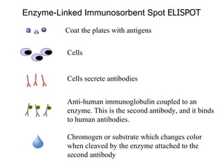
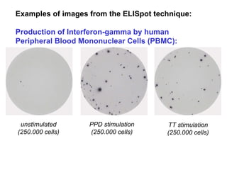
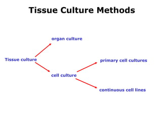
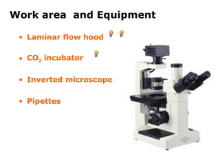
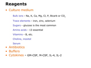
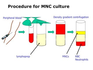
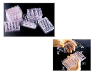
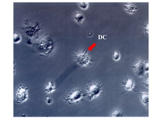
The document describes several immunology techniques:
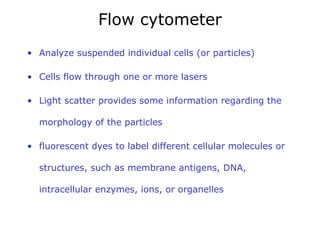
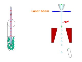
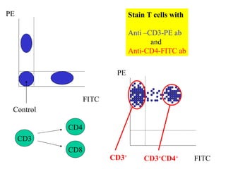
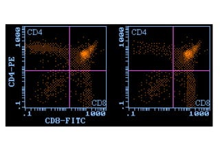
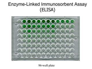
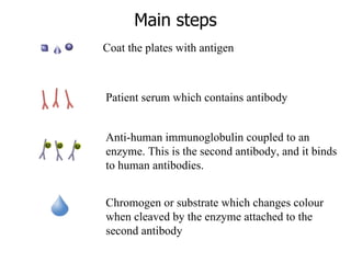
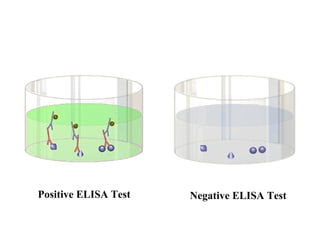
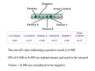
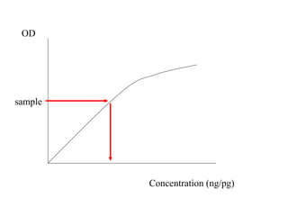
Flow cytometry analyzes individual cells or particles using laser light scattering and fluorescent dyes to label cellular molecules. ELISA uses antigen-coated plates and enzyme-linked antibodies to detect antibodies in serum samples. ELISPOT counts antibody-secreting cells by detecting individual antibody spots. Tissue culture methods grow cells in controlled conditions using culture medium, reagents, and equipment to maintain cells outside the body.