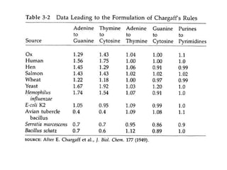
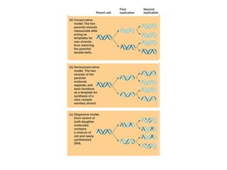
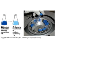
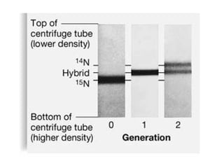
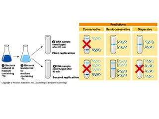
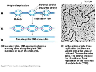
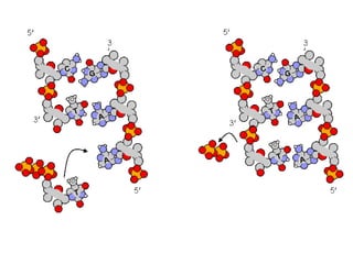
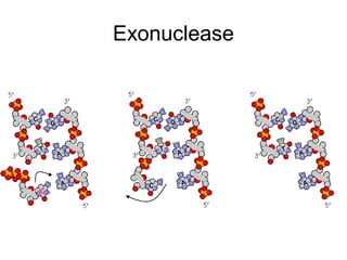
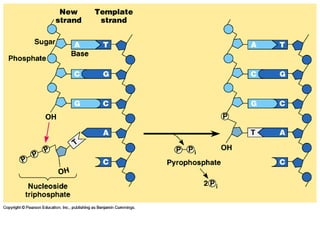
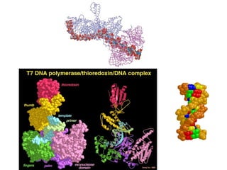
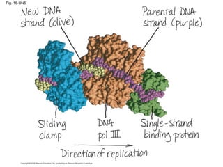
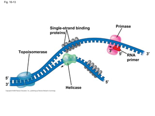
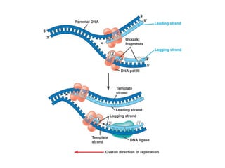
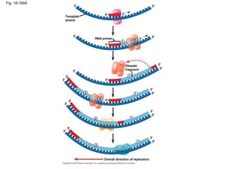
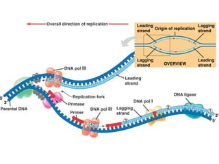
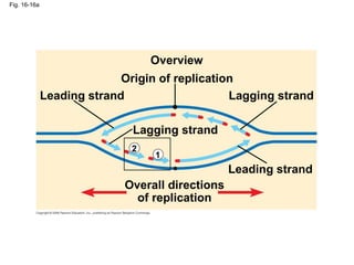
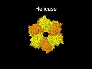
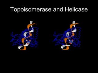
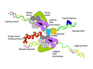
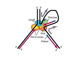
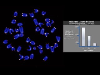
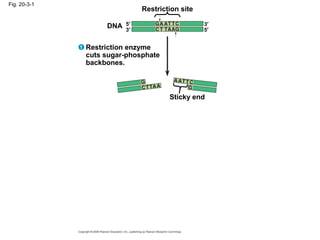
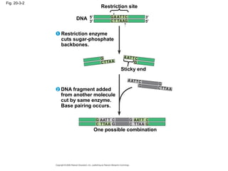
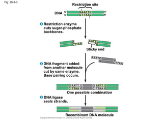
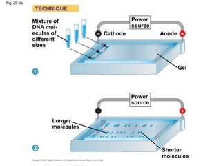
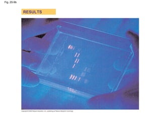
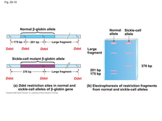
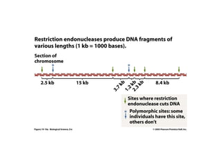
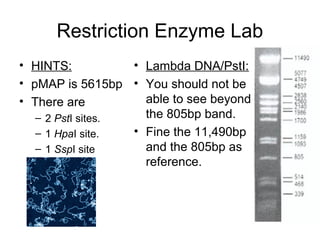
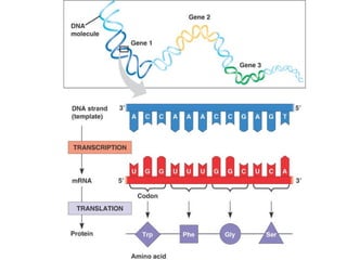
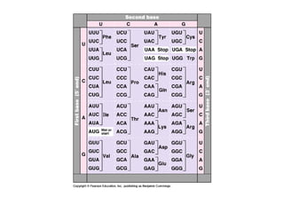
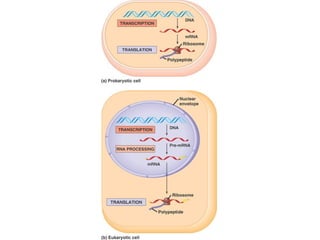
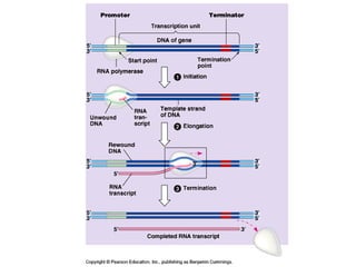
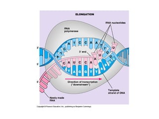
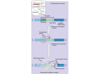
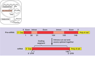
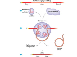
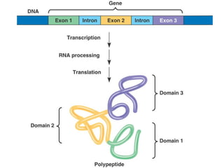
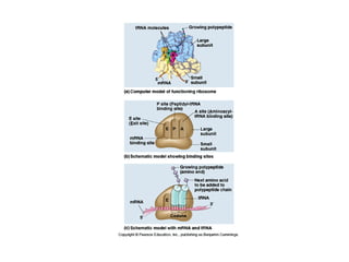
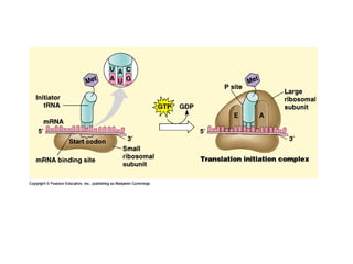
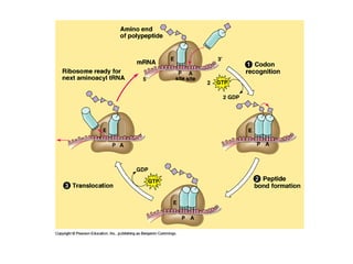
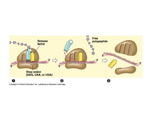
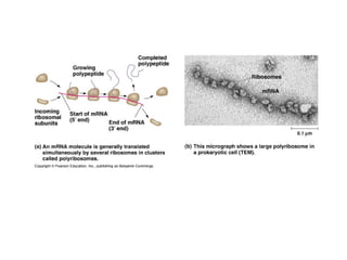
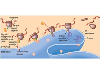
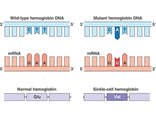
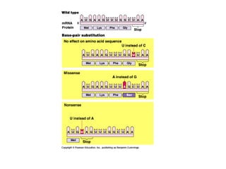
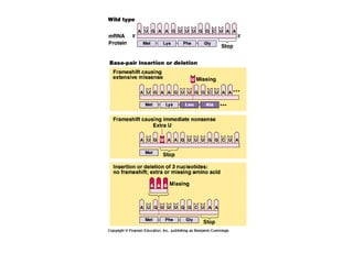
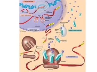
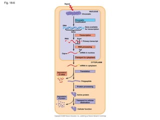
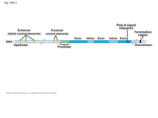
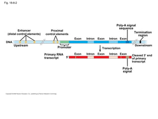
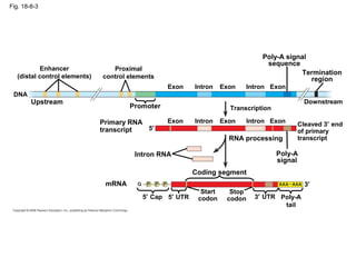
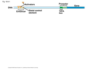
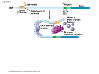
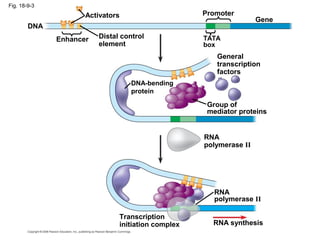
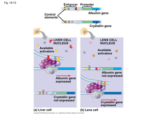
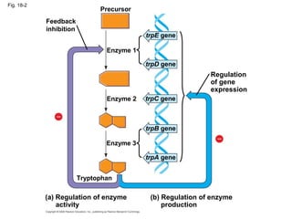
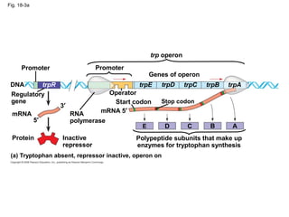
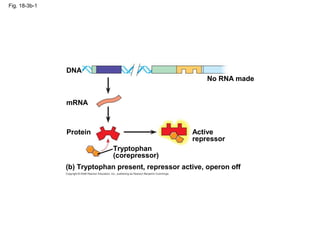
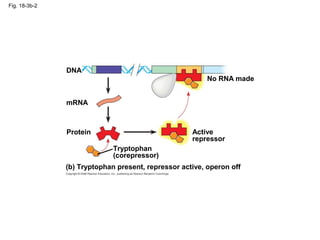
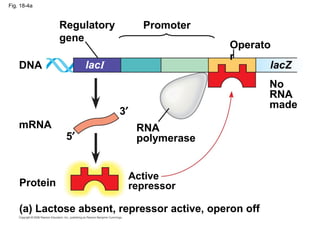
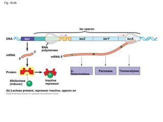
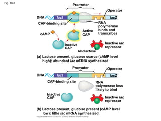
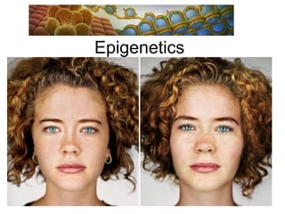
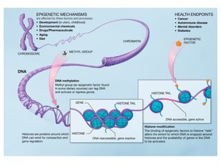
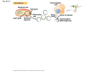
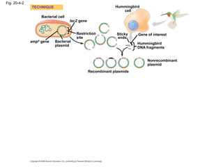
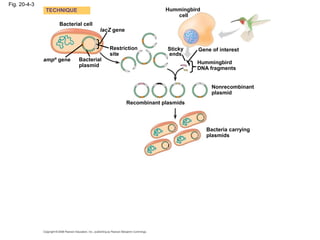
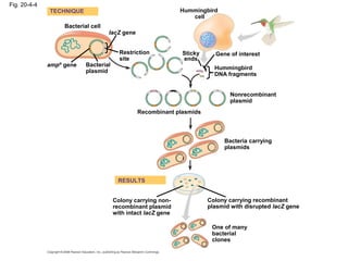
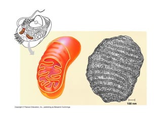
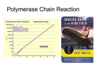
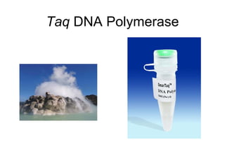
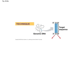
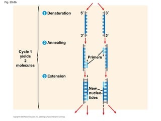
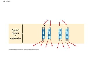
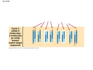
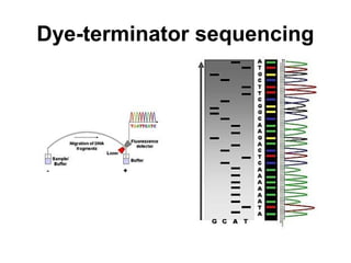
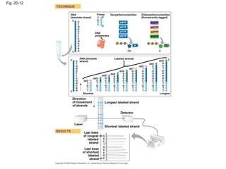
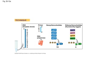
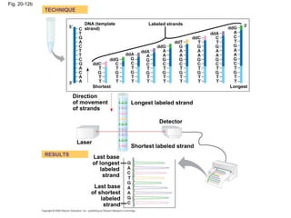
Molecular Biology
Honors Biology
Edgar
DNA Replication
Exonuclease
Fig. 16-UN5
Fig. 16-13
Topoisomerase
Helicase
PrimaseSingle-strand binding
proteins
RNA
primer
5′
5′
5′ 3′
3′
3′
Fig. 16-16b6
Template
strand
5′
5′3′
3′
RNA primer 3′
5′
5′
3′
1
1
3′
3′
5′
5′
Okazaki
fragment
12
3′
3′
5′
5′
12
3′
3′
5′
5′
1
2
5′
5′
3′
3′
Overall direction of replication
Fig. 16-16a
Overview
Origin of replication
Leading strand
Leading strand
Lagging strand
Lagging strand
Overall directions
of replication
1
2
Helicase
Topoisomerase and Helicase
Fig. 20-3-1
Restriction site
DNA
Sticky end
Restriction enzyme
cuts sugar-phosphate
backbones.
5′
3′
3′
5′
1
Fig. 20-3-2
Restriction site
DNA
Sticky end
Restriction enzyme
cuts sugar-phosphate
backbones.
5′
3′
3′
5′
1
DNA fragment added
from another molecule
cut by same enzyme.
Base pairing occurs.
2
One possible combination
Fig. 20-3-3
Restriction site
DNA
Sticky end
Restriction enzyme
cuts sugar-phosphate
backbones.
5′
3′
3′
5′
1
One possible combination
Recombinant DNA molecule
DNA ligase
seals strands.
3
DNA fragment added
from another molecule
cut by same enzyme.
Base pairing occurs.
2
Fig. 20-9a
Mixture of
DNA mol-
ecules of
different
sizes
Power
source
Longer
molecules
Shorter
molecules
Gel
AnodeCathode
TECHNIQUE
1
2
Power
source
– +
+–
Fig. 20-9b
RESULTS
Fig. 20-10
Normal
allele
Sickle-cell
allele
Large
fragment
(b) Electrophoresis of restriction fragments
from normal and sickle-cell alleles
201 bp
175 bp
376 bp
(a) DdeI restriction sites in normal and
sickle-cell alleles of β-globin gene
Normal β-globin allele
Sickle-cell mutant β-globin allele
DdeI
Large fragment
Large fragment
376 bp
201 bp175 bp
DdeIDdeI
DdeI DdeI DdeI DdeI
Restriction Enzyme Lab
• HINTS:
• pMAP is 5615bp
• There are
– 2 PstI sites.
– 1 HpaI site.
– 1 SspI site
• Lambda DNA/PstI:
• You should not be
able to see beyond
the 805bp band.
• Fine the 11,490bp
and the 805bp as
reference.
Transcription and Translation
To studying theTo studying the Wolbachia within?Wolbachia within?
Credit: Mark Taylor
16S rRNA (ribosomal RNA)16S rRNA (ribosomal RNA)
 Small ribosomal subunit involved in mRNA translation processSmall ribosomal subunit involved in mRNA translation process
 Ancient molecule, conserved function, universally distributedAncient molecule, conserved function, universally distributed
 Helps identify unknown bacterium to genus or species levelsHelps identify unknown bacterium to genus or species levels
 Present in bacteria; eukaryote has very divergent copy that is named 18S rRNA; present in all cellsPresent in bacteria; eukaryote has very divergent copy that is named 18S rRNA; present in all cells
 Plays a catalytic and structural role in the ribosomePlays a catalytic and structural role in the ribosome
DNA Barcoding
Cytochrome Oxidase (COI)
Bacteria 16S rRNA and EukaryoticBacteria 16S rRNA and Eukaryotic
18S rRNA – Similar!18S rRNA – Similar!
16S rRNA conservation (red)16S rRNA conservation (red)
Gene Regulation
Fig. 18-6
DNA
Signal
Gene
NUCLEUS
Chromatin
modification
Chromatin
Gene available
for transcription
Exon
Intron
Tail
RNA
Cap
RNA processing
Primary transcript
mRNA in nucleus
Transport to cytoplasm
mRNA in cytoplasm
Translation
CYTOPLASM
Degradation
of mRNA
Protein processing
Polypeptide
Active protein
Cellular function
Transport to cellular
destination
Degradation
of protein
Transcription
Gene Regulation Example 1
Activators, Enhancers and
Transcription Factors
Fig. 18-8-1
Enhancer
(distal control elements)
Proximal
control elements
Poly-A signal
sequence
Termination
region
Downstream
Promoter
Upstream
DNA
ExonExon ExonIntron Intron
Fig. 18-8-2
Enhancer
(distal control elements)
Proximal
control elements
Poly-A signal
sequence
Termination
region
Downstream
Promoter
Upstream
DNA
Exon Exon ExonIntronIntron
Cleaved 3′ end
of primary
transcript
Primary RNA
transcript
Poly-A
signal
Transcription
5′
ExonExon ExonIntron Intron
Fig. 18-8-3
Enhancer
(distal control elements)
Proximal
control elements
Poly-A signal
sequence
Termination
region
Downstream
Promoter
Upstream
DNA
ExonExon ExonIntron Intron
Exon Exon ExonIntronIntron
Cleaved 3′ end
of primary
transcript
Primary RNA
transcript
Poly-A
signal
Transcription
5′
RNA processing
Intron RNA
Coding segment
mRNA
5′ Cap 5′ UTR
Start
codon
Stop
codon 3′ UTR Poly-A
tail
3′
Fig. 18-9-1
Enhancer TATA
box
PromoterActivators
DNA
Gene
Distal control
element
Fig. 18-9-2
Enhancer TATA
box
PromoterActivators
DNA
Gene
Distal control
element
Group of
mediator proteins
DNA-bending
protein
General
transcription
factors
Fig. 18-9-3
Enhancer TATA
box
PromoterActivators
DNA
Gene
Distal control
element
Group of
mediator proteins
DNA-bending
protein
General
transcription
factors
RNA
polymerase II
RNA
polymerase II
Transcription
initiation complex RNA synthesis
Fig. 18-10
Control
elements
Enhancer
Available
activators
Albumin gene
(b) Lens cell
Crystallin gene
expressed
Available
activators
LENS CELL
NUCLEUS
LIVER CELL
NUCLEUS
Crystallin gene
Promoter
(a) Liver cell
Crystallin gene
not expressed
Albumin gene
expressed
Albumin gene
not expressed
Gene Regulation Example 2
The Operon
Fig. 18-2
Regulation
of gene
expression
trpE gene
trpD gene
trpC gene
trpB gene
trpA gene
(b) Regulation of enzyme
production
(a) Regulation of enzyme
activity
Enzyme 1
Enzyme 2
Enzyme 3
Tryptophan
Precursor
Feedback
inhibition
Fig. 18-3a
Polypeptide subunits that make up
enzymes for tryptophan synthesis
(a) Tryptophan absent, repressor inactive, operon on
DNA
mRNA 5′
Protein Inactive
repressor
RNA
polymerase
Regulatory
gene
Promoter Promoter
trp operon
Genes of operon
Operator
Stop codonStart codon
mRNA
trpA
5′
3′
trpR trpE trpD trpC trpB
ABCDE
Fig. 18-3b-1
(b) Tryptophan present, repressor active, operon off
Tryptophan
(corepressor)
No RNA made
Active
repressor
mRNA
Protein
DNA
Fig. 18-3b-2
(b) Tryptophan present, repressor active, operon off
Tryptophan
(corepressor)
No RNA made
Active
repressor
mRNA
Protein
DNA
Fig. 18-4a
(a) Lactose absent, repressor active, operon off
DNA
Protein
Active
repressor
RNA
polymerase
Regulatory
gene
Promoter
Operato
r
mRNA
5′
3′
No
RNA
made
lacI lacZ
Fig. 18-4b
(b) Lactose present, repressor inactive, operon on
mRNA
Protein
DNA
mRNA 5′
Inactive
repressor
Allolactose
(inducer)
5′
3′
RNA
polymerase
Permease Transacetylase
lac operon
β-
Galactosidase
lacYlacZ lacAlacI
Fig. 18-5
(b) Lactose present, glucose present (cAMP level
low): little lac mRNA synthesized
cAMP
DNA
Inactive lac
repressor
Allolactose
Inactive
CAP
lacI
CAP-binding site
Promoter
Active
CAP
Operator
lacZ
RNA
polymerase
binds and
transcribes
Inactive lac
repressor
lacZ
OperatorPromoter
DNA
CAP-binding site
lacI
RNA
polymerase less
likely to bind
Inactive
CAP
(a) Lactose present, glucose scarce (cAMP level
high): abundant lac mRNA synthesized
Gene Regulation Example 3
Epigenetics
Epigenetics
Epigenetics Intro
http://learn.genetics.utah.edu/content/epigenetics/intro/
Utah Epigenetics
http://
learn.genetics.utah.edu/content/epigenetics/intro/movies/epigenome
Gene Regulation Example 4
RNAi
RNAi
RNA
Induced
Silencing
Complex
Vascular Endothelial Growth Factor
Transformation – Recombinant
Organisms
Cloning Technologies
Fig. 20-4-1
Bacterial cell
Bacterial
plasmid
lacZ gene
Hummingbird
cell
Gene of interest
Hummingbird
DNA fragments
Restriction
site
Sticky
ends
ampR
gene
TECHNIQUE
Fig. 20-4-2
Bacterial cell
Bacterial
plasmid
lacZ gene
Hummingbird
cell
Gene of interest
Hummingbird
DNA fragments
Restriction
site
Sticky
ends
ampR
gene
TECHNIQUE
Recombinant plasmids
Nonrecombinant
plasmid
Fig. 20-4-3
Bacterial cell
Bacterial
plasmid
lacZ gene
Hummingbird
cell
Gene of interest
Hummingbird
DNA fragments
Restriction
site
Sticky
ends
ampR
gene
TECHNIQUE
Recombinant plasmids
Nonrecombinant
plasmid
Bacteria carrying
plasmids
Fig. 20-4-4
Bacterial cell
Bacterial
plasmid
lacZ gene
Hummingbird
cell
Gene of interest
Hummingbird
DNA fragments
Restriction
site
Sticky
ends
ampR
gene
TECHNIQUE
Recombinant plasmids
Nonrecombinant
plasmid
Bacteria carrying
plasmids
RESULTS
Colony carrying non-
recombinant plasmid
with intact lacZ gene
One of many
bacterial
clones
Colony carrying recombinant
plasmid with disrupted lacZ gene
mtDNA
Theories, Molecular Basis
and Real-World Application
“The Other Genome”
mtDNA
Endosymbiotic Theory
DNA Laboratory at
Milton Academy
• Isolate DNA
from cheek cells.
• Polymerase
Chair Reaction
• Electrophoresis
• Sequence DNA
mtDNA Control Region
Polymerase Chain Reaction
PCR
http://www.dnalc.org/resources/spotlight/index.html
Taq DNA Polymerase
Fig. 20-8a
5′
Genomic DNA
TECHNIQUE
Target
sequence
3′
3′ 5′
Fig. 20-8b
Cycle 1
yields
2
molecules
Denaturation
Annealing
Extension
Primers
New
nucleo-
tides
3′ 5′
3
2
5′ 3′1
Fig. 20-8c
Cycle 2
yields
4
molecules
Fig. 20-8d
Cycle 3
yields 8
molecules;
2 molecules
(in white
boxes)
match target
sequence
http://www.youtube.com/watch?v=CQEaX3MiDow
http://www.youtube.com/watch?v=x5yPkxCLads&feature=related
Gel Electrophoresis
DNA Sequencing
Chain Termination Methods
Sanger Methods
Dye-terminator sequencing
Fig. 20-12
DNA
(template strand)
TECHNIQUE
RESULTS
DNA (template
strand)
DNA
polymerase
Primer Deoxyribonucleotides
Shortest
Dideoxyribonucleotides
(fluorescently tagged)
Labeled strands
Longest
Shortest labeled strand
Longest labeled strand
Laser
Direction
of movement
of strands
Detector
Last base
of longest
labeled
strand
Last base
of shortest
labeled
strand
dATP
dCTP
dTTP
dGTP
ddATP
ddCTP
ddTTP
ddGTP
Fig. 20-12a
DNA
(template strand)
TECHNIQUE
DNA
polymerase
Primer Deoxyribonucleotides Dideoxyribonucleotides
(fluorescently tagged)
dATP
dCTP
dTTP
dGTP
ddATP
ddCTP
ddTTP
ddGTP
Fig. 20-12b
TECHNIQUE
RESULTS
DNA (template
strand)
Shortest
Labeled strands
Longest
Shortest labeled strand
Longest labeled strand
Laser
Direction
of movement
of strands
Detector
Last base
of longest
labeled
strand
Last base
of shortest
labeled
strand
Trace File
Amplification and clonal
selection
Kate Bator
Connor Johnson
High-throughput sequencing
Next-Gen Sequencing
mtDNA Sequence
http://www.dnalc.org/view/15979-A-mitochondrial-DNA-sequence.html

Honors ~ DNA 1213

Editor's Notes

  • #72 DNA-based Technologies Lecture - Dr. Seth Bordenstein 4 different prophage regions 3.2% of the total genome Low GC content similar to host chromosome dsDNA virus Icosohedral head 20.5Kb genome
  • #73 DNA-based Technologies Lecture - Dr. Seth Bordenstein
  • #78 DNA-based Technologies Lecture - Dr. Seth Bordenstein
  • #79 DNA-based Technologies Lecture - Dr. Seth Bordenstein