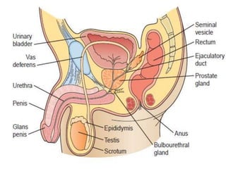
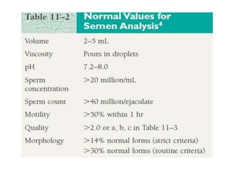
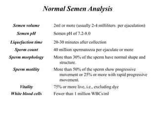
Semen analysis
Introduction
• A semen analysis measures the amount of semen a man
produces and determines the number and quality of
sperm in the semen sample.
• A semen analysis is usually one of the first tests done to
help determine whether a man has a problem fathering a
child (infertility).
• A problem with the semen or sperm affects more than
one-third of the couples who are unable to have
children (infertile).
• Sperm is the male gamete, that is, the male sex cell, which has the
capacity to fertilise an egg. Sperm are produced in
the seminiferous tubes of the testes. Germ cells for the production
of spermatozoa
• Specialized Sertoli cells provide support and nutrients for the
germ cells as they undergo mitosis and meiosis
(spermatogenesis).
• When spermatogenesis is complete, the immature sperm
(nonmotile) enter the epididymis.
• In the epididymis, the sperm mature and develop flagella.
• They remain stored in the epididymis until ejaculation.
• The ejaculatory ducts receive both the sperm from the ductus
deferens and fluid from the seminal vesicles.
Physiology
Fluid Fractions
1. Urethral glands (2-5%) are very small mucus
secreting glands.
2. Prostate: Approximately 20% to 30% of the semen
volume is acidic fluid produced by the prostate gland,
the secretion contains acid phosphatase and
proteolytic enzymes that act on the fluid from the
seminal vesicles, resulting in the coagulation and
liquefaction of the semen.
3. Seminal vesicles (produce about 46-80 % of the fluid
volume of semen) viscous, yellowish secretion is rich
in fructose, vitamin C, prostaglandin, and other
substances, which nourish and activate the sperm
passing through the tract.
4. Testis & Epididymis: (5%) Spermatozoa are
produced in the testis under the influence of
testosterone, and then the epididymis (is the first part
of the duct system) provides a temporary storage site
for the immature sperm that enter it from testis. This
fraction still in the inactive form until ejaculation.
Formation of the sperm cell
• Spermatogenesis is a cascade of cell divisions:
o Mitosis: spermatogonia to primary spermatocytes
o First meiotic division: secondary spermatocytes
o Second meiotic division: haploid spermatids
• This process takes 70 ± 4 days in the human.
• Spermiogenesis: differentiation of the round spermatid
into a spermatozoon
Sperm transport and seminal plasma
• “Testicular sperm” need to undergo more maturation
steps before they are ready to fertilize.
• Transported from the testes to the epididymis, where
they mature, and acquire the ability to swim.
• Then moved to the vas deferens, for storage.
• At ejaculation, the sperm are transported out of the
vas and mix with accessory gland secretions:
– prostatic fluid (pH slightly alkaline to neutral;
contains citric acid and zinc).
– seminal vesicle fluid (pH strongly alkaline;
contains fructose).
What the spermatozoon
• The human sperm cell is about 70 µm
long.
• The nucleus is in the head – contains
the 23 chromosomes.
• It is the head which binds to the egg at
fertilization.
• Mid-piece: the energy for motility is
generated.
• Tail: (motility the beat is initiated just
behind the mid-piece, and then
propagated along the tail).
What the purpose of the test
• Investigation of fertility
• Identify treatment options
 Surgical treatment.
 Medical treatment.
 Assisted conception treatment.
 Determine the suitability of semen for artificial insemination
or IVF.
Sample collection
• Specimen should be collected into pre warmed (21oC), sterile,
non-toxic, wide-mouth container, after a couple has abstain from
sexual activity for 2-3 days to not longer than 5 days.
• Specimens collected following prolonged abstinence tend to
have higher volumes and decreased motility.
• When performing fertility testing, two or three samples are
usually tested at 2-week intervals, with two abnormal samples
considered significant.
• The specimen should be delivered to the laboratory within 1
hour of collection and the laboratory personnel must record the
time of specimen collection and specimen receipt.
• The sample must be kept at 37°C until analysis, which begins
ideally within 30 min, but absolutely within 60 min, of
ejaculation
Methods of collection
1. Masturbation (the method of choice for all seminal fluid
tests).
2. By condom: it is not recommended for fertility testing
because the condoms may contain spermicidal agents.
3. By coitus interrupts: (withdrawal method).
4. TESA: Testicular sperm extraction (TESE)- Open Testicular
Biopsy: is a highly invasive, open surgical procedure
performed under general anaesthetic. The scrotum and testes
are cut open, before testicular tissues are cut away and
examined for sperm, which, if present can be extracted.
Label of sample
• Patient name
• Clinic number
• Date and time
• Laboratory request form
• The following should be recorded on the laboratory
analysis form:
– The period of abstinence (in days).
– If sample collection was complete or incomplete.
– The time interval from collection to analysis
Macroscopic examination
• There are several macroscopic evaluations which give useful
diagnostic information about the sample:
 Appearance
 Odour
 Liquefaction
 Volume
 Viscosity
 pH
Liquefaction
• A fresh semen specimen is clotted and should liquefy
within 30 to 60 minutes after collection; therefore,
recording the time of collection is essential for
evaluation of semen liquefaction.
• Analysis of the specimen cannot begin until after
liquefaction has occurred.
• If after 2 hours the specimen has not liquified
proteolytic enzymes such as alpha-chymotrypsin may
be added to allow the rest of the analysis to be
performed.
Macroscopic examination
 Volume: normal is (2-5 mL). Using disposable
volumetric pipette .
• WHO criteria specify that any volume greater than
2.0 mL is normal. Low volume may indicate partial
or complete blockage of the seminal vesicles, or that
the man was born without seminal vesicles.
• Viscosity: Estimate the viscosity of the semen by
aspirating the semen into the measuring pipette and
allowing the semen to drop by gravity and will not
appear clumped. Observe the length of the thread.
volumetric pipette
• The normal pH of semen is alkaline with a range of 7.0
to 8.0.
• Increased pH is indicative of infection within the
reproductive tract.
• A decreased pH is associated with increased prostatic
fluid.
pH
Microscopic examination
• The characteristics assessed are:
Motility.
Sperm aggregation (random clumping): “some” is normal, but
large clumps (each with hundreds of sperm) is abnormal.
Spermagglutination (between specific sites): could suggest the
presence of antisperm antibodies.
Epithelial cells: usually present in small numbers
Erythrocytes: should not be present
Bacteria and protozoa: presence indicates infection
Normal Semen Analysis
2ml or more (usually 2-4 milliliters per ejaculation)
Semen volume
Semen pH of 7.2-8.0
Semen pH
20-30 minutes after collection
Liquefaction time
40 million spermatozoa per ejaculate or more
Sperm count
More than 30% of the sperm have normal shape and
structure.
Sperm morphology
More than 50% of the sperm show progressive
movement or 25% or more with rapid progressive
movement.
Sperm motility
75% or more live, i.e., excluding dye
Vitality
Fewer than 1 million WBCs/ml
White blood cells
Microscopic examination
• Normal values for sperm concentration are commonly listed as
greater than 20 million sperm per milliliter, with concentrations
between 10 and 20 million per milliliter considered borderline.
• The total sperm count for the ejaculate can be calculated by
multiplying the sperm concentration by the specimen volume.
• Total sperm counts greater than 40 million per ejaculate are
considered normal (20 million per milliliter 2 mL).
Microscopic examination
Methods of measuring sperm concentration
• Hemacytometer: Sperm can be counted by make
dilution 1:20 in WBC pipette or by automatic pipette
(which is more accurate) with a solution containing
sodium bicarbonate (5g) and formalin (1ml)
(immobilize & preserve the spermatozoa), tap water
(100 ml) will suffice as a diluent.
Sperm concentration - haemocytometer
• The number of squares assessed
depends on the number of sperm
counted in the first large square:
–If < 10 counted, the whole
grid is assessed
–If 10-40 counted, 10 squares
are assessed
–If > 40 counted, 5 squares
are assessed
First large square counted
large central square.
This square is ruled into 25 small squares,
each of which is further divided into 16
Sperm concentration - calculations
• If the counts of the two chambers are not within 5% of their
average discard, remix the sample, and set it up again
• If the two counts are in agreement, then the sum of the two
counts is divided by the correction factor:
If 2 × 25 squares counted, divide their sum by 10
If 2 × 10 squares counter, divide their sum by 4
If 2 × 5 squares counted, divide their sum by 2
• This gives the sperm concentration in millions per ml
• Sperm count = concentration × total volume
Calculations
• Using a 1:20 dilution and four large WBC’s squares
counted
• The sperm concentration/ml = No of sperms counted
x 50,000
• Using a 1:20 dilution and five small RBC’s squares
counted
• The sperm concentration /ml = No of sperms
counted x 1,000,000
Sperm concentration – interpretation
• The WHO Reference values for:
Sperm concentration is > 20 × 106 sperm/ml
• Counts of less than 20 million per milliliter (<20
million/ml) are considered sub-fertile
• If a man has a sperm concentration < 5 × 10⁶ sperm/ml,
the WHO recommends assessment for numerical and
structural abnormalities of sex chromosomes
Direct smear or Wet preparation
Place 10µl of thoroughly mixed, liquefied semen on a
clean glass slide under a lightly applied glass cover
slip will allow visualization of the sperm in a
specimen of semen under HPF.
• The World Health Organization has a value of 50%
and this must be measured within 60 minutes of
collection.
• A man can have a total number of sperm far over the
limit of 20 million sperm cells per milliliter, but still
have bad quality because too few of them are motile.
• The other way around, a man can have a sperm count
far less than 20 million sperm cells per millilitre and
still have good motility, if more than 60% of those
observed sperm cells show good forward movement.
Motility
Motility of sperm are divided into four different grades
• Grade 4: Sperm with progressive motility. These are the
strongest and swim fast in a straight line. Sometimes it is also
denoted motility a.
• Grade 3: (non-linear motility): These also move forward but
tend to travel in a curved or crooked motion. Sometimes also
denoted motility b.
• Grade 2: These have non-progressive motility because they do
not move forward despite the fact that they move their tails.
• Grade 1: These are immotile and fail to move at all.
Sperm morphology
• Morphology is even more important than motility and
concentration
• Because of the small size of the human sperm head, must use an
air-dried smear which has been stained
• Prepared samples are assessed using a 100× oil-immersion
objective under bright field optics
• The WHO recommends that 200 spermatozoa are counted per
sample
• Fields for counting must be selected at random
• When counting, remember about the normal distribution
Abnormal morphology
Sperm Vitality
• Usually performed using a vital stain, such as eosin Y,
with a counterstain (nigrosin) to differentiate live
(unstained) and dead (stained) spermatozoa.
Agglutination
• Agglutination of spermatozoa means that motile spermatozoa
stick to each other, head to head, mid-piece to mid-piece, tail to
tail, or mixed, e.g. mid-piece to tail.
• The adherence of either immotile or motile spermatozoa to
mucus threads, to cells other than spermatozoa, or to debris is not
considered agglutination and should not be recorded as such.
• The presence of agglutination is suggestive of an immunological
factor of infertility.
Other cells in semen
• Leukocytes: normally (1-4/HPF), increase number
(leukocytospermia) indicates reproductive tract infection
• Epithelial cells: normally (1-2/HPF)
• Spermatocytes: (Immature germ cells) 1-2/HPF.
• Erythrocytes: (1-2/HPF). Increased number may indicate a
reproductive tract infection or damage to a small capillary
during sample production.
• Note: Some protozoan such as Trichomonas vaginalis are
uncommon in human semen but their presence is indicative of
possible male reproductive tract infection and should be
reported to the referring doctor for further evaluation.
Semen biochemistry
• Acid phosphatase: marker for prostatic function
• Citric acid: can indicate prostatic function – low
levels may indicate dysfunction or a prostatic duct
obstruction
• Zinc: marker for prostatic function – colorimetric
assay (WHO)
• Fructose: marker for seminal vesicle function,
and is a substrate for sperm metabolism –
spectrophotometric assay (WHO)
Terms
• Aspermia: absence of semen
• Azoospermia: describe a total absence of spermatozoa
in semen. (After centrifuge sperm count is zero/HPF).
• Oligozoospermia: refers to a reduced number of
spermatozoa in semen and is usually used to describe a
sperm concentration of less than 20 million/ml. Sperm
count 5-10 sperm/HPF.
• Severe oligospermia: sperm count 1-2 sperm/HPF.
• Polyzoospermia: denotes an increased number of
spermatozoa in semen and is usually refers to a sperm
concentration in excess of 350 million/ml.
• Asthenozoospermia: refers to a man who produces a
greater proportion of sperm which are immotile or
have reduced motility, compared to the WHO
reference values.
• Teratozoospermia: sperm carry more morphological
defects than usual
TZI
• The Teratozoospermic Index is an expression of the average
number of abnormalities per abnormal sperm
• Each sperm cell is assessed for an abnormality in the head,
neck/mid-piece, or tail, and for a cytoplasmic droplet
• If it does not have any of these abnormalities, it is “normal”
• If it does have an abnormality, it is “abnormal”, and we score
each abnormality. So, if a cell has an abnormal head and tail,
it is counted as 1 cell, and 2 abnormalities
• Then, (total # abnormalities) / (total # sperm) = TZI
• A TZI > 1.80 has been associated with poor sperm fertilizing
ability
infertility
What is infertility?
• Infertility means not being able to get pregnant after one year of
trying. Or, women who can get pregnant but are unable to stay
pregnant may also be infertile.
• Pregnancy: is the result of a process that has many steps. To get
pregnant:
 A woman must release an egg from one of her ovaries
(ovulation).
 The egg must go through Fallopian tube toward the uterus
(womb).
 A man's sperm must join with (fertilize) the egg along the way.
 The fertilized egg must attach to the inside of the uterus
(implantation).
 Infertility can happen if there are problems with any of these
steps.
• High temperature of testicles (testes). Sperm are made in the
testes which are in the scrotum. This is the body's way of
keeping the testes slightly cooler than the rest of the body,
which is best for making sperm.
• Smoking can affect the sperm count. If you smoke, you
should stop completely for optimum sperm production.
• Alcohol: equivalent eight pints of normal strength beer or
sixteen small glasses of wine, may interfere with optimum
fertility.
• Medicines and drugs. Most do not interfere with sperm
production such as tetracyclines, colchicine, allopurinol.
What increases a man's risk of infertility?
What things increase a woman's risk of
infertility?
 Age
 Stress
 Poor diet
 Being overweight or underweight
 Smoking
 Excess alcohol use
 Sexually transmitted infections (STIs)
 Health problems that cause hormonal changes, such as
polycystic ovarian syndrome and primary ovarian
insufficiency
assisted reproduction techniques
(ART)
• ART includes all fertility treatments in which both eggs and
sperm are handled. In general, ART procedures involve surgically
removing eggs from a woman’s ovaries, combining them with
sperm in the laboratory, and returning them to the woman’s body.
or procedures in which a woman takes medicine only to stimulate
egg production without the intention of having eggs retrieved.
• The different types of assisted reproductive technology
(ART)?
1. In vitro fertilization (IVF) means fertilization outside of the
body. IVF is the most effective ART. It is often used when a
woman's Fallopian tubes are blocked or when a man produces
too few sperm.
2. Zygote intrafallopian transfer (ZIFT): is similar to IVF.
Fertilization occurs in the laboratory. Then the very young
embryo is transferred to the Fallopian tube instead of the
uterus.
3. Gamete intrafallopian transfer (GIFT) involves transferring
eggs and sperm into the woman's Fallopian tube. So
fertilization occurs in the woman's body.
4. Intracytoplasmic sperm injection (ICSI) is often used for
couples in which there are serious problems with the sperm.
Sometimes it is also used for older couples or for those with
failed IVF attempts. In ICSI, a single sperm is injected into a
mature egg. Then the embryo is transferred to the uterus or
Fallopian tube
Sperm preparation
• Discontinuous density gradient: is a technique in
which all sperm in the seminal fluid are separated by
centrifugation.
• Swim up: is a technique in which the most motile
sperm are selected from the ejaculate. Fresh semen is
covered with a medium and placed in a cylinder which
is laid at a 45oC angle. Motile sperm in the ejaculate
will then swim up to the top of the tube, leaving
immotile sperm and debris in the lower part of the
cylinder.

Final-Seminal-fluid-analysis-Alaqsa.ppt

  • 1.
  • 2.
    Introduction • A semenanalysis measures the amount of semen a man produces and determines the number and quality of sperm in the semen sample. • A semen analysis is usually one of the first tests done to help determine whether a man has a problem fathering a child (infertility). • A problem with the semen or sperm affects more than one-third of the couples who are unable to have children (infertile).
  • 3.
    • Sperm isthe male gamete, that is, the male sex cell, which has the capacity to fertilise an egg. Sperm are produced in the seminiferous tubes of the testes. Germ cells for the production of spermatozoa • Specialized Sertoli cells provide support and nutrients for the germ cells as they undergo mitosis and meiosis (spermatogenesis). • When spermatogenesis is complete, the immature sperm (nonmotile) enter the epididymis. • In the epididymis, the sperm mature and develop flagella. • They remain stored in the epididymis until ejaculation. • The ejaculatory ducts receive both the sperm from the ductus deferens and fluid from the seminal vesicles. Physiology
  • 4.
    Fluid Fractions 1. Urethralglands (2-5%) are very small mucus secreting glands. 2. Prostate: Approximately 20% to 30% of the semen volume is acidic fluid produced by the prostate gland, the secretion contains acid phosphatase and proteolytic enzymes that act on the fluid from the seminal vesicles, resulting in the coagulation and liquefaction of the semen.
  • 5.
    3. Seminal vesicles(produce about 46-80 % of the fluid volume of semen) viscous, yellowish secretion is rich in fructose, vitamin C, prostaglandin, and other substances, which nourish and activate the sperm passing through the tract. 4. Testis & Epididymis: (5%) Spermatozoa are produced in the testis under the influence of testosterone, and then the epididymis (is the first part of the duct system) provides a temporary storage site for the immature sperm that enter it from testis. This fraction still in the inactive form until ejaculation.
  • 8.
    Formation of thesperm cell • Spermatogenesis is a cascade of cell divisions: o Mitosis: spermatogonia to primary spermatocytes o First meiotic division: secondary spermatocytes o Second meiotic division: haploid spermatids • This process takes 70 ± 4 days in the human. • Spermiogenesis: differentiation of the round spermatid into a spermatozoon
  • 10.
    Sperm transport andseminal plasma • “Testicular sperm” need to undergo more maturation steps before they are ready to fertilize. • Transported from the testes to the epididymis, where they mature, and acquire the ability to swim. • Then moved to the vas deferens, for storage. • At ejaculation, the sperm are transported out of the vas and mix with accessory gland secretions: – prostatic fluid (pH slightly alkaline to neutral; contains citric acid and zinc). – seminal vesicle fluid (pH strongly alkaline; contains fructose).
  • 12.
    What the spermatozoon •The human sperm cell is about 70 µm long. • The nucleus is in the head – contains the 23 chromosomes. • It is the head which binds to the egg at fertilization. • Mid-piece: the energy for motility is generated. • Tail: (motility the beat is initiated just behind the mid-piece, and then propagated along the tail).
  • 14.
    What the purposeof the test • Investigation of fertility • Identify treatment options  Surgical treatment.  Medical treatment.  Assisted conception treatment.  Determine the suitability of semen for artificial insemination or IVF.
  • 15.
    Sample collection • Specimenshould be collected into pre warmed (21oC), sterile, non-toxic, wide-mouth container, after a couple has abstain from sexual activity for 2-3 days to not longer than 5 days. • Specimens collected following prolonged abstinence tend to have higher volumes and decreased motility. • When performing fertility testing, two or three samples are usually tested at 2-week intervals, with two abnormal samples considered significant. • The specimen should be delivered to the laboratory within 1 hour of collection and the laboratory personnel must record the time of specimen collection and specimen receipt. • The sample must be kept at 37°C until analysis, which begins ideally within 30 min, but absolutely within 60 min, of ejaculation
  • 16.
    Methods of collection 1.Masturbation (the method of choice for all seminal fluid tests). 2. By condom: it is not recommended for fertility testing because the condoms may contain spermicidal agents. 3. By coitus interrupts: (withdrawal method). 4. TESA: Testicular sperm extraction (TESE)- Open Testicular Biopsy: is a highly invasive, open surgical procedure performed under general anaesthetic. The scrotum and testes are cut open, before testicular tissues are cut away and examined for sperm, which, if present can be extracted.
  • 18.
    Label of sample •Patient name • Clinic number • Date and time • Laboratory request form • The following should be recorded on the laboratory analysis form: – The period of abstinence (in days). – If sample collection was complete or incomplete. – The time interval from collection to analysis
  • 19.
    Macroscopic examination • Thereare several macroscopic evaluations which give useful diagnostic information about the sample:  Appearance  Odour  Liquefaction  Volume  Viscosity  pH
  • 21.
    Liquefaction • A freshsemen specimen is clotted and should liquefy within 30 to 60 minutes after collection; therefore, recording the time of collection is essential for evaluation of semen liquefaction. • Analysis of the specimen cannot begin until after liquefaction has occurred. • If after 2 hours the specimen has not liquified proteolytic enzymes such as alpha-chymotrypsin may be added to allow the rest of the analysis to be performed.
  • 22.
    Macroscopic examination  Volume:normal is (2-5 mL). Using disposable volumetric pipette . • WHO criteria specify that any volume greater than 2.0 mL is normal. Low volume may indicate partial or complete blockage of the seminal vesicles, or that the man was born without seminal vesicles. • Viscosity: Estimate the viscosity of the semen by aspirating the semen into the measuring pipette and allowing the semen to drop by gravity and will not appear clumped. Observe the length of the thread. volumetric pipette
  • 23.
    • The normalpH of semen is alkaline with a range of 7.0 to 8.0. • Increased pH is indicative of infection within the reproductive tract. • A decreased pH is associated with increased prostatic fluid. pH
  • 24.
    Microscopic examination • Thecharacteristics assessed are: Motility. Sperm aggregation (random clumping): “some” is normal, but large clumps (each with hundreds of sperm) is abnormal. Spermagglutination (between specific sites): could suggest the presence of antisperm antibodies. Epithelial cells: usually present in small numbers Erythrocytes: should not be present Bacteria and protozoa: presence indicates infection
  • 25.
    Normal Semen Analysis 2mlor more (usually 2-4 milliliters per ejaculation) Semen volume Semen pH of 7.2-8.0 Semen pH 20-30 minutes after collection Liquefaction time 40 million spermatozoa per ejaculate or more Sperm count More than 30% of the sperm have normal shape and structure. Sperm morphology More than 50% of the sperm show progressive movement or 25% or more with rapid progressive movement. Sperm motility 75% or more live, i.e., excluding dye Vitality Fewer than 1 million WBCs/ml White blood cells
  • 26.
  • 27.
    • Normal valuesfor sperm concentration are commonly listed as greater than 20 million sperm per milliliter, with concentrations between 10 and 20 million per milliliter considered borderline. • The total sperm count for the ejaculate can be calculated by multiplying the sperm concentration by the specimen volume. • Total sperm counts greater than 40 million per ejaculate are considered normal (20 million per milliliter 2 mL). Microscopic examination
  • 28.
    Methods of measuringsperm concentration • Hemacytometer: Sperm can be counted by make dilution 1:20 in WBC pipette or by automatic pipette (which is more accurate) with a solution containing sodium bicarbonate (5g) and formalin (1ml) (immobilize & preserve the spermatozoa), tap water (100 ml) will suffice as a diluent.
  • 29.
    Sperm concentration -haemocytometer • The number of squares assessed depends on the number of sperm counted in the first large square: –If < 10 counted, the whole grid is assessed –If 10-40 counted, 10 squares are assessed –If > 40 counted, 5 squares are assessed First large square counted large central square. This square is ruled into 25 small squares, each of which is further divided into 16
  • 30.
    Sperm concentration -calculations • If the counts of the two chambers are not within 5% of their average discard, remix the sample, and set it up again • If the two counts are in agreement, then the sum of the two counts is divided by the correction factor: If 2 × 25 squares counted, divide their sum by 10 If 2 × 10 squares counter, divide their sum by 4 If 2 × 5 squares counted, divide their sum by 2 • This gives the sperm concentration in millions per ml • Sperm count = concentration × total volume
  • 31.
    Calculations • Using a1:20 dilution and four large WBC’s squares counted • The sperm concentration/ml = No of sperms counted x 50,000 • Using a 1:20 dilution and five small RBC’s squares counted • The sperm concentration /ml = No of sperms counted x 1,000,000
  • 32.
    Sperm concentration –interpretation • The WHO Reference values for: Sperm concentration is > 20 × 106 sperm/ml • Counts of less than 20 million per milliliter (<20 million/ml) are considered sub-fertile • If a man has a sperm concentration < 5 × 10⁶ sperm/ml, the WHO recommends assessment for numerical and structural abnormalities of sex chromosomes
  • 33.
    Direct smear orWet preparation Place 10µl of thoroughly mixed, liquefied semen on a clean glass slide under a lightly applied glass cover slip will allow visualization of the sperm in a specimen of semen under HPF.
  • 34.
    • The WorldHealth Organization has a value of 50% and this must be measured within 60 minutes of collection. • A man can have a total number of sperm far over the limit of 20 million sperm cells per milliliter, but still have bad quality because too few of them are motile. • The other way around, a man can have a sperm count far less than 20 million sperm cells per millilitre and still have good motility, if more than 60% of those observed sperm cells show good forward movement. Motility
  • 35.
    Motility of spermare divided into four different grades • Grade 4: Sperm with progressive motility. These are the strongest and swim fast in a straight line. Sometimes it is also denoted motility a. • Grade 3: (non-linear motility): These also move forward but tend to travel in a curved or crooked motion. Sometimes also denoted motility b. • Grade 2: These have non-progressive motility because they do not move forward despite the fact that they move their tails. • Grade 1: These are immotile and fail to move at all.
  • 36.
    Sperm morphology • Morphologyis even more important than motility and concentration • Because of the small size of the human sperm head, must use an air-dried smear which has been stained • Prepared samples are assessed using a 100× oil-immersion objective under bright field optics • The WHO recommends that 200 spermatozoa are counted per sample • Fields for counting must be selected at random • When counting, remember about the normal distribution
  • 37.
  • 39.
    Sperm Vitality • Usuallyperformed using a vital stain, such as eosin Y, with a counterstain (nigrosin) to differentiate live (unstained) and dead (stained) spermatozoa.
  • 40.
    Agglutination • Agglutination ofspermatozoa means that motile spermatozoa stick to each other, head to head, mid-piece to mid-piece, tail to tail, or mixed, e.g. mid-piece to tail. • The adherence of either immotile or motile spermatozoa to mucus threads, to cells other than spermatozoa, or to debris is not considered agglutination and should not be recorded as such. • The presence of agglutination is suggestive of an immunological factor of infertility.
  • 41.
    Other cells insemen • Leukocytes: normally (1-4/HPF), increase number (leukocytospermia) indicates reproductive tract infection • Epithelial cells: normally (1-2/HPF) • Spermatocytes: (Immature germ cells) 1-2/HPF. • Erythrocytes: (1-2/HPF). Increased number may indicate a reproductive tract infection or damage to a small capillary during sample production. • Note: Some protozoan such as Trichomonas vaginalis are uncommon in human semen but their presence is indicative of possible male reproductive tract infection and should be reported to the referring doctor for further evaluation.
  • 42.
    Semen biochemistry • Acidphosphatase: marker for prostatic function • Citric acid: can indicate prostatic function – low levels may indicate dysfunction or a prostatic duct obstruction • Zinc: marker for prostatic function – colorimetric assay (WHO) • Fructose: marker for seminal vesicle function, and is a substrate for sperm metabolism – spectrophotometric assay (WHO)
  • 43.
    Terms • Aspermia: absenceof semen • Azoospermia: describe a total absence of spermatozoa in semen. (After centrifuge sperm count is zero/HPF). • Oligozoospermia: refers to a reduced number of spermatozoa in semen and is usually used to describe a sperm concentration of less than 20 million/ml. Sperm count 5-10 sperm/HPF. • Severe oligospermia: sperm count 1-2 sperm/HPF. • Polyzoospermia: denotes an increased number of spermatozoa in semen and is usually refers to a sperm concentration in excess of 350 million/ml.
  • 44.
    • Asthenozoospermia: refersto a man who produces a greater proportion of sperm which are immotile or have reduced motility, compared to the WHO reference values. • Teratozoospermia: sperm carry more morphological defects than usual
  • 45.
    TZI • The TeratozoospermicIndex is an expression of the average number of abnormalities per abnormal sperm • Each sperm cell is assessed for an abnormality in the head, neck/mid-piece, or tail, and for a cytoplasmic droplet • If it does not have any of these abnormalities, it is “normal” • If it does have an abnormality, it is “abnormal”, and we score each abnormality. So, if a cell has an abnormal head and tail, it is counted as 1 cell, and 2 abnormalities • Then, (total # abnormalities) / (total # sperm) = TZI • A TZI > 1.80 has been associated with poor sperm fertilizing ability
  • 46.
  • 47.
    What is infertility? •Infertility means not being able to get pregnant after one year of trying. Or, women who can get pregnant but are unable to stay pregnant may also be infertile. • Pregnancy: is the result of a process that has many steps. To get pregnant:  A woman must release an egg from one of her ovaries (ovulation).  The egg must go through Fallopian tube toward the uterus (womb).  A man's sperm must join with (fertilize) the egg along the way.  The fertilized egg must attach to the inside of the uterus (implantation).  Infertility can happen if there are problems with any of these steps.
  • 48.
    • High temperatureof testicles (testes). Sperm are made in the testes which are in the scrotum. This is the body's way of keeping the testes slightly cooler than the rest of the body, which is best for making sperm. • Smoking can affect the sperm count. If you smoke, you should stop completely for optimum sperm production. • Alcohol: equivalent eight pints of normal strength beer or sixteen small glasses of wine, may interfere with optimum fertility. • Medicines and drugs. Most do not interfere with sperm production such as tetracyclines, colchicine, allopurinol. What increases a man's risk of infertility?
  • 49.
    What things increasea woman's risk of infertility?  Age  Stress  Poor diet  Being overweight or underweight  Smoking  Excess alcohol use  Sexually transmitted infections (STIs)  Health problems that cause hormonal changes, such as polycystic ovarian syndrome and primary ovarian insufficiency
  • 50.
  • 51.
    • ART includesall fertility treatments in which both eggs and sperm are handled. In general, ART procedures involve surgically removing eggs from a woman’s ovaries, combining them with sperm in the laboratory, and returning them to the woman’s body. or procedures in which a woman takes medicine only to stimulate egg production without the intention of having eggs retrieved. • The different types of assisted reproductive technology (ART)? 1. In vitro fertilization (IVF) means fertilization outside of the body. IVF is the most effective ART. It is often used when a woman's Fallopian tubes are blocked or when a man produces too few sperm.
  • 52.
    2. Zygote intrafallopiantransfer (ZIFT): is similar to IVF. Fertilization occurs in the laboratory. Then the very young embryo is transferred to the Fallopian tube instead of the uterus. 3. Gamete intrafallopian transfer (GIFT) involves transferring eggs and sperm into the woman's Fallopian tube. So fertilization occurs in the woman's body. 4. Intracytoplasmic sperm injection (ICSI) is often used for couples in which there are serious problems with the sperm. Sometimes it is also used for older couples or for those with failed IVF attempts. In ICSI, a single sperm is injected into a mature egg. Then the embryo is transferred to the uterus or Fallopian tube
  • 53.
    Sperm preparation • Discontinuousdensity gradient: is a technique in which all sperm in the seminal fluid are separated by centrifugation. • Swim up: is a technique in which the most motile sperm are selected from the ejaculate. Fresh semen is covered with a medium and placed in a cylinder which is laid at a 45oC angle. Motile sperm in the ejaculate will then swim up to the top of the tube, leaving immotile sperm and debris in the lower part of the cylinder.