Issues in brainmapping...EEG and brainmap spectral profiles in cortical lesions, subcortical white matter lesions and subcortical gray matter (diencephalic) lesions
The tropical cone snail hunts fish using a venomous harpoon-like tooth. Its venom acts extremely quickly, paralyzing prey within seconds through a mixture of toxins that disrupt neuronal signaling. Neurons communicate through electrical and chemical signals, and the cone snail venom interferes with both types of signaling. This prevents the prey's neurons from controlling locomotion and respiration, resulting in its inability to escape or survive the venom's effects.
This document summarizes recent findings on the mechanisms that govern neuronal migration during development. It discusses:
1) Neuronal migration involves leading process dynamics and somal translocation that allow neurons to move through the brain. Different neuron types adjust this process depending on their migratory pathway.
2) Real-time imaging has revealed distinct leading process morphologies in different neuron types that influence their directional guidance. Tangentially migrating neurons display more elaborate branching than radially migrating pyramidal cells.
3) Guidance cues influence the branching and orientation of leading processes to allow rapid changes in migratory direction without reorienting existing branches. This is a different mechanism than growth cone steering in axon guidance
The document discusses the organization of nervous systems and how scientists study brain activity. It begins by explaining how early techniques like brain imaging allowed researchers to monitor brain activity during tasks. More recently, a technique called "brainbow" uses colored proteins to uniquely label individual neurons in the brain of mice, enabling scientists to map neural connections between brain regions. The document concludes by stating that neuroscientists hope to develop detailed maps of brain connections using this brainbow technology.
Hormones communicate regulatory messages throughout the body via the bloodstream. There are two main pathways for cellular responses to hormones. Water-soluble hormones bind to cell surface receptors and trigger intracellular signaling cascades, sometimes altering gene expression. Lipid-soluble hormones diffuse into cells and bind intracellular receptors, directly activating gene transcription and cellular responses. Hormones regulate processes like metabolism, development, behavior, and homeostasis.
Neural oscillations occur throughout the central nervous system and can be measured at different scales: microscopic (single neuron), mesoscopic (local groups of neurons), and macroscopic (between brain regions). At the microscopic level, neurons generate action potentials that form rhythmic spike trains. Groups of synchronized neurons give rise to local field potential oscillations. Interactions between brain areas also produce large-scale oscillations measured by EEG. Different neural oscillations have been linked to cognitive functions like perception and memory.
The basal ganglia and cerebellum work together to modify movement on a minute-to-minute basis. The basal ganglia are a collection of nuclei that provide inhibitory output and help apply "brakes" to movement. The cerebellum compares intended and actual movements to coordinate them and learns motor skills; it provides excitatory output. Disturbances in either system cause movement disorders like Parkinson's and Huntington's diseases.
A ‘neural’ net that can be seen with the naked eye - by Andrew PackardPim Piepers
1. The skin of cephalopods like squid and octopus contains an array of pigmented spots called chromatophores that can change color and contract. These spots form interconnected networks within the skin.
2. When the nerve connecting to one side of an animal is severed, the spots on that side begin pulsating and changing color in waves due to the intrinsic connectivity of the myogenic (muscle-based) networks.
3. These waves provide insights into the properties and behavior of complex interconnected biological networks under myogenic rather than neurogenic (nerve-based) control. The role of the nervous system is to modulate the networks and control the intrinsic chaotic behavior.
Function of Thalamus & thalamic syndromeDr Sara Sadiq
The thalamus is located in the middle of the brain between the cerebral cortex and midbrain. It acts as a relay station for all sensory pathways and regulates states of sleep and wakefulness. The thalamus is divided into sensory, motor, limbic, and association nuclei that receive and project different types of sensory and motor information to various parts of the cerebral cortex. Damage to the thalamus can lead to a condition called thalamic syndrome, also known as Dejerine-Roussy syndrome, which involves loss of fine sensation, exaggerated pain sensation, and motor problems due to disturbances in thalamocortical connections.
The thalamus acts as a relay center, routing sensory and motor information between subcortical structures and the cerebral cortex. It contains several nuclei that can be categorized as relay, association, or intralaminar/midline nuclei based on their input-output connections. Relay nuclei receive specific sensory inputs and project to defined cortical areas, transmitting information. Association nuclei connect subcortical areas to cortical association regions. Intralaminar and midline nuclei are involved in arousal, movement, and limbic functions. The reticular nucleus provides inhibitory input regulating thalamocortical signaling.
This document discusses cholinergic and noradrenergic neurons. Cholinergic neurons are excitatory or inhibitory and include motor neurons, all autonomic preganglionic neurons, and postganglionic parasympathetic neurons. The basal forebrain complex provides cholinergic innervation to the hippocampus and neocortex and is involved in regulating arousal, sleep-wake cycles, and learning and memory. The pontomesencephalotegmental complex projects to the thalamus and regulates sensory relay nuclei. Noradrenergic neurons are located in the locus coeruleus and project widely throughout the central nervous system, regulating attention, arousal, sleep-wake cycles, learning, mood, and brain metabolism
This document discusses the resting membrane potential in neurons. It explains that the plasma membrane of a resting neuron is polarized with negatively charged fluid inside and positively charged fluid outside, resulting in a resting membrane potential of around -70 mV. This polarization is maintained by the sodium-potassium pump and concentration gradients of sodium and potassium ions. When an action potential is triggered, the permeability to sodium ions increases, causing depolarization and propagating the nerve impulse down the axon.
This document discusses neurotransmitters and how they function in the nervous system. It describes how neurotransmitters are synthesized and stored in neurons before being released into the synaptic cleft upon receiving an action potential. The neurotransmitters then bind to receptors on the adjacent cell, either opening ion channels to continue the nerve impulse or closing ion channels to inhibit the impulse. Different classes of neurotransmitters are identified, including amino acids, monoamines, peptides, and purines. Specific neurotransmitters like acetylcholine and GABA are highlighted for their roles in the parasympathetic and central nervous systems. Abnormalities in neurotransmitter levels and activity are also linked to neurological and psychiatric disorders.
Neuroplasticity refers to the brain's ability to change and reorganize itself in response to experience. The brain forms new connections and pathways when we learn new skills or have new experiences. Neuroplasticity allows the brain to compensate for injury and disease and is responsible for development throughout childhood. Promising therapies that may enhance neuroplasticity include brain stimulation, cognitive training, and certain drugs. Assessing neuroplasticity in humans through biomarkers can help predict treatment response and monitor recovery. Harnessing neuroplasticity is key for rehabilitation from brain injuries and disorders.
This document provides an overview of sleep, its stages, circadian rhythms, and relationship to anesthesia. It defines sleep and describes its typical stages: non-rapid eye movement (NREM) sleep including stages 1-3, and rapid eye movement (REM) sleep. NREM sleep involves slow brain waves and restoration, while REM involves dreaming and autonomic instability. Circadian rhythms regulate daily cycles of activity and rest and are generated by biological clocks in cells. Perioperative sleep deprivation and sleep disorders like sleep apnea can impact patient outcomes, making sleep important for anesthesia.
The Role Of The Cytoskeleton During Neuronal PolarizationDalí Mb
This document summarizes recent advances in understanding the intracellular mechanisms underlying neuronal polarization, with a focus on the role of the cytoskeleton. It discusses how local actin instability in the future axonal growth cone allows microtubule protrusion and formation of multiple axons. Key regulators of this process include Rho-GTPases and their downstream targets, which influence actin dynamics. The Ena/VASP family of proteins also promotes neurite formation by antagonizing actin capping and facilitating bundling. Recent studies further reveal microtubule stabilization as another mechanism in neuronal polarization, complementing actin dynamics.
This document discusses the physiology of higher nervous activity. It describes behavior as interactions with the external environment accompanied by vegetative reactions. Higher nervous activity involves human adjustment to changing external conditions and involves cortical and sub-cortical structures. Behavior can be inborn, involving unconditioned reflexes, or acquired through conditioned reflexes. Conditioned reflexes are individually acquired and adjusted to changing external environments. The document also examines the formation of conditioned reflexes and different types of inhibition.
Neurochemistry involves the study of neurochemicals that influence neuronal function and neural networks. Key developments include the discovery that messages are sent chemically between neurons rather than electrically. Molecular neuroscience applies concepts of molecular biology to the nervous system. Main components of neuroanatomy include the brain, neurons, synapses, and neuroglia. Neurons have a cell body, dendrites that receive inputs, and an axon that transmits signals. Synapses allow communication between neurons. Neurotransmitters are the chemical messengers released at synapses to affect another neuron.
The document summarizes key information about the structure and function of the human cerebral cortex. It discusses the layers of the cortex, methods used to study cortical functions including stimulation and electrophysiological procedures like EEG. Specific topics covered include sensory and motor areas of the cortex, sleep centers and stages of sleep, and the influence of the reticular formation on cortical activity.
This document summarizes evidence that some synaptic contacts become "silent" during postnatal synapse elimination in muscle, retaining the ability to release acetylcholine. The authors investigated whether blocking certain molecular pathways could induce the functional recruitment of these silent synapses. They found that blocking muscarinic acetylcholine autoreceptors, calcium channels, or protein kinase C, or applying brain-derived neurotrophic factor, increased the number of functional inputs per neuromuscular junction. This suggests silent synapses may be recruited through these molecular mechanisms before being eliminated. The balance between trkB and muscarinic signaling pathways may regulate synaptic suppression during development.
Neurotransmission involves the release of neurotransmitters from the axon terminal of one neuron that bind to and react with receptors on another neuron. Nerve signals travel as electrical nerve impulses along neurons. A neuron consists of a cell body, dendrites that receive signals, and an axon that transmits signals. When a neuron is stimulated, sodium ions enter the cell causing an action potential to propagate along the axon. At synaptic junctions, neurotransmitters are released from vesicles and bind to receptors, causing excitation or inhibition of the downstream neuron. Neurotransmitters are then removed from the synapse to terminate signaling.
This document provides an overview of neuropharmacology and neurotransmission. It defines neuropharmacology and describes the two main branches. It explains what neurotransmission is and how it works, describing the role of neurons, neurotransmitters, and the mechanism of neurotransmission. It discusses different types of neurons, neurotransmitters like acetylcholine and dopamine, and conditions they are involved in like Alzheimer's and Parkinson's disease. The document also provides interesting facts about neurons and neurotransmitters. It concludes with a recent discovery about how endocannabinoids travel in the brain to reach receptors.
The human nervous system develops from the neural tube during early embryogenesis. Neurons differentiate and grow axons that connect with other neurons. The central nervous system develops from the neural tube, with the brain and spinal cord forming. The brain is made up of several specialized regions including the cerebral hemispheres, cerebellum, and brainstem. The cerebral cortex has evolved in humans to have extensive folding and the largest surface area, allowing for more complex functions.
This document describes the structure and function of neurons and synapses. It discusses the key parts of neurons including the soma, dendrites, axon, and myelin sheath. It classifies neurons as multipolar, bipolar, or pseudounipolar. Synapses are described as the junction between an axon and dendrite or cell body. The stages of synaptic transmission and types of mediators are outlined. The principles of spatial and temporal summation are introduced as ways neurons integrate multiple inputs. Reflexes are defined as automatic sensory-motor responses mediated by the central nervous system. Inhibition is described as a process that suppresses excitation.
This document summarizes the key components and functions of the nervous system. It describes the central nervous system consisting of the brain and spinal cord, and the peripheral nervous system containing sensory and motor neurons. It explains the roles of neurons and glial cells, and how neurons communicate with each other through electrical and chemical signals across synapses using neurotransmitters. Specifically, it outlines the anatomy of neurons including dendrites, cell bodies, axons, and myelin sheaths. It also discusses how action potentials are generated and propagated in neurons and the functions of common neurotransmitters like acetylcholine and endorphins.
This document discusses neurological complications that can occur during hematopoietic cell transplantation (HCT). It describes complications that occur during different stages of HCT: 1) During conditioning regimens, complications include encephalopathy, seizures, and cerebral infarction caused by chemotherapy drugs or medical procedures. 2) During bone marrow depletion, complications include encephalopathy, seizures, cerebral infarctions, and hemorrhages due to metabolic issues, drugs, or infections. 3) During chronic immunosuppression in allogeneic HCT, complications include infections by viruses and opportunistic organisms. 4) Late complications include relapse of the original disease, neurological graft-versus-host disease, and second neoplasms.
This document summarizes the pathology of pituitary tumors, including:
1) Pituitary adenomas are benign neoplasms originating from pituitary cells and are the most common tumors of the sella region.
2) Pituitary tumors are currently classified based on their hormonal content and ultrastructural morphology, with 14 recognized subtypes.
3) Growth hormone-secreting adenomas are associated with acromegaly or gigantism and can have various histologic appearances, most commonly densely or sparsely granulated somatotroph adenomas.
This document is a case publication from January 2008 edited by Professor Yasser Metwally about acute disseminated encephalomyelitis (ADEM). It describes an 18-year-old female patient who presented with disturbed consciousness, seizures, optic neuritis and meningism. MRI images show lesions in the brain consistent with ADEM. The publication discusses differentiating ADEM from other conditions like multiple sclerosis, infections and metabolic disorders based on clinical features and test results. It provides guidance on the diagnosis and management of ADEM cases.
A 40-year-old patient presented with relapsing-remitting spinal multiple sclerosis. MRI revealed asymptomatic brain involvement and recent progression of myelopathy. The document discusses the diagnosis of spinal MS and differentiating it from transverse myelitis using MRI characteristics such as lesion location and contrast enhancement. Figures show examples of MS plaques and diffuse abnormalities on MRI of the spinal cord.
A 66-year-old male patient presented with non-specific lower back pain. MRI images showed signs of lumbar spondylosis including disc degeneration and bulging in the lower lumbar spine. The document provides figures showing disc herniation and details on accessing additional materials on the author's website, including updates versions of the case publication.
This document is a short case publication about a 60-year-old male patient presenting with symptoms of cerebellopontine angle syndrome affecting the 7th, 5th, and bulbar cranial nerves, as well as cerebellar deficits and right ear tinnitus. Imaging revealed a fusiform aneurysm of the vertebrobasilar system with mural thrombosis. Fusiform aneurysms commonly involve the vertebrobasilar system, causing arteries to be diffusely dilated, tortuous, and prolonged with frequent mural thrombosis. They rarely rupture but can cause ischemic manifestations or pressure effects from mass effect. The publication is periodically updated on the author's website.
A 16-year-old female patient presented with diminished vision, back pain, seizures, and poor school performance. Examination revealed optic nerve damage, cafe au lait spots on the skin, and skin growths. She was diagnosed with neurofibromatosis type 1 based on these findings. The document describes this genetic disorder and includes figures showing abnormalities in the patient's optic nerves, spine, and brain white matter seen on imaging. It also provides information on typical MRI findings in neurofibromatosis type 1 patients and references for further reading.
A 40-year-old male patient was found unconscious on a toilet with a ruptured anterior communicating artery aneurysm and subarachnoid hemorrhage. CT scans showed interfrontal subarachnoid hemorrhage breaking into the ventricles with blood in the occipital horns and bilateral anterior cerebral artery infarctions, more severe on the right side. The document describes images of the ruptured anterior communicating artery aneurysm and associated hemorrhaging and edema. It also provides information on accessing updated case publications on the author's website.
1) Brainmapping provides a quantitative analysis of EEG data by measuring frequency bands and topographic distribution, allowing objective characterization beyond visual analysis.
2) Studies have found focal increases in beta band energy can localize epileptic foci, while decreased alpha/theta with increased beta may indicate surrounding gliosis.
3) Brainmapping uncovered covert epilepsy and subclinical foci, with patterns of diminished and augmented activity suggesting regions of atrophy surrounded by epileptogenic cortex.
A 32-year-old male patient presented with a single grand mal seizure. Imaging showed extradural masses in the left parietal and frontal regions. Biopsy revealed non-Hodgkin B-cell lymphoma. The CNS manifestations were initially diagnosed as secondary CNS lymphoma. The patient was referred for oncology treatment. The document discusses epidural lymphomatous deposits and provides information on accessing additional case publications and archives on the author's website.
This document discusses a case of subarachnoid hemorrhage and bilateral anterior cerebral artery infarctions caused by a ruptured anterior communicating artery aneurysm in a 40-year-old male patient found unconscious. It provides radiological images showing the location of the aneurysm and resulting hemorrhage. The diagnosis is given as a ruptured anterior communicating artery aneurysm. The discussion section covers complications of cerebral aneurysms including hematoma formation and patterns associated with different aneurysm locations. It describes how aneurysm ruptures can lead to subarachnoid or intracerebral hemorrhage and secondary effects like vasospasm and infarction.
A 40-year-old female presented with headache, seizures, and altered mental status. MRI showed widespread cerebral venous thrombosis resulting in venous hypertension and congestion. This led to dilated veins and edema in the brain, seen as hyperintensities on T2-weighted MRI. The document discusses the pathophysiology of cerebral venous thrombosis, noting that increased venous pressure can cause edema, hemorrhage, and potentially infarction if pressure exceeds arterial pressure long enough. It describes three stages: initial sinus changes only, early encephalopathy with reversible edema on MRI, and later irreversible infarction if not resolved.
The document describes a study that classified 21 patients with brain stem gliomas into 5 groups based on clinical features and radiological findings. Group 1 had diffuse gliomas and a relatively better prognosis. Group 2 also had diffuse gliomas but a worse prognosis. Group 3 consisted of patients with focal pontine or midbrain gliomas. Group 4 contained patients with cervicomedullary gliomas. The study analyzed each group's clinical features, treatment responses, and outcomes to better understand patterns in brain stem gliomas.
This document summarizes a case study of spinal multiple sclerosis seen on MRI imaging. It includes the following key points:
1) MRI images of the patient show multiple well-defined pencil-shaped lesions occupying 2-3 spinal segments that appear hypointense on T1-weighted images and hyperintense on T2-weighted images.
2) The lesions are characteristic of multiple sclerosis and located peripherally within the spinal cord.
3) The imaging and clinical presentation lead to a diagnosis of spinal multiple sclerosis.
This document summarizes a case study of a 37-year-old female patient who presented with Parinaud syndrome. She had first noticed eye movement abnormalities at age 12 but did not seek medical care until age 37. An MRI revealed a tectal plate glioma (focal midbrain glioma). The tumor enhanced with contrast and appeared hyperintense on T2-weighted MRI. The patient's condition remained stable with follow-up MRIs every two years.
This document summarizes a case report of a 22-year-old male patient diagnosed with Friedreich's ataxia. The patient presented with bilateral cerebellar ataxia, pyramidal signs, loss of tendon reflexes, peripheral neuropathy, enlarged nerves, kyphoscoliosis, and pes cavus. Nerve biopsy revealed demyelinating neuropathy. MRI showed cervical spinal cord atrophy. The clinical diagnosis was Friedreich's ataxia based on the progressive nature of symptoms starting at age 12. The document provides figures of MRI images and references for further information on the case.
This document provides an overview of how to conduct a neurological examination, including taking a thorough patient history and performing a physical exam. Some key points:
1. Taking a thorough history is important for localizing lesions and making a differential diagnosis. Leading questions should be asked about symptoms, onset/progression, relieving/precipitating factors, and associated symptoms.
2. Common complaints warranting detailed history include headache, dizziness/vertigo, sensory symptoms, cognitive decline, speech disorders, weakness, and visual abnormalities.
3. The physical exam follows a standardized pattern but can be tailored based on pertinent findings. It includes tests of consciousness, cognition, cranial nerves, motor function, sensory function
This case report describes subdural empyema in a 26-year-old male patient presenting with seizures, fever, headache, and nausea. MRI scans showed multiple oval-shaped cystic lesions located between the dura mater and arachnoid mater along the falx cerebri and tentorium cerebelli, consistent with subdural empyema. Subdural empyema is a potentially life-threatening infection most often caused by bacterial spread from sinusitis or mastoiditis. It is important to promptly diagnose and treat subdural empyema surgically in addition to antibiotics due to its rapid spread and high mortality rate if left untreated.
The document describes a case of acute disseminated encephalomyelitis (ADEM) in an 18-year-old female patient. She presented with disturbed consciousness, seizures, optic neuritis, and signs of meningismus. MRI showed multifocal white matter lesions characteristic of ADEM. She was diagnosed with ADEM and recovered fully after two weeks of hospitalization and treatment. ADEM is an immune-mediated inflammatory demyelinating disorder of the CNS that typically follows infections or vaccinations and presents with polysymptomatic neurological dysfunction.
Case record... Acute disseminated encephalomyelitis
Similar to Issues in brainmapping...EEG and brainmap spectral profiles in cortical lesions, subcortical white matter lesions and subcortical gray matter (diencephalic) lesions
The document describes a new fever affecting an unknown number of people. It was written by Aneesa Naadira Khan, who calls herself "crazy" for having a new idea about the fever. The fever is referred to as a "strange fever" and the author aims to briefly describe it. The cause and symptoms of the fever are not provided in the 3 sentences.
Neural oscillations occur throughout the central nervous system and can be measured at different scales: microscopic (single neuron), mesoscopic (local groups of neurons), and macroscopic (between brain regions). At the microscopic level, neurons generate action potentials that form rhythmic spike trains. Groups of synchronized neurons give rise to local field potential oscillations. Interactions between brain areas also produce large-scale oscillations measured by EEG. Different neural oscillations are linked to cognitive functions like perception and memory.
Tognoli & Kelso, Society for Neuroscience 2009, diversity of 10Hz rhythms in ...EmmanuelleTognoli
This document discusses the heterogeneity of 10Hz rhythms seen in EEG data and proposes guidelines for their proper measurement and analysis. It presents a tentative dictionary of various 10Hz rhythms distinguished by their spatial distribution, frequency localization, and functional significance. It also puts forth a theory relating EEG spectral peaks to instantaneous brain oscillation patterns, and how the time scale of analysis impacts which patterns appear as peaks. Analyzing 10Hz rhythms at a fine spectral resolution and temporal scale can provide insights into distinct brain processes and functions.
This document discusses different types of neural oscillations observed in the brain, including alpha waves, delta waves, theta rhythm, mu waves, beta waves, and gamma waves. It provides details on the frequency ranges and functions of each type of wave. For example, it states that alpha waves originate from the occipital lobe during relaxation with closed eyes, delta waves are associated with deep sleep, and gamma waves may be implicated in creating unified conscious perception. The document also discusses how neural oscillations are involved in cognitive functions and can be used to transmit information across brain regions in a radio-like fashion on different frequencies.
COGS 107B - Winter 2010 - Lecture 12 - control and function of sleep-wake st...Tim Mullen
1. The document discusses homeostasis and functional anatomy in the context of sleep and how brain activity varies across sleep states.
2. It covers the differences between REM and NREM sleep, noting that both are actively induced by specific brain mechanisms to regulate processes like memory, metabolism, and development.
3. Key factors that influence sleep-wake cycles are discussed like the activity levels of different neurotransmitter systems and populations of neurons in regions like the pons, thalamus, and hypothalamus across states.
Issues in brain mapping...Polymorphic delta activity and its brainmap counter...Professor Yasser Metwally
This document discusses various brain mapping terminologies used in EEGs including absolute power, relative power, coherence, symmetry, and polymorphic delta activity (PDA). PDA consists of arrhythmic slow waves that vary in frequency, amplitude, and morphology. It is commonly seen with subcortical white matter lesions and indicates deafferentation of cortical neurons from a subcortical pacemaker due to a destructive focal lesion interrupting thalamocortical pathways. PDA is nonspecific but suggests an underlying structural abnormality.
This document discusses different types of brain waves and neural oscillations, including their frequencies and functions. It provides information on alpha, delta, theta, mu, beta, and gamma waves. Key points include:
- Different types of brain waves are associated with different cognitive states, like relaxation vs concentration. They facilitate processes like memory and perception.
- Neural oscillations occur throughout the nervous system and can be measured by EEG. Their synchronization is linked to cognitive functions.
- Frequencies of gamma oscillations route information flow in the hippocampus. The brain uses different wave frequencies to transmit different kinds of information between regions.
The reticular formation is a network of neurons located in the brainstem that serves important functions. It extends from the spinal cord up through the midbrain. The reticular formation receives input from various areas of the brain and spinal cord and sends output to many regions including the thalamus and cerebral cortex. It is involved in arousal, motor control, sensory processing, sleep-wake cycles and other vital functions through the ascending and descending reticular activating systems. Damage to or disruption of the reticular formation can impact consciousness, muscle tone, learning, and circadian rhythms.
PP para pais e professores para que compartilhem com filhos e alunos. É como o cérebro funciona e como as drogas que causam dependência funcionam no cérebro. Essa é a primeira parte.
"Theta waves" redirects here. For the System of a Down song, see Steal This Album!
An EEG theta wave
Theta waves generate the theta rhythm, a neural oscillation in the brain that underlies various aspects of cognition and behavior, including learning, memory, and spatial navigation in many animals.[1][2] It can be recorded using various electrophysiological methods, such as electroencephalogram (EEG), recorded either from inside the brain or from electrodes attached to the scalp.
At least two types of theta rhythm have been described. The hippocampal theta rhythm is a strong oscillation that can be observed in the hippocampus and other brain structures in numerous species of mammals including rodents, rabbits, dogs, cats, and marsupials. "Cortical theta rhythms" are low-frequency components of scalp EEG, usually recorded from humans. Theta rhythms can be quantified using quantitative electroencephalography (qEEG) using freely available toolboxes, such as, EEGLAB or the Neurophysiological Biomarker Toolbox (NBT).
The following power point presentation talks about neural control and coordination in humans. In this, we study about neurons, the conduction of nerve impulse, about Central Nervous System and also about sense organs
The document provides an overview of the nervous system:
1. It describes the nervous system as a network of billions of nerve cells that functions as the control center of the body, integrating homeostasis, movement, and other functions.
2. The peripheral nervous system communicates between the central nervous system and the rest of the body, and can be divided into sensory and motor divisions.
3. Within neurons, the cell body contains organelles and receives inputs, while the axon conducts electrical signals to transmit outputs to other neurons.
The document provides an overview of the nervous system, including its basic functions, organization, and components. Key points:
1) The nervous system is a network of nerve cells that functions as the control center of the body, integrating homeostasis, movement, and other functions.
2) It has two main divisions - the central nervous system (CNS) comprising the brain and spinal cord, and the peripheral nervous system outside of the CNS.
3) Neurons are the basic functional units that conduct electrical signals to transmit information via chemical neurotransmitters at synapses.
The document summarizes key aspects of the nervous system including its organization, cells, nerve impulses, synapses, and reflexes. It describes the central and peripheral nervous systems, types of neurons, action potentials, and synaptic transmission. It also presents data from a neuromuscular reflex lab experiment showing involuntary reflexes have faster reaction times than voluntary movements.
Sleep slow oscillation as traveling wave massimini tononi jns2004Bernard Baars
1) The study used high-density EEG recordings in humans to show that each cycle of the slow oscillation during NREM sleep is a traveling wave that originates at a specific site in the brain and propagates across the scalp.
2) Waves most frequently originated in prefrontal-orbitofrontal regions and propagated in an anteroposterior direction.
3) The rate of occurrence of these waves increased progressively during NREM sleep, reaching almost once per second as sleep deepened.
This document provides an overview of spasticity including:
- Definitions of spasticity and tone as the continuous partial contraction of muscles at rest.
- Pathophysiology involving an imbalance between excitatory and inhibitory signals to motor neurons due to lack of descending inhibition.
- Characteristics including increased resistance to passive movement and hyperactive stretch reflexes.
- Factors that can aggravate spasticity like pain, stress, and infection.
- A spectrum of care including oral drugs, rehabilitation, intrathecal baclofen, and neurosurgery to manage spasticity based on individual needs.
The document discusses several types of action potentials recorded from nerve and muscle cells, including:
- Monophasic and biphasic action potentials recorded extracellularly from nerve fibers. Biphasic potentials have two peaks that are mirror images of each other.
- Compound action potentials recorded from stimulating multiple nerve fibers simultaneously. This results in a larger potential that is the algebraic sum of individual fiber potentials.
- Cardiac muscle action potentials have a prolonged plateau phase mediated by calcium channels, prolonging depolarization.
- Skeletal muscle contraction can be tetanized if stimulated at a high frequency, fusing individual twitches into a sustained contraction through calcium summation.
Driving Slow-Oscillations (1 Hz) in rats with optical readout via two-photon microscopy.
Alternative download link: https://dl.dropboxusercontent.com/u/6757026/slideShare/neuromodFUS_v2016.pdf
The document discusses several topics related to action potentials in nerve and muscle cells:
- Compound action potentials recorded from nerves are the summed action potentials of many fibers in the nerve bundle.
- Cardiac muscle action potentials have a prolonged plateau phase due to slow calcium channels, which causes sustained depolarization.
- Tetanic contraction occurs when muscle is stimulated at a high frequency, causing individual twitches to fuse into a sustained contraction.
- Tetany is involuntary muscle contraction that can be caused by conditions increasing the firing frequency of motor neurons.
basic nervous system-CNS-PNS -cell bodie- axon-dendron-grye matter- white mat...shailesh sangle
The nervous system is a complex network of cells, tissues, and organs that coordinates and regulates the body's responses to internal and external stimuli. It is responsible for the control and coordination of all the body's functions, including movement, sensation, thought, and behavior.
The nervous system can be divided into two main parts: the central nervous system (CNS) and the peripheral nervous system (PNS). The CNS consists of the brain and spinal cord, while the PNS consists of all the nerves that extend from the CNS to the rest of the body.
The nervous system is made up of different types of cells, including neurons and glial cells. Neurons are specialized cells that transmit signals through the body in the form of electrical impulses. Glial cells, on the other hand, support and protect the neurons and help maintain the proper functioning of the nervous system.
The nervous system is responsible for many vital functions, including:
Sensory processing: The nervous system receives sensory information from the environment and the body's internal organs, and processes and interprets this information to generate appropriate responses.
Motor control: The nervous system controls the muscles and other organs of the body to produce movement and other responses.
Cognitive functions: The nervous system is responsible for the processes of learning, memory, language, and other complex mental activities.
Autonomic functions: The nervous system regulates the body's automatic functions, such as breathing, heart rate, digestion, and other bodily processes that are not under conscious control.
Overall, the nervous system is a complex and intricate system that plays a critical role in maintaining the body's homeostasis and overall well-being.
IF YOU ARE INTRESTED TO SHOP MAMA EARTH'S PRODUCTES ....
Mamaearth is an Indian personal care brand that offers natural, toxin-free, and sustainable products for all age groups. Their product range includes skincare, haircare, baby care, and men's grooming products. The brand is focused on promoting a healthy and sustainable lifestyle, and all their products are cruelty-free and environmentally friendly.
COPY THAT LINK AND PASTE ON SEARCH BAR OF GOOGLE
https://ekaro.in/enkr20230328s23089700
CHOOSE YOUR AND ORDER NOW.......
thank you....
Similar to Issues in brainmapping...EEG and brainmap spectral profiles in cortical lesions, subcortical white matter lesions and subcortical gray matter (diencephalic) lesions (20)
The document discusses the benefits of exercise for mental health. It states that regular physical activity can help reduce anxiety and depression and improve mood and cognitive function. Exercise causes chemical changes in the brain that may help alleviate symptoms of mental illness.
The document discusses the benefits of exercise for mental health. Regular physical activity can help reduce anxiety and depression and improve mood and cognitive functioning. Exercise causes chemical changes in the brain that may help protect against mental illness and improve symptoms.
The document discusses the benefits of exercise for mental health. Regular physical activity can help reduce anxiety and depression and improve mood and cognitive function. Exercise causes chemical changes in the brain that may help protect against mental illness and improve symptoms.
The document discusses the benefits of exercise for mental health. Regular physical activity can help reduce anxiety and depression and improve mood and cognitive function. Exercise causes chemical changes in the brain that may help protect against mental illness and improve symptoms for those who already suffer from conditions like depression and anxiety.
The document discusses the benefits of exercise for mental health. Regular physical activity can help reduce anxiety and depression and improve mood and cognitive function. Exercise causes chemical changes in the brain that may help protect against mental illness and improve symptoms.
This document discusses the radiological pathology of seizure disorders. It describes various developmental anomalies, neoplasms, infections, immune-mediated disorders, cerebrovascular diseases, and trauma that can cause seizures. Specific conditions mentioned include cortical dysplasia, tuberous sclerosis, Sturge-Weber syndrome, neuronal migration disorders, vascular malformations, infections, and immune-mediated Rasmussen's encephalitis. The document provides detailed descriptions of the histopathological findings and MRI/CT appearances of different lesions that can underlie seizure disorders.
This document discusses cerebral haemorrhage (ICH), which accounts for 10-15% of strokes. ICH can result from several mechanisms, including hypertension (47-66% of cases), cerebral amyloid angiopathy (CAA), and vascular malformations. CAA typically affects the elderly and causes lobar ICH that is often recurrent or involves multiple simultaneous haemorrhages. Vascular malformations like arteriovenous malformations (AVMs) and cavernous angiomas are a common cause of ICH in young, non-hypertensive patients. Imaging techniques like CT and MRI can identify vascular malformations and help determine the underlying cause of ICH.
Cerebral amyloid angiopathy (CAA) refers to the deposition of β-amyloid in the arteries of the cerebral cortex. It is commonly seen in Alzheimer's disease but can also occur in healthy elderly individuals. CAA can cause intracerebral hemorrhage, dementia, or transient neurological symptoms. The deposition damages blood vessels and increases the risk of hemorrhage. Imaging such as CT scans can detect hemorrhages characteristic of CAA, which are often lobar and cortical. Genetic factors like the ApoE genotype can influence the severity and presentation of CAA.
Cerebral microbleeds are small brain hemorrhages detected by MRI that are caused by leakage of blood from damaged small vessel walls. They are increasingly recognized in patients with cerebrovascular disease, Alzheimer's disease, vascular cognitive impairment, and normal elderly populations. Microbleeds in lobar regions may indicate cerebral amyloid angiopathy and link vascular and amyloid neuropathologies, while deep or infratentorial microbleeds often reflect hypertensive vasculopathy. Detection of microbleeds provides insight into cerebral small vessel disease and its relationship to cognitive impairment and dementia.
The document discusses the benefits of exercise for mental health. Regular physical activity can help reduce anxiety and depression and improve mood and cognitive functioning. Exercise causes chemical changes in the brain that may help protect against mental illness and improve symptoms.
A 57-year-old male patient presented with left lower limb weakness that had progressed over three months. MRI images showed bilateral, symmetrical lesions in the posterior parieto-occipital white matter, which had scalloped margins and did not enhance or cause mass effect. Based on the clinical presentation and MRI findings, the patient was diagnosed with progressive multifocal leukoencephalopathy (PML), a demyelinating disease caused by JC virus reactivation that predominantly affects immunocompromised individuals. PML lesions are typically multifocal and located in the white matter of the brain, most often in the parieto-occipital region.
Issues in radiological pathology: Radiological pathology of watershed infarct...Professor Yasser Metwally
The document discusses border zone or watershed infarcts, which occur at the junction between two main arterial territories and constitute approximately 10% of all brain infarcts. There are two types - external (cortical) and internal (subcortical). External infarcts are often embolic in nature while internal infarcts are mainly caused by hemodynamic compromise. Advanced imaging can help identify areas of low perfusion and distinguish the two types. The document then examines the classification, imaging appearance, causal mechanisms, and clinical course of both external and internal border zone infarcts in more detail.
How to Fix the Import Error in the Odoo 17Celine George
An import error occurs when a program fails to import a module or library, disrupting its execution. In languages like Python, this issue arises when the specified module cannot be found or accessed, hindering the program's functionality. Resolving import errors is crucial for maintaining smooth software operation and uninterrupted development processes.
How to Add Chatter in the odoo 17 ERP ModuleCeline George
In Odoo, the chatter is like a chat tool that helps you work together on records. You can leave notes and track things, making it easier to talk with your team and partners. Inside chatter, all communication history, activity, and changes will be displayed.
Biological screening of herbal drugs: Introduction and Need for
Phyto-Pharmacological Screening, New Strategies for evaluating
Natural Products, In vitro evaluation techniques for Antioxidants, Antimicrobial and Anticancer drugs. In vivo evaluation techniques
for Anti-inflammatory, Antiulcer, Anticancer, Wound healing, Antidiabetic, Hepatoprotective, Cardio protective, Diuretics and
Antifertility, Toxicity studies as per OECD guidelines
Thinking of getting a dog? Be aware that breeds like Pit Bulls, Rottweilers, and German Shepherds can be loyal and dangerous. Proper training and socialization are crucial to preventing aggressive behaviors. Ensure safety by understanding their needs and always supervising interactions. Stay safe, and enjoy your furry friends!
This presentation was provided by Steph Pollock of The American Psychological Association’s Journals Program, and Damita Snow, of The American Society of Civil Engineers (ASCE), for the initial session of NISO's 2024 Training Series "DEIA in the Scholarly Landscape." Session One: 'Setting Expectations: a DEIA Primer,' was held June 6, 2024.
Strategies for Effective Upskilling is a presentation by Chinwendu Peace in a Your Skill Boost Masterclass organisation by the Excellence Foundation for South Sudan on 08th and 09th June 2024 from 1 PM to 3 PM on each day.
Your Skill Boost Masterclass: Strategies for Effective Upskilling
Issues in brainmapping...EEG and brainmap spectral profiles in cortical lesions, subcortical white matter lesions and subcortical gray matter (diencephalic) lesions
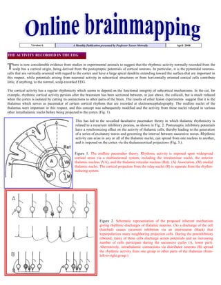
1. Version 4. A Monthly Publication presented by Professor Yasser Metwally April 2008
THE ACTIVITY RECORDED IN THE EEG
scalp
now considerable evidence from
from
in
postsynaptic potentials
suggest that the rhythmic activity
is the
recorded from the
There ishas a cortical origin, being derivedstudies theexperimental animals toof cortical neurons. In particular, itnormallypyramidal neurons-
cells that are vertically oriented with regard to the cortex and have a large apical dendrite extending toward the surface-that are important in
this respect, while potentials arising from neuronal activity in subcortical structures or from horizontally oriented conical cells contribute
little, if anything, to the normal, scalp-recorded EEG.
The cortical activity has a regular rhythmicity which seems to depend on the functional integrity of subcortical mechanisms. In the cat, for
example, rhythmic cortical activity persists after the brainstem has been sectioned between, or just above, the colliculi, but is much reduced
when the cortex is isolated by cutting its connections to other parts of the brain. The results of other lesion experiments suggest that it is the
thalamus which serves as pacemaker of certain cortical rhythms that are recorded at electroencephalography. The midline nuclei of the
thalamus were important in this respect, and this concept was subsequently modified and the activity from these nuclei relayed in various
other intrathalamic nuclei before being projected to the cortex (Fig. 1).
This has led to the so-called facultative pacemaker theory in which thalamic rhythmicity is
related to a recurrent inhibitory process, as shown in Fig. 2. Postsynaptic inhibitory potentials
have a synchronizing effect on the activity of thalamic cells, thereby leading to the generation
of a series of excitatory waves and governing the interval between successive waves. Rhythmic
activity can arise in any or all of the thalamic nuclei, can spread from one nucleus to another,
and is imposed on the cortex via the thalamocortical projections (Fig. 3.).
Figure 1. The midline pacemaker theory. Rhythmic activity is imposed upon widespread
cortical areas via a multineuronal system, including the intralaminar nuclei, the anterior
thalamic nucleus (VA), and the thalamic reticular nucleus (Ret). (A) Association, (M) medial
thalamic nuclei. The cortical projection from the relay nuclei (R) is separate from the rhythm-
inducing system
Figure 2. Schematic representation of the proposed inherent mechanism
giving rhythmic discharges of thalamic neurons. (A) a discharge of the cell
(hatched) causes recurrent inhibition via an interneuron (black) that
hyperpolarizes many neighboring projection cells. During the postinhibitory
rebound, many of these cells discharge action potentials and an increasing
number of cells participate during the successive cycles (A, lower part).
Alternatively, intrathalamic connections via distributor neurons (B) spread
the rhythmic activity from one group to other parts of the thalamus (from-
left-to-right group )
2. Figure 3. Diagrammatic representation of the facultative pacemaker theory.
Rhythmic activity is assumed to be an inherent property of groups of cells in all
thalamic nuclei. The rhythm (series of arrows) is imposed upon the cortex in a
topographic pattern determined by the specific thalamocortical fibers from the relay
and association nuclei. The arrows between the thalamic nuclei indicate mutual
connections between various thalamic parts. These connections determine the
degree of synchrony of the rhythmic activity in the thalamus and cortex.
Alpha Rhythm
Alpha rhythm has a frequency of between 8 and 13 Hz, is found posterior portions of the head during wakefulness, and is best seen when the
patient is resting with eyes closed. It is attenuated or abolished by visual attention, and transiently by other sensory stimuli. Alpha activity is
well-formed and prominent in some normal subjects, while in others it is relatively inconspicuous. Its precise frequency is usually of little
diagnostic significance, unless information is available about its frequency on earlier occasions.
Slowing occurs with advancing age, as a consequence of certain medication such as anticonvulsant drugs, and in patients with clouding of
consciousness, metabolic disorders, or virtually any type of cerebral pathology. The alpha activity may increase in frequency in children as they
mature, and in older subjects who are thyrotoxic. A slight asymmetry is often present between the two hemispheres with regard to the amplitude
of alpha activity and the degree to which it extends anteriorly. In particular, alpha rhythm may normally be up to 50 percent greater in amplitude
over the non-dominant hemisphere. A more marked asymmetry of its amplitude may have lateralizing significance but is difficult to interpret
unless other EEG abnormalities are present, because either depression or enhancement may occur on the side of a hemisphere lesion. Similarly, a
persistent difference in alpha frequency of more than I Hz between the two hemispheres is generally regarded as abnormal, but it is usually
difficult to be certain which is the abnormal side unless other abnormalities are also found.
Beta Activity
EEG CHANGES DUE TO A PURELY CORTICAL LESION NOT INVOLVING SUBCORTICAL WHITE MATTER
Figure 4. A purely cortical lesion not deafferentating the cortical neurons or interrupting
the thalamocortical circuitry
A purely cortical lesions can be due to extraaxial tumors like meningiomas, cortical atrophy
and others. In this situation cortical neurons are unhealthy, diseased (By edema, compression
of ischemia...etc.) yet not deafferentated from the subcortical pace-maker.
EEG activity recorded from diseased, well afferentated cortical neurons (that are not cut
from the pace-maker) are not as chaotic and as slow as EEG activity recorded from
deafferentated healthy neurons. If the later is called polymorphic delta activity, the former is
better called polymorphic theta activity.
Polymorphic theta activity is generated from neurons that are still paced by the subcortical
pace making neurons, these cortical neurons will fire at a faster rate (theta range) compared
with completely deafferentated neurons and because they are still driven by the subcortical
pace maker, their activity is faster, more regular and more synchronous (compared with
deafferentated neurons) with less variability in shape, duration and amplitude of recorded
EEG waves. While neurons that are completely deafferentated and cut from the deep
Purely cortical lesion
3. subcortical pace-making neurons will fire at their own slow rhythm (delta range) and because they are no longer driven by the subcortical pace
maker, their activities is more chaotic, irregular and less synchronous with much more variability in shape, duration and amplitude of recorded
EEG waves. (Polymorphic delta activity).
Unhealthy, diseased cortical neurons, that are uncut from the incoming arousal stimuli but still viable, will not respond to the incoming arousal
stimuli by degenerating alpha spindles (which is the normal situation), but rather their response will be down-shifted to the theta range
(Polymorphic theta acivity). The generated theta waves will not be as organized as the alpha spindles but, at the same time, will not be a chaotic,
irregular and variable as the polymorphic delta activity that are generated by deafferentated neurons.
Polymorphic theta activity maps exactly the diseased neurons and
points to the anatomical site of the lesion (picked up from electrodes
that overly the lesion), this is in contrast to polymorphic delta activity
which is projected to a much wider cortical area and has no localizing
significance in so far as the anatomical site of the lesion is concerned.
However it should be mentioned that the theta focus is totally non-
specific and tells us noting about the nature of the cortical pathology.
In general a purely cortical destructive lesion of any etiology produces
focal increase of percentage theta activity (that is lateralised to the
anatomical site of the destructive lesion and exactly maps it). The
theta focus is not accompanied with any increase in the percentage
delta activity. Alpha maps might be normal or might show occipital
reduction of the percentage Alpha activity ipsilateral to the cortical
destructive lesion. Beta percentage activity maps might show a focal
increase (that is lateralised to the anatomical site of the cortical lesion
and might be present in the same electrodes that show the theta
focus). The beta focus (when present) probably indicates the existence
of cortical irritability. Figure 5. Extra-axial meningioma producing a left fronto-temporal
theta focus with normal delta percentage activity map. A purely
A SUBCORTICAL WHITE MATTER DESTRUCTIVE LESION cortical destructive lesion produces increase of the percentage theta
activity that is is not surrounded by diffuse delta activity and not
associated with any increase of delta activity
Polymorphic delta activity (PDA)
Polymorphic delta activity (PDA) consists of arrhythmic slow waves that vary in
frequency, amplitude, and morphology. PDA can occur in either a focal or
generalized distribution. Continuous PDA is indicative of abnormalities involving
subcortical white matter. One of the shortcomings of standard scalp EEG
recordings is their limited spatial resolution. This holds true for the relationship of
PDA to an underlying structural abnormality. Not only is the inherent localizing
ability of the scalp EEG limited, but also the PDA of a structural lesion is referable
not to the lesion itself but to the surrounding brain tissue. Because of this
limitation, the area of a lesion is indicated not by the maximal amplitude of PDA
but rather by a region of relatively low-amplitude slowing. Continuous, rather than
intermittent, PDA is associated with large lesions, mass effect, and impairment of
consciousness.
Persistent polymorphic delta activity may not precisely match the true location of
the lesion, particularly since it presumably arises from physiological deranged
neurons often lying on the margin of the destructive lesion. Persistent polymorphic
delta activity is aetiologically nonspecific and is seen in a variety of subcortical
(while matter) destructive lesions including neoplasms, infarctions, abscesses,
trauma, and haemorrhage. It can also be seen in reversible processes such as focal ischemia in transient ischemic attacks or focal depression
from a recent seizure.
Pathophysiology and clinical significance of polymorphic delta activity
The diencephalon, as the pacemaker of EEG activity, sends arousal stimuli (through the thalamocortical pathways) to the cortical neurons which
respond by generating the EEG waves. There are always a good degree of coherence and bilateral symmetry of the generated EEG waves
because cortically neurons are paced by the subcortical pacemaker). Any subcortical destructive focal lesion (like infarction, abscess,
tumors...etc.) interrupting the thalamocortical pathway will deprive the cortical neurons, in a particular cortical area, from the arousal stimuli
resulting in cortical neuronal physiological dysfunctions. The neurons in that area, being deafferentated and deprived form the thalamocortical
4. arousal stimuli, will fall out of synchrony and coherence and will start to
generate waves in a chaotic pattern and at a much slower rate than
normal, these waves are irregular in shape, amplitude and duration. and
shows little variation with change in the physiological state of the
patient. Cortical neurons, in the deafferentated cortical strip, are no
longer paced (being cut from the pace maker by the subcortical
destructive focal lesion) and each neuron will fire at its own rhythm
resulting in polymorphic delta activity. Polymorphic delta activity is due
to deafferentation of physically normal cortical neurons by a subcortical
destructive white matter focal lesion. Only subcortical white matter focal
lesions can produce Polymorphic delta activity (PDA) and purely
cortical lesions do not produce PDA.
Please note
1- Cortical neurons that generate PDA are physically normal, however
they are simply deafferentated, physiologically deranged and deprived Figure 6. Lateralised polymorphic delta activity
from arousal stimuli.
2- PDA poorly localizes the destructive subcortical area and is generated from a much wider cortical area overlying the subcortical destructive
focal lesion which is spatially much smaller than the affected cortical area that generates the PDA. The subcortical destructive focal lesions
simply act by deafferentation of the cortical strip which contains the physiological deranged neurons that generate the PDA.
3- A cortical lesion that organically and physically affects cortical neurons (either a primary cortical lesion, a subcortical focal lesion extending
to the cortical neurons or an extra-axial focal lesion compression or extending to the cortical neurons) does not produce PDA. Only normal,
physiological deafferentated, cortical neurons are capable of producing PDA.
Figure 7. Lateralised polymorphic delta activity
Brainmapping Correlates of PDA
1. Theta percentage activity is focally increased, the
theta focus exactly maps and matches the
anatomical site of the lesion.
2. Delta percentage activity is Diffusely increased
and is projected to the cortical area dysfunctioned
by the subcortical destructive lesion. The delta
percentage activity is seen surrounding the theta
focus and mapping the deafferentated cortical electrodes areas that showed PDA in conventional EEG. The delta wave projection, spatial
extension, distribution, and percentage activity correlates nicely with the clinical picture and the degree of clinical disability.
3. The alpha and beta percentage activity are reduced in the affected hemisphere. Alpha is maximally reduced in the delta area while the beta
percentage activity is maximally reduced in the theta focus area.
The pattern described above is aetiologically non-specific and occurs due to any destructive subcorticl lesion of any aetiology.
The percentage delta activity maps the dysfunctioned cortical zone and exactly correlates with the clinical picture. The patient in figure 8 map
presented clinically with left sided weakness and expressive aphasia due to a subcortical lacunar infarction. The delta activity is lateralzed to the
left frontal region in electrodes FP1, F7. The same electrodes area showed polymorphic delta activity on conventional EEG. That is to say delta
activity, in this patient, is projected to the dysfunctioned cortical area and exactly correlates with the clinical picture. As is the case with
polymorphic delta activity, delta activity was generated by deafferentated, physically normal cortical neurons.
In this patient theta maps showed focal increase in the percentage activity that was recorded from the cortical area that exactly overlies the deep
destructive lesion. The theta focus, in this patient, showed a fairly good anatomical localization of the subcortical destructive lesion that exactly
correlated with MRI study (theta band points to the site of the lesion). Probably this focal theta activity was generated by neurons in the cortical
5. strip that directly overlies the deep lesion, these neurons are probably
physically abnormal and is directly involved by the pathological
process through the effect of edema or ischemia. These neurons are not
deafferentated by the subcortical destructive lesions but rather they are
directly affected by the pathological process. Because of their close
proximity to the lesion, these neurons are probably directly implicated
by the subcortical lesion by edema or ischemia.
Careful inspection of the conventional EEG data recorded at the same
time from the electrodes that generated the focal theta activity by
brainmapping showed polymorphic theta activity, however this theta
Figure 8. A brainmap study of a patient with a left deep frontoparietal
infarction producring right sided hemiplegia and expressive aphasia.
Notice the theta focus surrounded by diffuse delta activity projected to
the dysfunctioned area. Alpha is maximally reduced in the delta
area and beta is maximally reduduced at the region of the theat focus
activity was not as disorganized as the polymorphic delta activity,
although it showed variability in wave morphology, amplitude and
duration. EEG wave recording from healthy, yet deafferentated cortical
neurons is more likely to produce EEG activity that is much slower
and more disorganized than EEG recording from diseased , yet pace-
maker connected neurons.
Percentage alpha activity is almost invariably reduced in the affected
hemisphere. The alpha activity is particularly markedly reduced at
electrodes which show maximum delta activity. Alpha visual reactivity
is impaired (diminished or reversed). Alpha percentage activity
correlates inversely with the degree of clinical disability (The less
alpha, the more of the clinical disability). Alpha percentage activity is
an indicator of a more global hemispherical impairment than delta or
theta activities which probably indicate a more focal or regional
impairment restricted to regions of maximum theta or delta activity. Beta percentage activity is commonly focally reduced at the electrodes which
show focal theta activity. Beta activity might be increased in some patients, a pattern which probably indicates the existence of cortical irritability.
Subcortical diencephalic lesion
Subcortical gray matter lesions: Intermittent rhythmic slow wave activity
Subcortical gray matter lesion involving the EEG
pace-maker generates intermittent delta activity
which has the following characteristic
Consists of sinusoidal waveforms of
approximately 2.5 Hz that occur intermittently
in the EEG recording. It is most often
symmetric but can be lateralized.
In adults, the delta activity has a frontal
predominance (frontal intermittent rhythmic
delta activity [FIRDA]). In children, it is
Figure 9. The intermittent rhythmic delta
maximal posteriorly (occipital intermittent
activity
rhythmic delta activity [OIRDA])
The intermittent rhythmic delta activity shows visual reactivity and is commonly suppressed
in the eye open state unless the patient is comatose.
Intermittent rhythmic delta activity is associated with structural lesions, most commonly diencephalic, infratentorial or intraventricular
tumors, or with diffuse encephalopathies.
FIRDA occurring in patients with a normal EEG background suggests that the pattern is due to a structural lesion; when associated with
EEG background abnormalities, it is likely to be due to encephalopathy.
OIRDA is associated with absence epilepsy in children aged 6-10 years.
6. Brainmap correlate of intermittent rhythmic slow wave activity
1-Intermittent rhythmic slow wave activity is seen in diffuse encephalopathic
process (except hepatic encephalopathy which has a different brainmap spectral
profile as will be explained in a different issue) and in middle line thalamic, or
huge parasellar or infratentorial tumors. It was also seen by the author in
vertebro-basilar insufficiency and migraine.
2-There is reduction of alpha percentage activity, and reduced or reversed alpha
visual reactivity. The degree of reduction of alpha percentage activity correlates
with prognosis. The alpha topography is normal and retains its middle line
topography with posterior maximum and is more reduced anteriorly than
posteriorly. There is a fairly good degree of symmetry, synchrony and
coherence in alpha percentage activity maps. however it is not very infrequent
to see some degree of asymmetry with more alpha percentage activity seen on
one side than the other.
3- The alpha land (the middle line electrodes) is now occupied by theta and
delta percentage activity, which is now maximum in the middle line electrodes
at F3,F4,O2,O1. The maximum middle line percentage activity is now shifted
down to the theta rage in most patients and occasionally to the delta rage
depending on the severity of the encephalopathic process and the state of the
patient. Combined theta and delta percentage activity maps clearly
demonstrates the midline maximum. The topography of theta percentage
activity is identical to that of normal alpha percentage activity maps in the eye
closed state. Slow wave activity commonly show visual reactivity exactly as
alpha percentage activity behaves under normal physiological conditions.
4- There is a fairly good degree of symmetry, synchrony and coherence in theta
Figure 10. Two studies showing the brainmap counterpart of percentage activity. However it is not very infrequent to see some degree of
intermittent rhythmic slow wave activity.. Notice the bilateral asymmetry with more theta percentage activity seen on one side than the other.
symmetrical theta activity occupying the same topography of
the alpha activity with middle maximum. Some degree of 5- Beta percentage activity is reduced in the midline frontal, central/parietal
visual reactivity is observed in the lower study. Some degree of and occipital electrodes.
asymmetry is observed in the lower maps
Because the intermittent
rhythmic slow wave activity
occupies the same electrode regions that are normally occupied by the alpha activity, so it
looks like that both (the intermittent rhythmic slow wave activity and alpha spindles) share a
common generator. Similarities between both activities are:
1. Both are sinusoidal activity that waxes and wanes (intermittent)
2. Both show visual reactivity
4. Both are maximum in the middle line electrodes
So it looks like that the generator that is responsible for the generation of the intermittent
rhythmic slow wave activity is the same generator that is responsible for the generation of
alpha spindles under normal conditions, however in the former condition the generator (the
pace-maker) is diseased and firing at a lower rate.
A dysfunctioned pacemaker (subcortical gray matter, diencephalon) is probably necessary for
the intermittent slow wave activity to be seen on EEG.
Arousal stimuli in the thalamo-cortical circuitry are responsible for the production of alpha Figure 11. The brainmap counterpart of
spindles under normal physiological conditions. However when a diffuse encephalopathic intermittent rhythmic slow wave activity.
process involves the diencephalic neurons bilaterally, the dysfunctioned diencephalic neurons Notice the Bifrontal middle line theta
fire at a slower rate and instead of producing alpha spindles they produce theta or delta maximum with reduced alpha and beta activity.
spindles that retain the same characteristics of alpha spindles in spatial topography and visual Maximum theta activity is seen at F3,F4,FZ
reactivity. electrodes