Downloaded 401 times


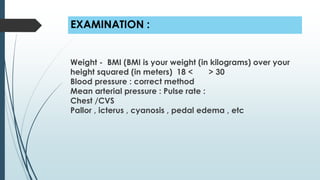

![MEASURING MEAN ARTERIAL PRESSURE
METHODOLOGY :The BP was taken by automated devices (3BTO-A2,
Microlife),which were calibrated before and at regular intervals during the
study. The recordings were made by doctors who had received
appropriate training on the use of these machines. The women were
in the seated position, their arms were supported at the level of the heart,
and a small (22-cm), normal (22- to 32-cm), or large (33- to 42-cm) adult
cuff was used depending on the midarm circumference.26 After rest for 5
minutes, BP was measured in both arms simultaneously, and a series of
recordings were made at 1-minute intervals until variations between
consecutive readings fell within 10 mm Hg in systolic and 6 mm Hg in
diastolic BP in both arms.When this point of stability was reached, we
calculated the MAP of each arm as the average of the last 2 stable
measurements, and, as recommended, we took the arm with the highest
final MAP for the subsequent analysis of results.
MAP = MAP = [(2 x diastolic)+systolic] / 3 CUTT OFF : > 90 mm Hg](https://image.slidesharecdn.com/focusedapproachtoantenatalcare-151003230516-lva1-app6891/85/Focused-approach-to-antenatal-care-First-trimester-screening-71-320.jpg)
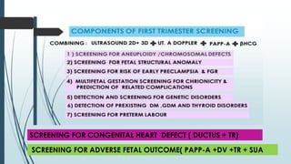

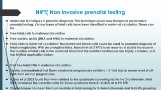
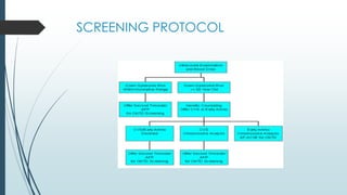
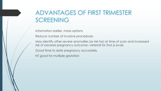
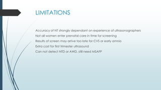

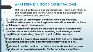
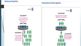
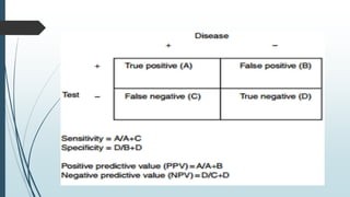
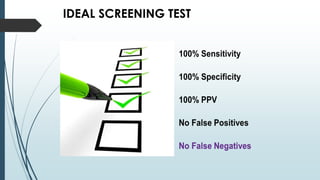
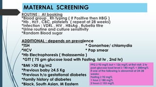
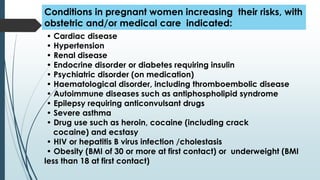
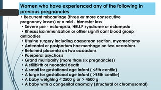
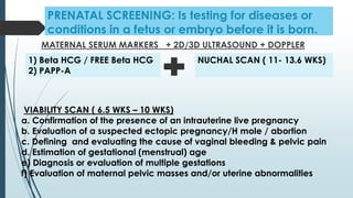
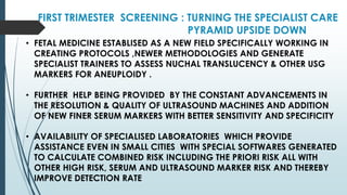
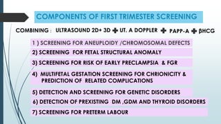
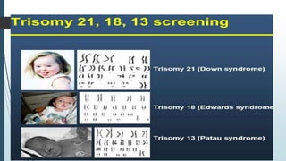
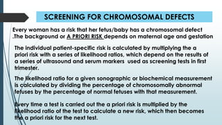
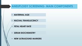
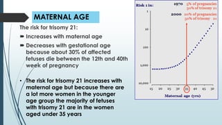
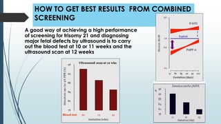
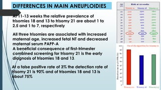
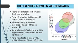
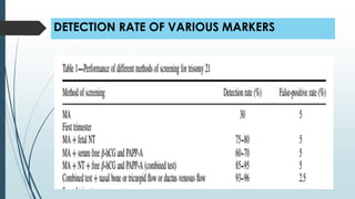
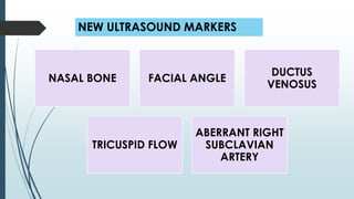
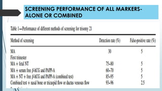
This document discusses focused antenatal care and first trimester screening. It describes the essential elements of antenatal care including targeted assessments based on individual risk factors. First trimester screening aims to detect conditions like aneuploidy through measuring the nuchal translucency, analyzing maternal serum markers, and assessing fetal heart rate between 11-13 weeks of gestation. Screening tests are evaluated based on their sensitivity, specificity, and rates of false positives and negatives.


![Antenatal_Care[1].pptx](https://cdn.slidesharecdn.com/ss_thumbnails/antenatalcare1-221127110720-810db45a-thumbnail.jpg?width=640&height=640&fit=bounds)


