The circulationof oxygenated
blood, de-oxygenated blood,
nutritive material etc in the fetus is
termed as
‘Fetal circulation’
3.
By thethird month of development, all
major blood vessels are present and
functioning.
Fetus must have blood flow to placenta.
Resistance to blood flow is high in
lungs.
4.
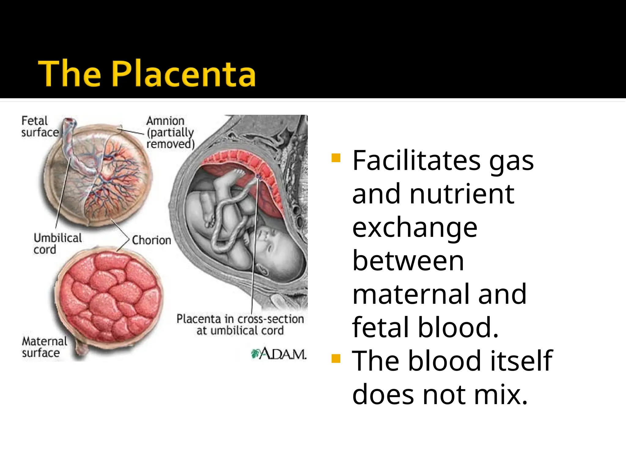
Facilitates gas
andnutrient
exchange
between
maternal and
fetal blood.
The blood itself
does not mix.
5.
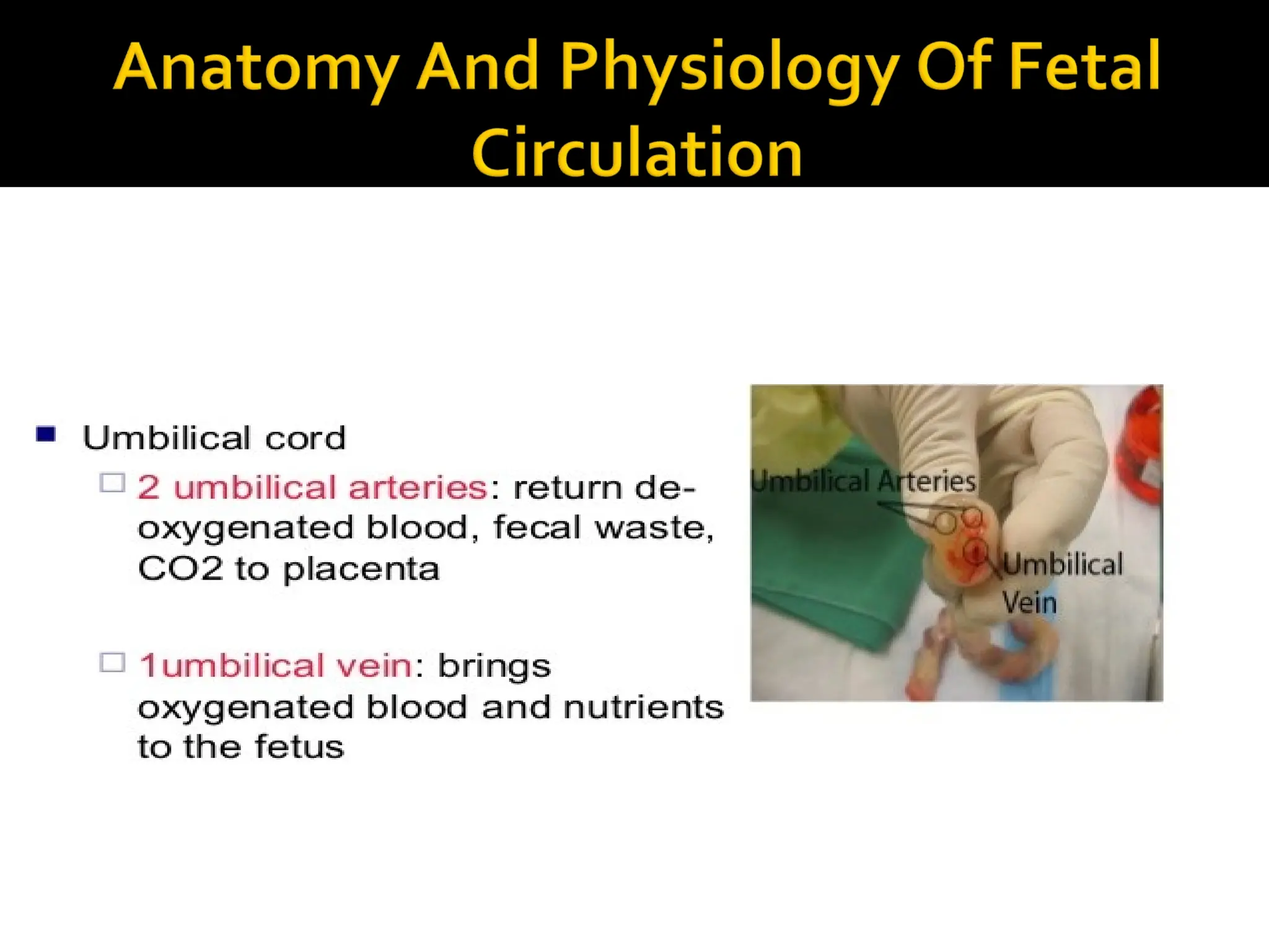
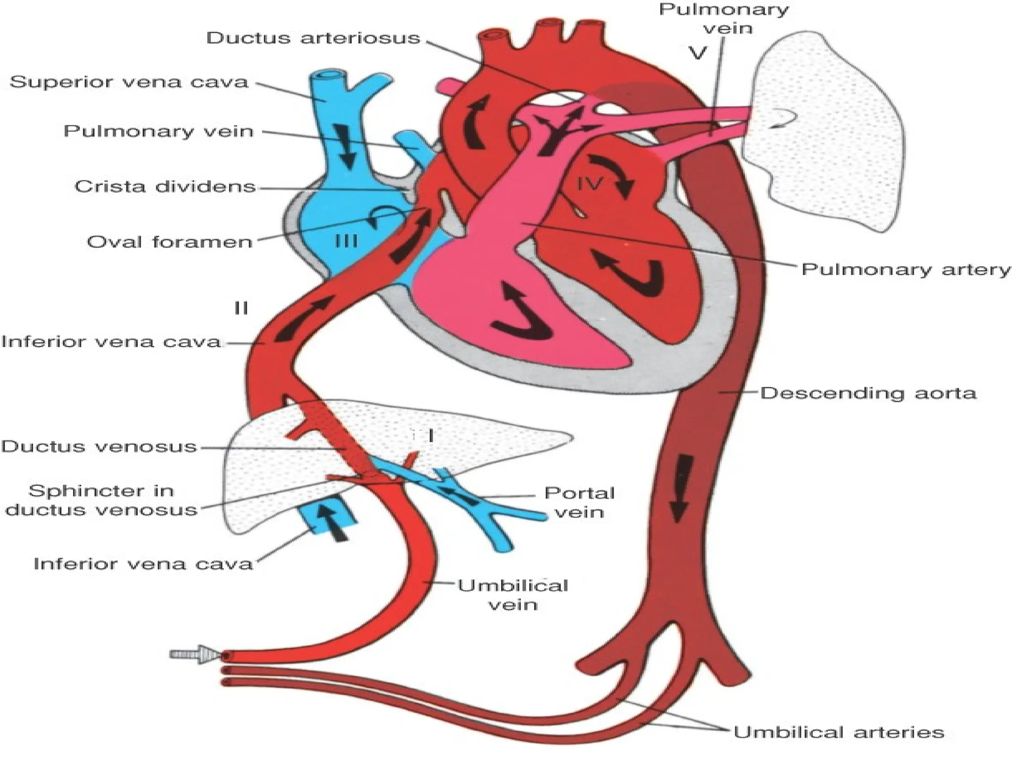
Pair ofumbilical arteries carry
deoxygenated blood & wastes to
placenta.
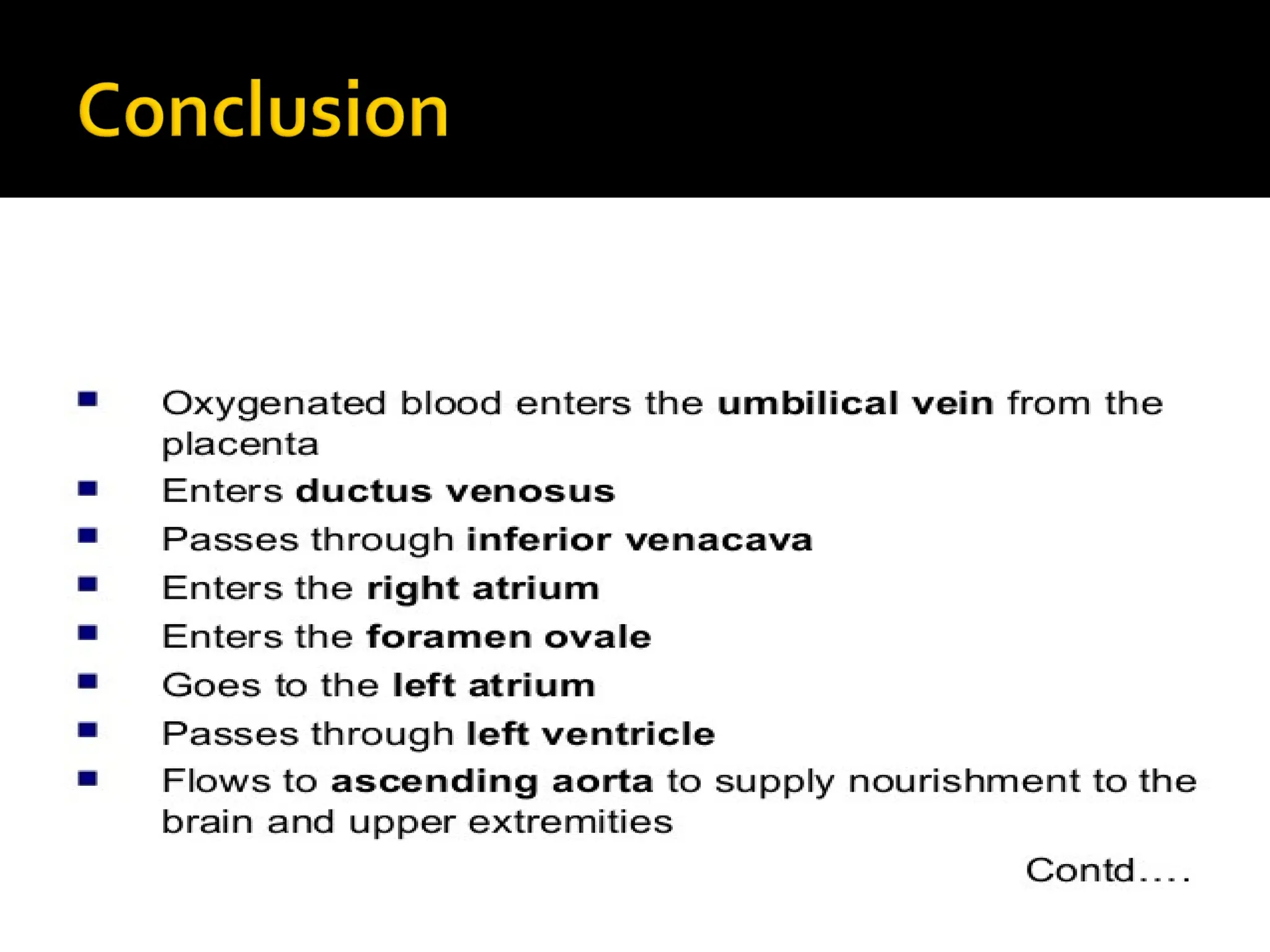
Umbilical vein carries oxygenated blood
and nutrients from the placenta.
12.
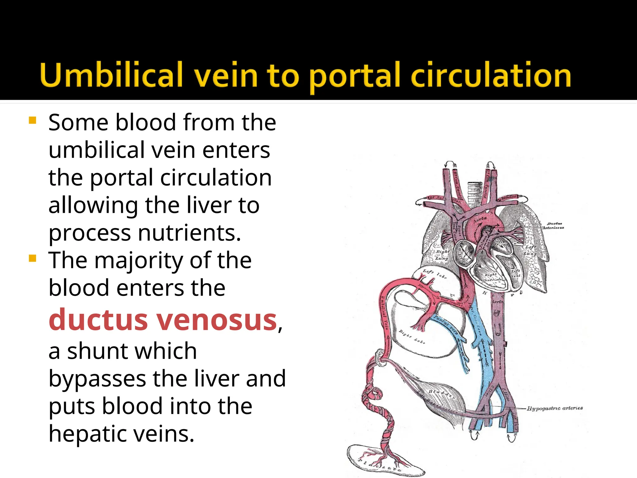
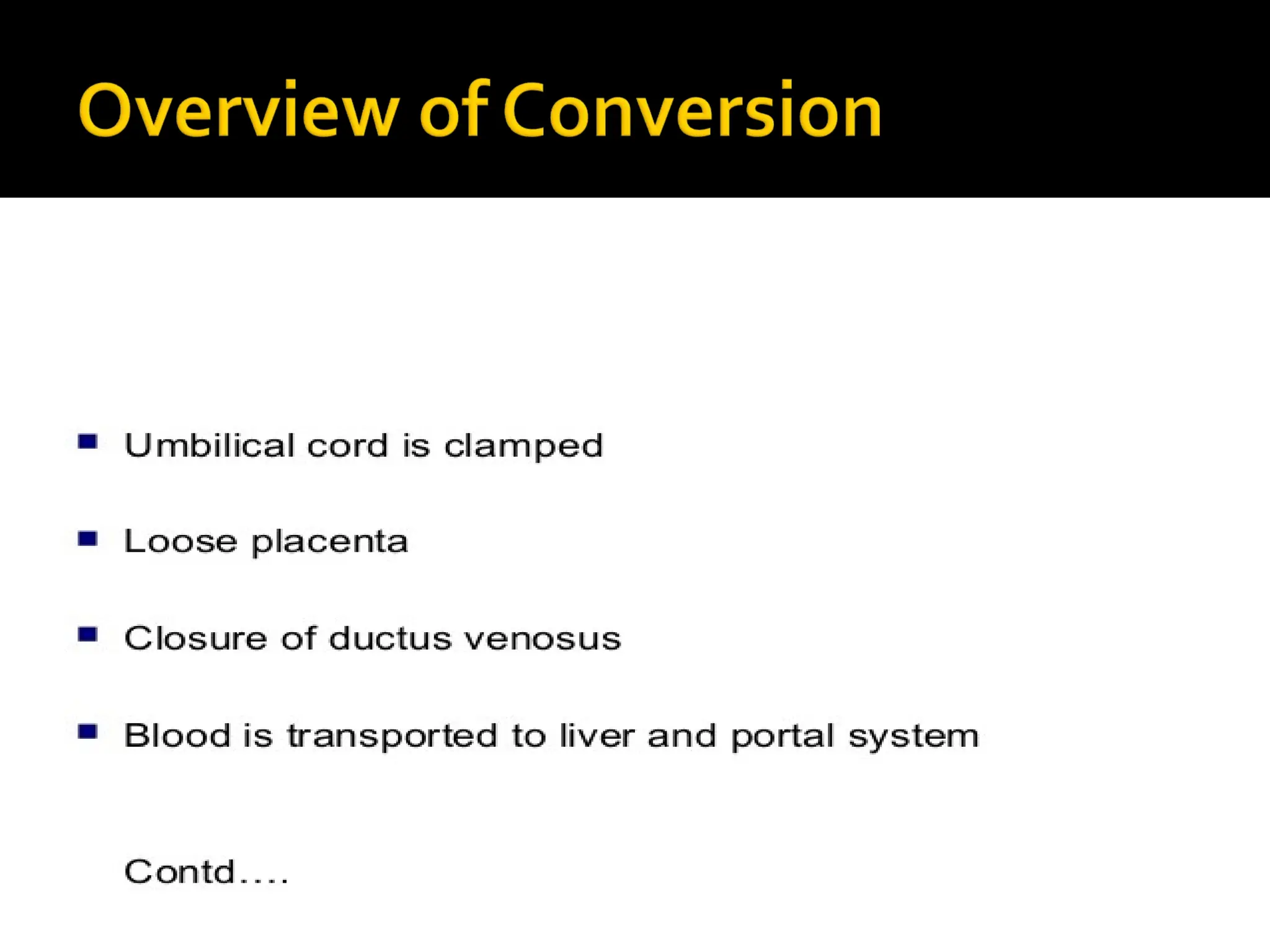
Some bloodfrom the
umbilical vein enters
the portal circulation
allowing the liver to
process nutrients.
The majority of the
blood enters the
ductus venosus,
a shunt which
bypasses the liver and
puts blood into the
hepatic veins.
13.
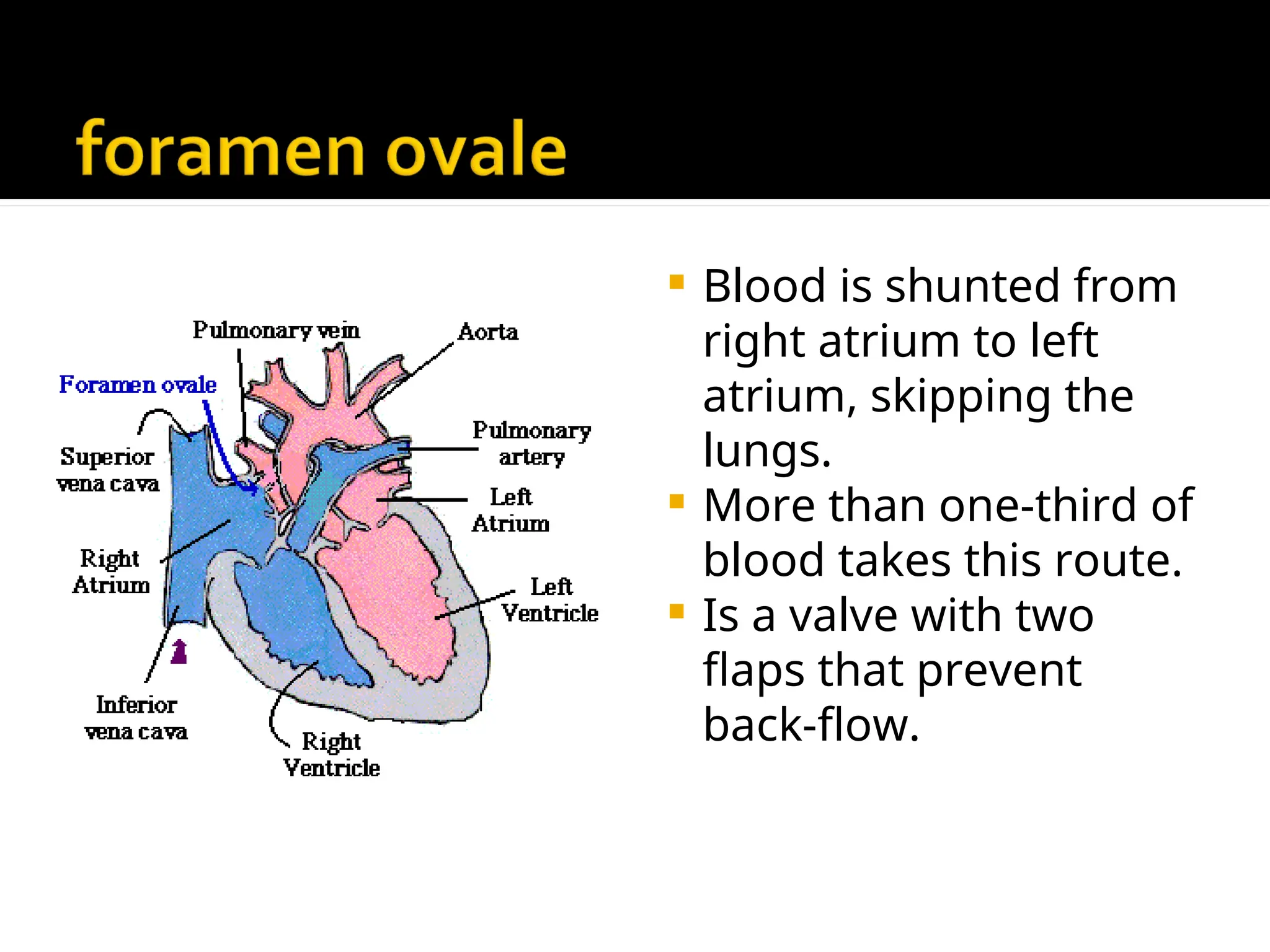
Blood isshunted from
right atrium to left
atrium, skipping the
lungs.
More than one-third of
blood takes this route.
Is a valve with two
flaps that prevent
back-flow.
14.
The bloodpumped from the right
ventricle enters the pulmonary trunk.
Most of this blood is shunted into the
aortic arch through the ductus
arteriousus.
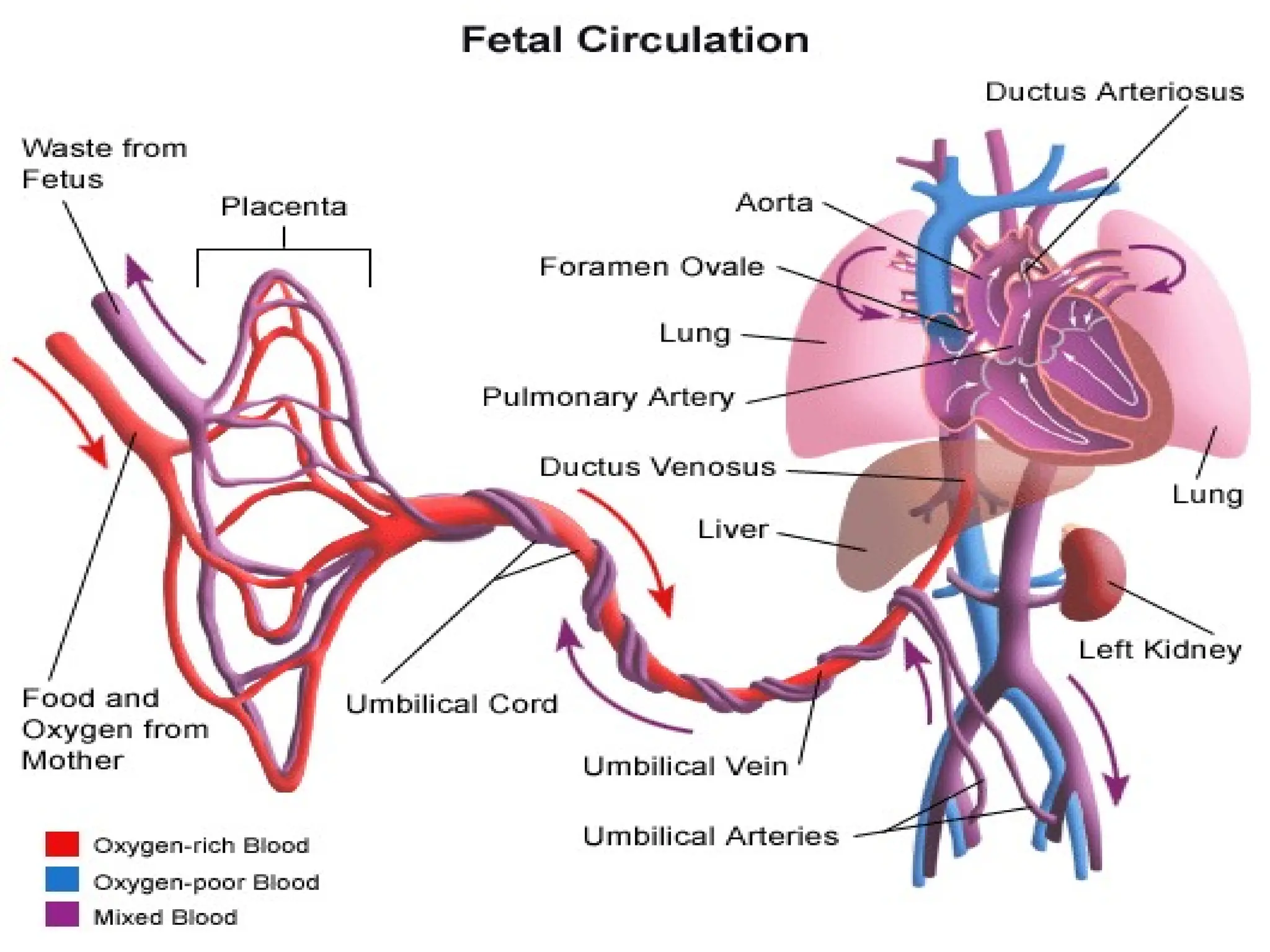
16.
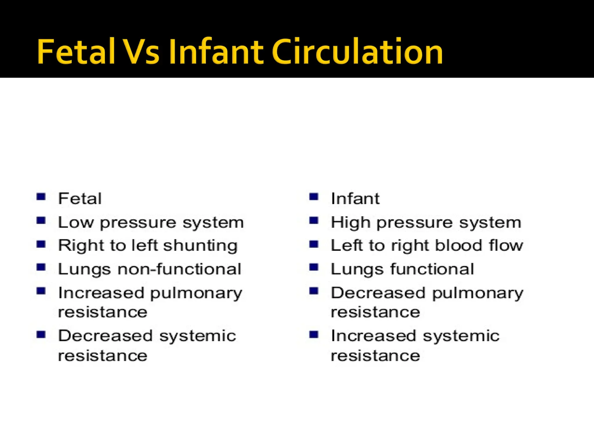
During pregnancy,the fetal circulatory
system works differently than after birth:
The fetus is connected by the umbilical cord
to the placenta, the organ that develops and
implants in the mother's uterus during
pregnancy.
Through the blood vessels in the umbilical
cord, the fetus receives all the necessary
nutrition, oxygen, and life support from the
mother through the placenta.
17.
Waste productsand carbon dioxide from
the fetus are sent back through the
umbilical cord and placenta to the mother's
circulation to be eliminated.
Blood from the mother enters the fetus
through the vein in the umbilical cord. It
goes to the liver and splits into three
branches. The blood then reaches the
inferior vena cava, a major vein connected
to the heart.
18.
Inside thefetal heart:
Blood enters the right atrium, the chamber
on the upper right side of the heart. Most of
the blood flows to the left side through a
special fetal opening between the left and
right atria, called the foramen ovale.
Blood then passes into the left ventricle
(lower chamber of the heart) and then to
the aorta, (the large artery coming from the
heart).
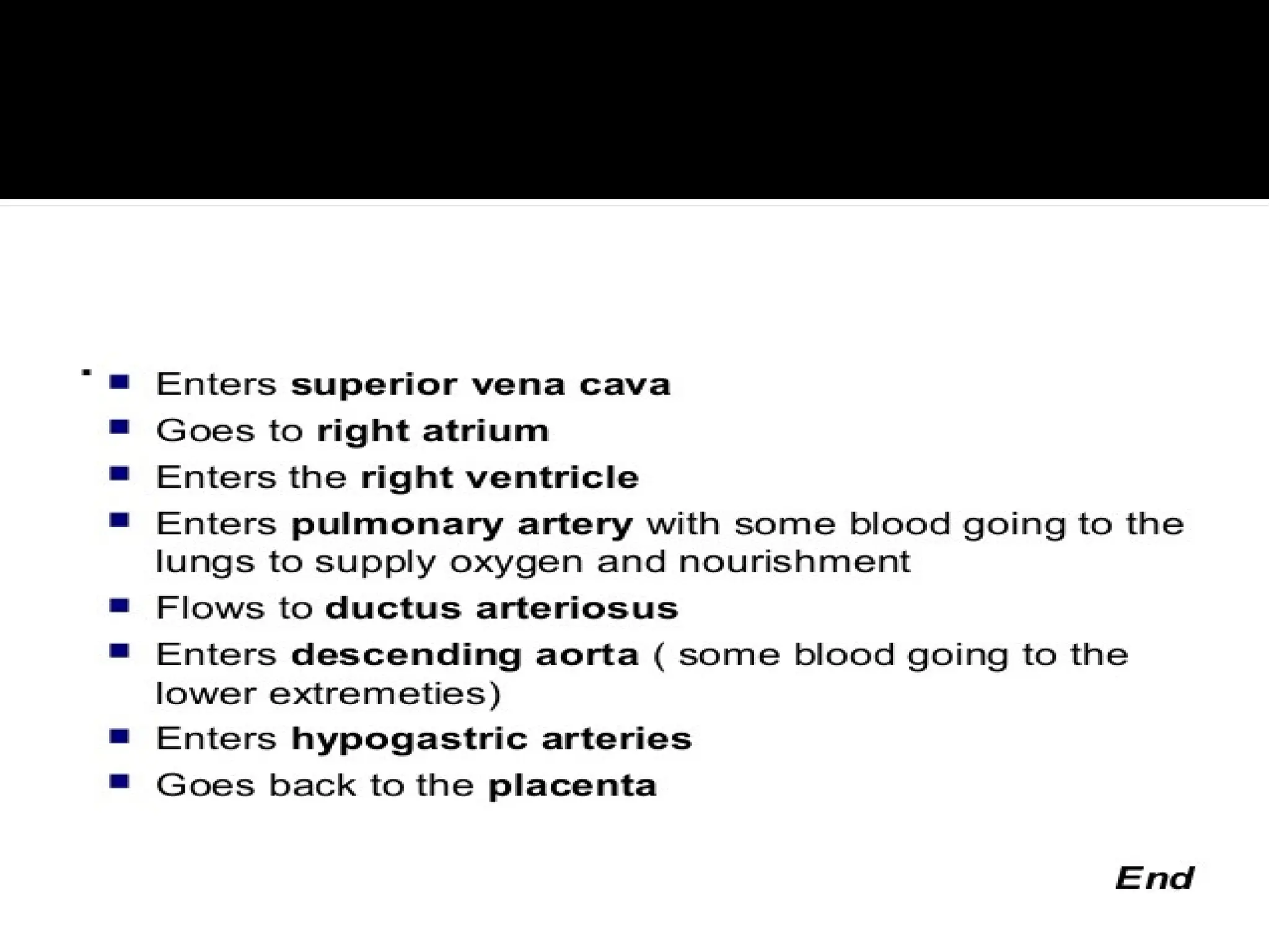
19.
From theaorta, blood is sent to the head
and upper extremities. After circulating
there, the blood returns to the right atrium
of the heart through the superior vena
cava.
About one third of the blood entering the
right atrium does not flow through the
foramen ovale, but, instead, stays in the
right side of the heart, eventually flowing
into the pulmonary artery.
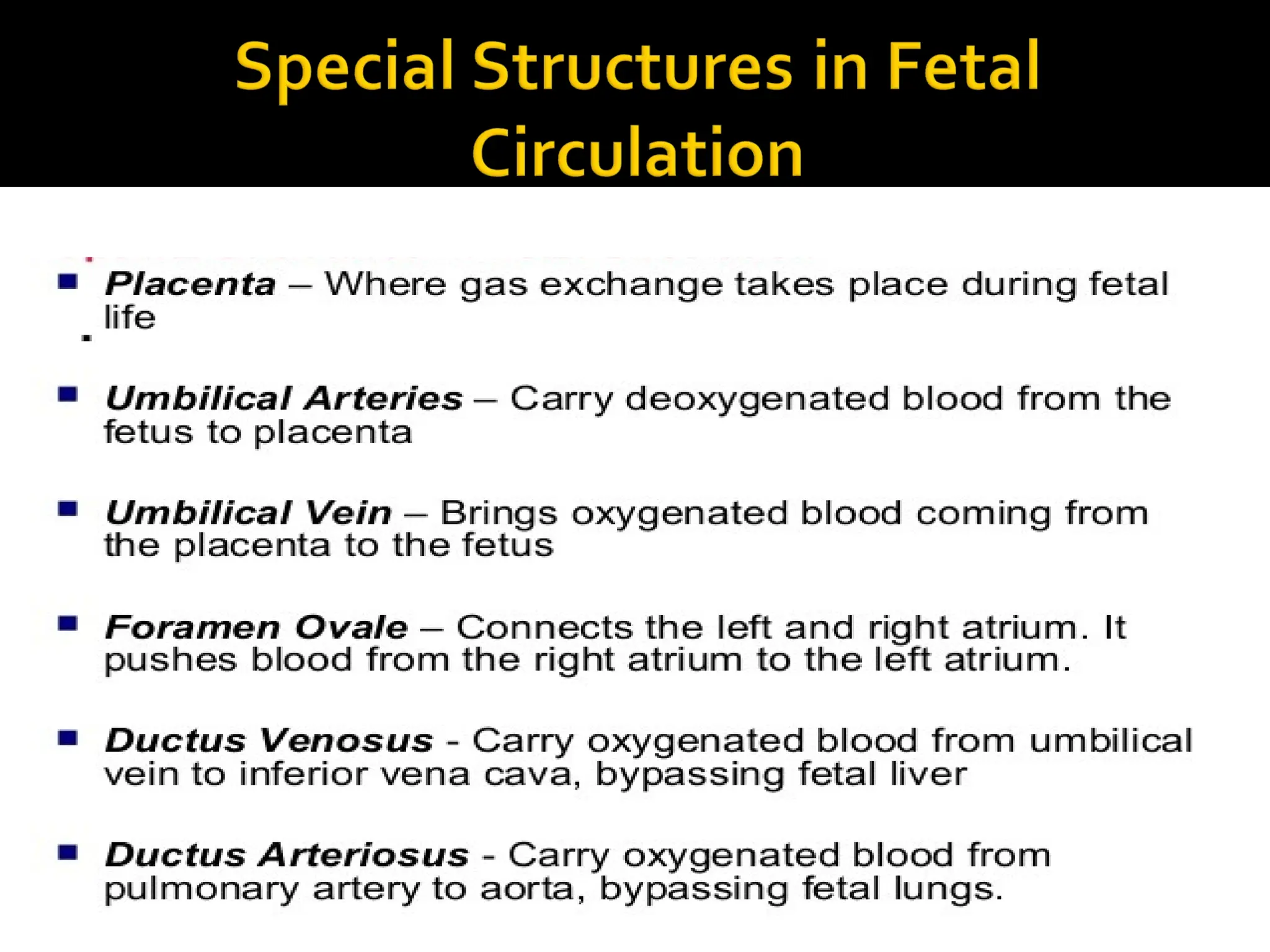
20.
Because theplacenta does the work of
exchanging oxygen (O2) and carbon dioxide
(CO2) through the mother's circulation, the fetal
lungs are not used for breathing. Instead of
blood flowing to the lungs to pick up oxygen and
then flowing to the rest of the body, the fetal
circulation shunts (bypasses) most of the blood
away from the lungs. In the fetus, blood is
shunted from the pulmonary artery to the aorta
through a connecting blood vessel called the
ductus arteriosus.
21.
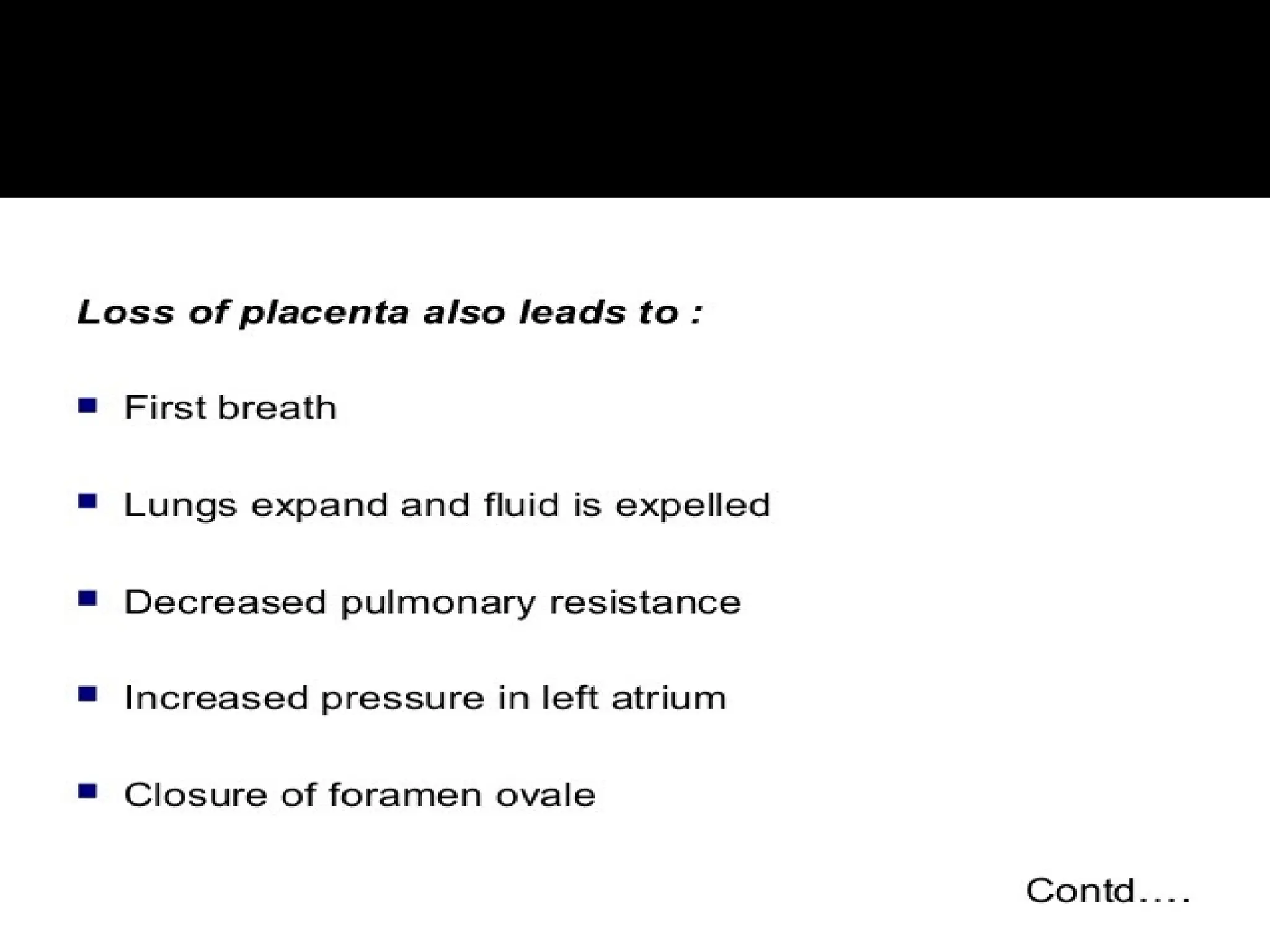
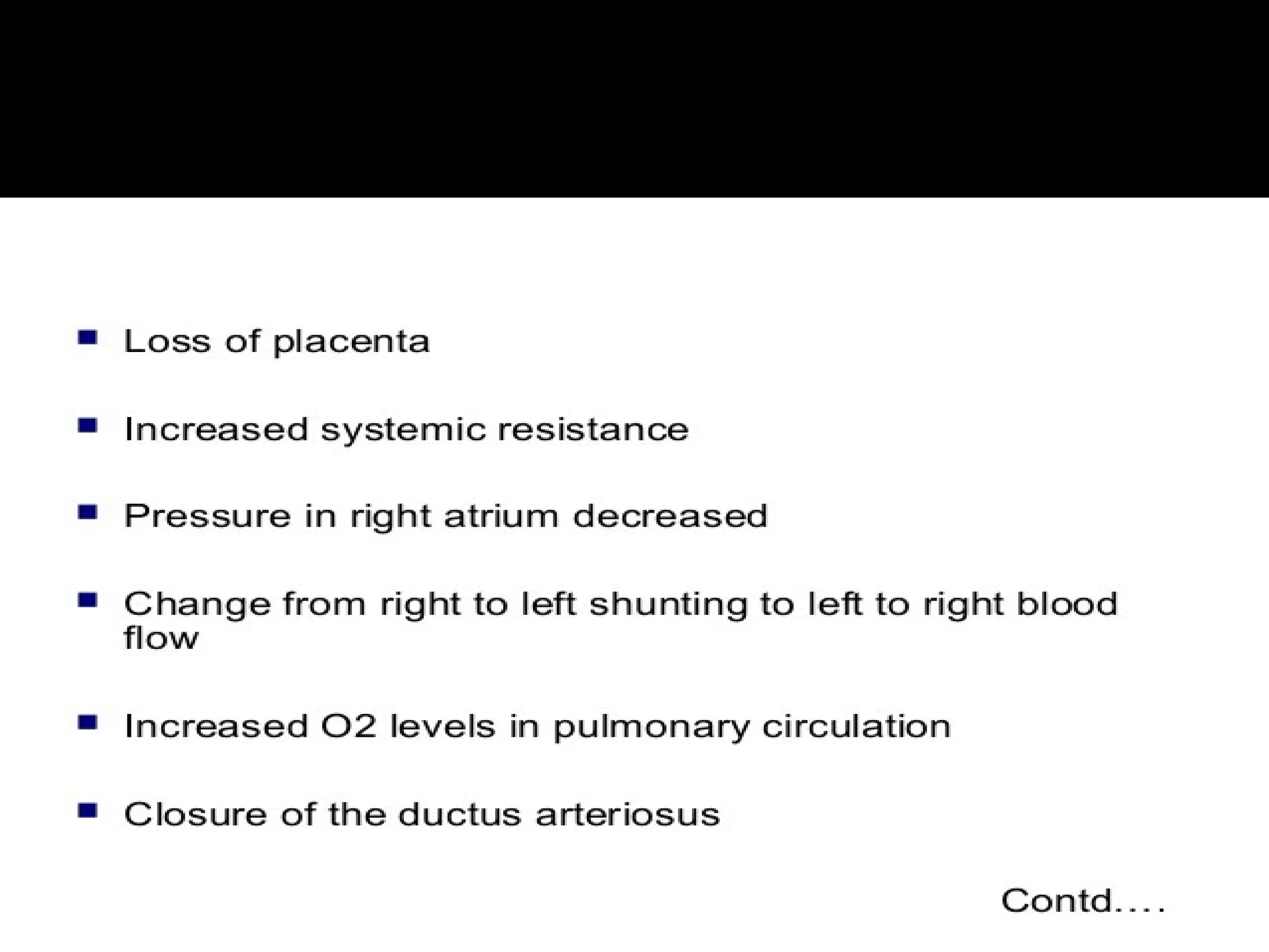
With thefirst breaths of air the baby takes at
birth, the fetal circulation changes. A larger
amount of blood is sent to the lungs to pick up
oxygen.
Because the ductus arteriosus is no longer
needed, it begins to wither and close off.
The circulation in the lungs increases and more
blood flows into the left atrium of the heart.
This increased pressure causes the foramen
ovale to close and blood circulates normally.
22.
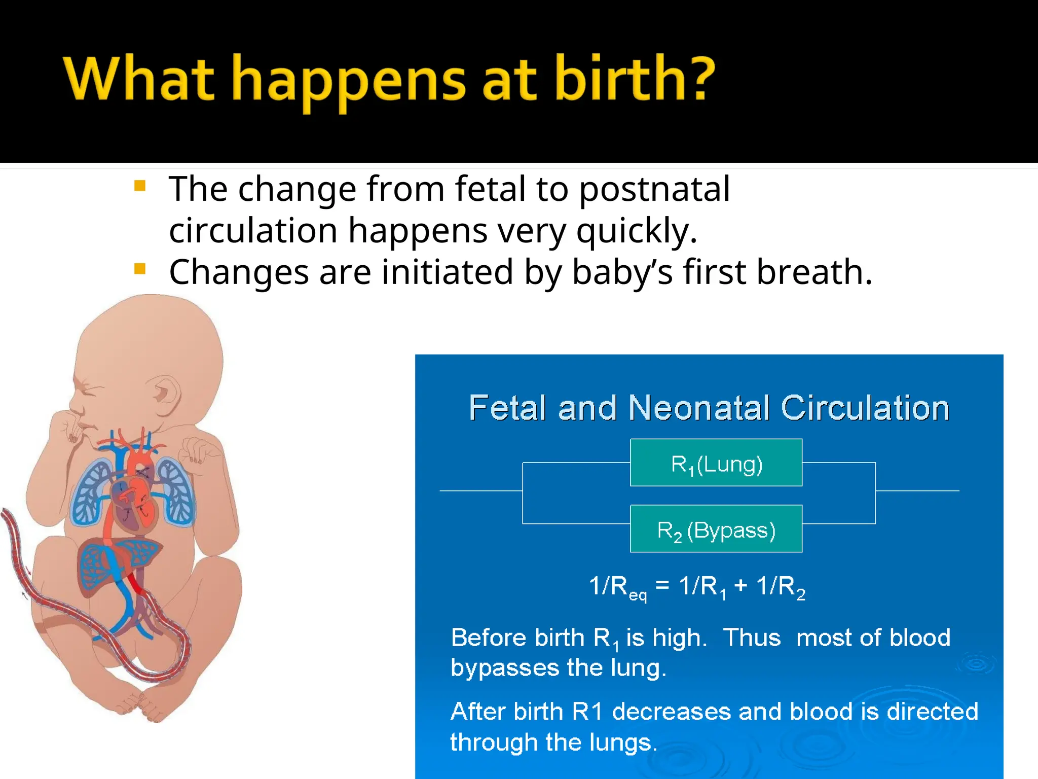
The changefrom fetal to postnatal
circulation happens very quickly.
Changes are initiated by baby’s first breath.
23.
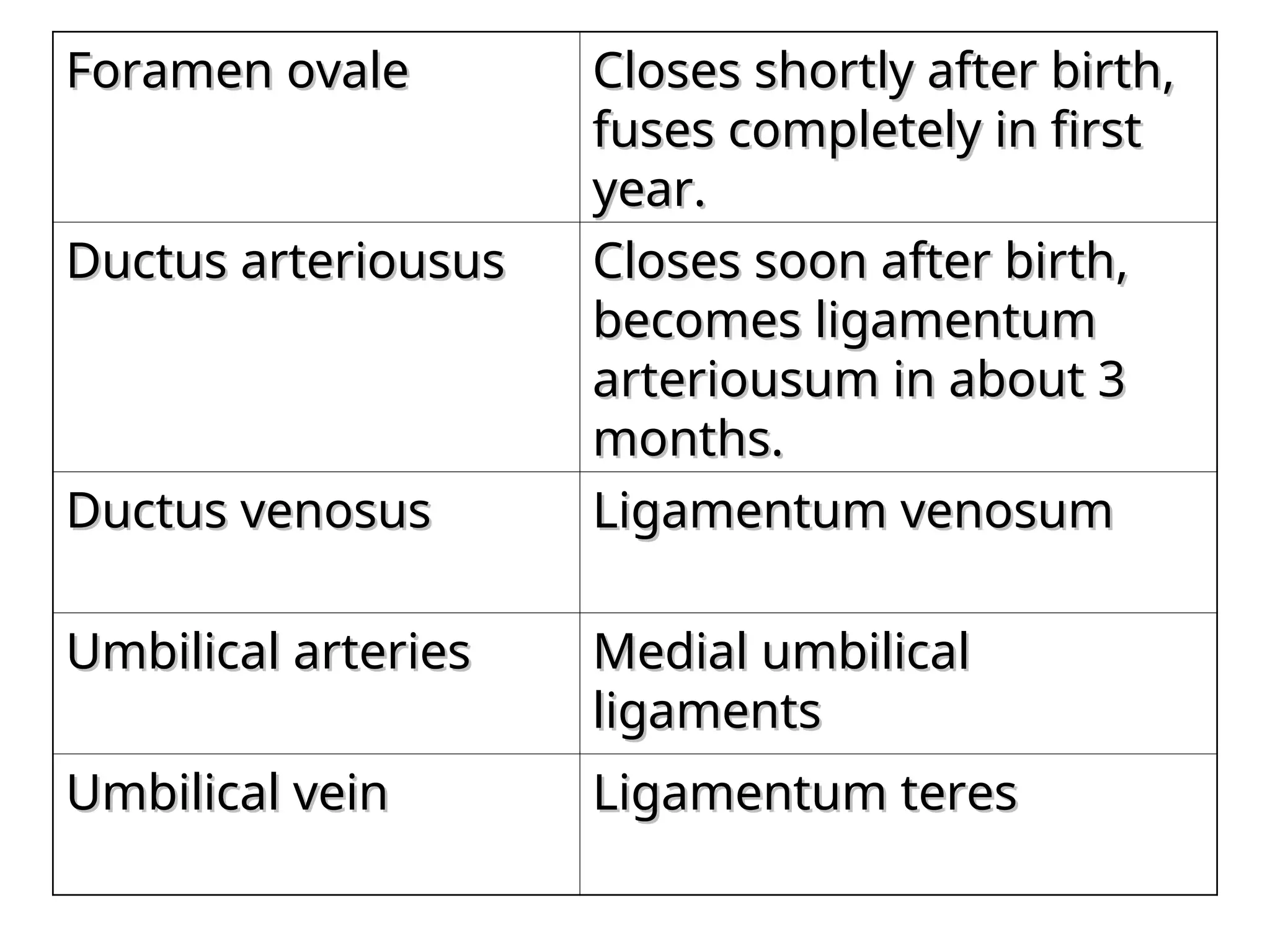
Foramen ovale
Foramen ovaleCloses shortly after birth,
Closes shortly after birth,
fuses completely in first
fuses completely in first
year.
year.
Ductus arteriousus
Ductus arteriousus Closes soon after birth,
Closes soon after birth,
becomes ligamentum
becomes ligamentum
arteriousum in about 3
arteriousum in about 3
months.
months.
Ductus venosus
Ductus venosus Ligamentum venosum
Ligamentum venosum
Umbilical arteries
Umbilical arteries Medial umbilical
Medial umbilical
ligaments
ligaments
Umbilical vein
Umbilical vein Ligamentum teres
Ligamentum teres
27.
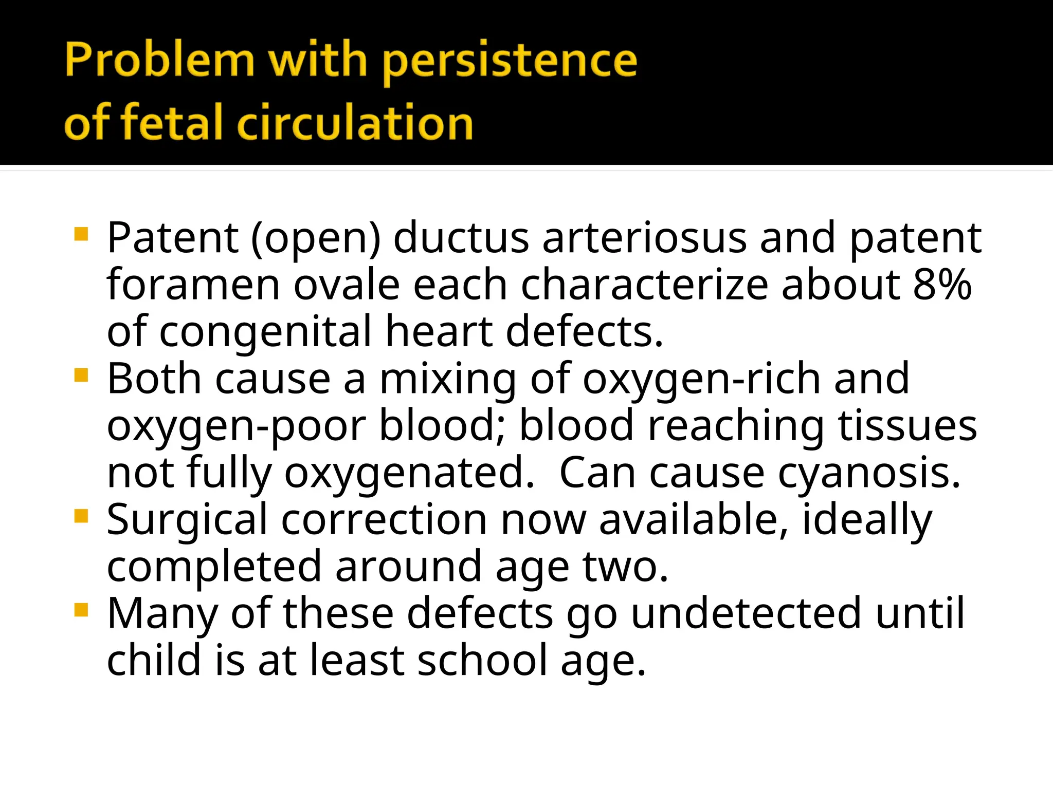
Patent (open)ductus arteriosus and patent
foramen ovale each characterize about 8%
of congenital heart defects.
Both cause a mixing of oxygen-rich and
oxygen-poor blood; blood reaching tissues
not fully oxygenated. Can cause cyanosis.
Surgical correction now available, ideally
completed around age two.
Many of these defects go undetected until
child is at least school age.