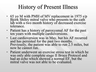
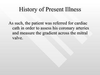
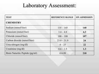
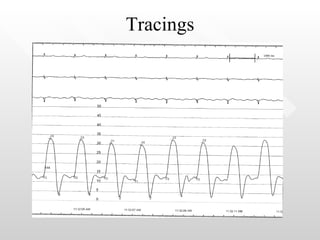
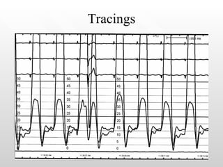
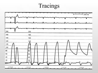
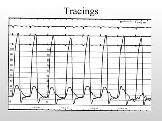
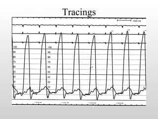
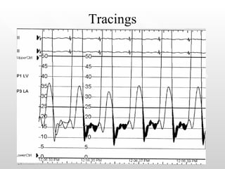
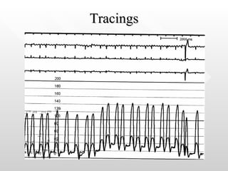
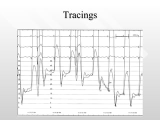
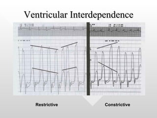
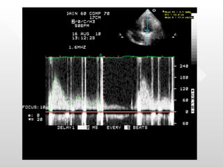
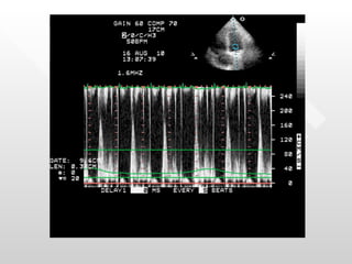
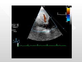
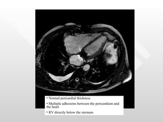
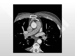
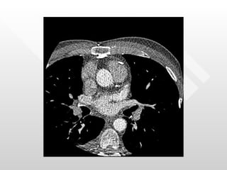
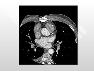
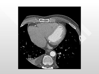
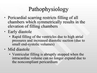
DC Fellows Forum Raj Khandwalla M.D. Georgetown/Washington Hospital Center
History of Present Illness 65 yo M with PMH of MV replacement in 1975 s/p Bjork Shiley mitral valve who presents to the cath lab with a two month history of decreased exercise tolerance.  Patient has a history of paroxysmal AF for the past ten years with multiple cardioversions.  Last cardioversion was in May, but his AF recurred and has persisted for the past two months. Previously, the patient was able to run 2.5 miles, but now he cannot run.  Patient underwent an exercise stress test in which he was only able complete stage II Bruce Protocol and had an echo which showed a normal EF, but the mitral valve was not able to be evaluated.
History of Present Illness As such, the patient was referred for cardiac cath in order to assess his coronary arteries and measure the gradient across the mitral valve.
History of Present Illness PMHX:  as above, GERD MEDS:  Propafenone, Warfarin ALL:  NKDA SOCHX:  social drinker, quit smoking 40 years ago FH:  denies
Physical Exam Gen: no apparent distress, appears younger than stated age VS: T 98.0  BP129/77  HR 60  RR 16  98% RA Neck: JVD elevated to angle of jaw Chest: Well healed sternotomy scar Heart: irreg, irreg, +valve clicks appreciated Lungs: clear to auscultation Abdomen: soft, NT, ND, + hepatojugular reflex Ext: 2+ pitting edema to the shins
Laboratory Assessment: 1.6 11.3 – 13.3 INR 33.6 22.1 – 35.1 Partial-thromboplastin time (sec) 118 150,000 – 300,000 Platelet Count (per mm 3 ) 4.5 0 – 8 Eosinophils 17.6 4 – 11 Monocytes 37.5 22 – 44 Lymphocytes 40 40 – 70 Neutrophils Differential Count (%) 4.0 4,500 – 11,000 White-cell count (per mm 3 ) 36.2 41.0 – 53.0 Hematocrit (%) 12.3 13.5 – 17.5 Hemoglobin (g/dl) HEMATOLOGY ON ADMISSION REFERENCE RANGE TEST
Laboratory Assessment: 218 0.0-99 Brain Naturitic Peptide (pg/ml) 1.3 0.6 – 1.5 Creatinine (mg/dl) 22 8 – 25 Urea nitrogen (mg/dl) 24 23.0 – 31.9 Carbon dioxide (mmol/liter) 107 100 – 108 Chloride (mmol/liter) 4.3 3.4 – 4.8 Potassium (mmol/liter) 140 135 – 145 Sodium (mmol/liter) CHEMISTRY ON ADMISSION REFERENCE RANGE TEST
 
Tracings
Tracings
Tracings
Tracings
Tracings
Tracings
What is the differential diagnosis for these tracings? How do we make the diagnosis?
Tracings
Tracings
Ventricular Interdependence Restrictive   Constrictive


Normal pericardial thickness Multiple adhesions between the pericardium and the heart RV directly below the sternum


Constrictive Pericarditis Represents the end stage of an inflammatory process  Can occur in months, but usually takes years to develop Etiologies Idiopathic Irradiation Postsurgical Infectious Neoplastic Autoimmune disorder Uremia Posttraumatic Sarcoid Methysergide therapy Implantable defibrillator patches
Pathophysiology Pericardial scarring restricts filling of all chambers which symmetrically results in the elevation of filling chambers Early diastole Rapid filling of the ventricles due to high atrial pressures and increased diastolic suction (due to small end-systolic volumes) Mid diastole Ventricular filling is abruptly stopped when the intracardiac volume can no longer expand due to the noncompliant pericardium
Hemodynamics Constrictive pericarditis RV infarct Tamponade Restrictive cardiac  disease Pulses paradoxus < 1/3 Occasional Frequent Rare RA waveforms Prominent y descent Prominent  y descent  Prominent x descent Insp.   Variable y descent Equalization of diastolic pressures Frequent Frequent Frequent Rare “ Square root” sign Frequent Frequent Absent Variable
 

Fellows Conference

  • 1.
    DC Fellows ForumRaj Khandwalla M.D. Georgetown/Washington Hospital Center
  • 2.
    History of PresentIllness 65 yo M with PMH of MV replacement in 1975 s/p Bjork Shiley mitral valve who presents to the cath lab with a two month history of decreased exercise tolerance. Patient has a history of paroxysmal AF for the past ten years with multiple cardioversions. Last cardioversion was in May, but his AF recurred and has persisted for the past two months. Previously, the patient was able to run 2.5 miles, but now he cannot run. Patient underwent an exercise stress test in which he was only able complete stage II Bruce Protocol and had an echo which showed a normal EF, but the mitral valve was not able to be evaluated.
  • 3.
    History of PresentIllness As such, the patient was referred for cardiac cath in order to assess his coronary arteries and measure the gradient across the mitral valve.
  • 4.
    History of PresentIllness PMHX: as above, GERD MEDS: Propafenone, Warfarin ALL: NKDA SOCHX: social drinker, quit smoking 40 years ago FH: denies
  • 5.
    Physical Exam Gen:no apparent distress, appears younger than stated age VS: T 98.0 BP129/77 HR 60 RR 16 98% RA Neck: JVD elevated to angle of jaw Chest: Well healed sternotomy scar Heart: irreg, irreg, +valve clicks appreciated Lungs: clear to auscultation Abdomen: soft, NT, ND, + hepatojugular reflex Ext: 2+ pitting edema to the shins
  • 6.
    Laboratory Assessment: 1.611.3 – 13.3 INR 33.6 22.1 – 35.1 Partial-thromboplastin time (sec) 118 150,000 – 300,000 Platelet Count (per mm 3 ) 4.5 0 – 8 Eosinophils 17.6 4 – 11 Monocytes 37.5 22 – 44 Lymphocytes 40 40 – 70 Neutrophils Differential Count (%) 4.0 4,500 – 11,000 White-cell count (per mm 3 ) 36.2 41.0 – 53.0 Hematocrit (%) 12.3 13.5 – 17.5 Hemoglobin (g/dl) HEMATOLOGY ON ADMISSION REFERENCE RANGE TEST
  • 7.
    Laboratory Assessment: 2180.0-99 Brain Naturitic Peptide (pg/ml) 1.3 0.6 – 1.5 Creatinine (mg/dl) 22 8 – 25 Urea nitrogen (mg/dl) 24 23.0 – 31.9 Carbon dioxide (mmol/liter) 107 100 – 108 Chloride (mmol/liter) 4.3 3.4 – 4.8 Potassium (mmol/liter) 140 135 – 145 Sodium (mmol/liter) CHEMISTRY ON ADMISSION REFERENCE RANGE TEST
  • 8.
  • 9.
  • 10.
  • 11.
  • 12.
  • 13.
  • 14.
  • 15.
    What is thedifferential diagnosis for these tracings? How do we make the diagnosis?
  • 16.
  • 17.
  • 18.
  • 19.
  • 20.
  • 21.
  • 22.
  • 23.
    Normal pericardial thicknessMultiple adhesions between the pericardium and the heart RV directly below the sternum
  • 24.
  • 25.
  • 26.
  • 27.
  • 28.
    Constrictive Pericarditis Representsthe end stage of an inflammatory process Can occur in months, but usually takes years to develop Etiologies Idiopathic Irradiation Postsurgical Infectious Neoplastic Autoimmune disorder Uremia Posttraumatic Sarcoid Methysergide therapy Implantable defibrillator patches
  • 29.
    Pathophysiology Pericardial scarringrestricts filling of all chambers which symmetrically results in the elevation of filling chambers Early diastole Rapid filling of the ventricles due to high atrial pressures and increased diastolic suction (due to small end-systolic volumes) Mid diastole Ventricular filling is abruptly stopped when the intracardiac volume can no longer expand due to the noncompliant pericardium
  • 30.
    Hemodynamics Constrictive pericarditisRV infarct Tamponade Restrictive cardiac disease Pulses paradoxus < 1/3 Occasional Frequent Rare RA waveforms Prominent y descent Prominent y descent Prominent x descent Insp.  Variable y descent Equalization of diastolic pressures Frequent Frequent Frequent Rare “ Square root” sign Frequent Frequent Absent Variable
  • 31.