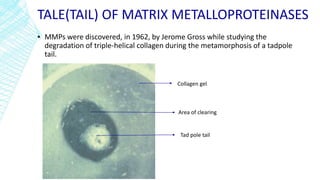
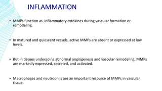
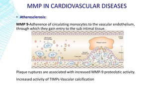
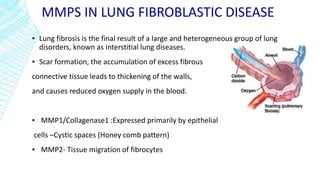
MATRIX
METALLOPROTEINASES
IN HEALTH AND
DISEASE
OVERVIEW
▪ Introduction
▪ Definition
▪ Anatomy
▪ Types
▪ Role in maintaining health
▪ Role in diseases and malignancy
INTRODUCTION
▪ Living tissues are not just accumulations of tightly packed cells.
▪ Much of a tissue's volume is made up of extracellular space .This void is filled with a
complex meshwork called the extracellular matrix.
▪ If cells were seeds then the extracellular matrix (ECM) would be the soil.
▪ In addition to providing structural support for the cells embedded within a tissue, the
extracellular matrix guides their division, growth, and development.
▪ Hence ,the extracellular matrix largely determines how a tissue looks and functions.
▪ CONTENTS OF ECM
Collagen Elastin Fibronectin Laminin Growth factors
and
Metalloproteinases
CONTENTS OF ECM
▪ Collagen-Main structural protein in the extracellular space.
Type I: skin, tendon, bone Type II: cartilage
Type III: main component of reticular fibers, commonly found alongside type I.
Type IV: basement membrane Type V: cell surfaces, hair and placenta
▪ Elastin-Highly elastic protein in connective tissue and allows many tissues in the body to
resume their shape after stretching or contracting.
CONTENTS OF ECM
▪ Fibronectin-located within the basement membrane and key role in cell
adhesion and wound healing response to injury.
▪ Laminins-are among the first proteins of the ECM to appear in embryos ,role in
cell differentiation and migration
▪ Growth Factors-The ECM has been referred to as a ‘reservoir’ for GFs. VEGF,
FGF and TGF are tied to the ECM through heparan sulphate.
▪ Matrix Metalloproteinases (MMPs)
TALE(TAIL) OF MATRIX METALLOPROTEINASES
▪ MMPs were discovered, in 1962, by Jerome Gross while studying the
degradation of triple-helical collagen during the metamorphosis of a tadpole
tail.
Tad pole tail
Area of clearing
Collagen gel
METALLOPROTEINASES
▪ The timely breakdown of extracellular matrix (ECM) is essential for embryonic
development, morphogenesis, reproduction, and tissue resorption and remodeling.
▪ Metalloproteinases /Matrixins are a large group of zinc-dependent proteases
responsible for cleaving and rebuilding connective tissue.
N-terminal propeptide domain-MMPs are
initially synthesized as inactive
zymogens with a pro-peptide domain that
must be removed before the enzyme is active
-Cysteine switch
C-Terminal Domain-Structurally similar
to proteins of the hemopexin family. The
domain has a relatively large surface
area for protein-protein interactions.
Catalytic Domain-Contains two zinc ions and
two or three calcium ions. The first Zn ion
present in the active site directly participates in
catalytic processes. Calcium ions are necessary
to stabilize the domain structure
TYPES
▪ Collagenases
▪ Gelatinases
▪ Stromelysins and
▪ Membrane-type metalloproteinases.
COLLAGENASES
▪ Collagenases are enzymes that break the peptide bonds in collagen.
▪ MMP-1, MMP-8, and MMP-13 are the enzymes with the capacity to cleave
the triple helix of fibrillar collagen.
▪ Interstitial collagenase(MMP-1) has preferential activity against type III
collagen.
▪ MMP-1 is produced and secreted predominantly by cells prevalent in the
post acute stages of wound healing (fibroblasts, endothelial cells)
COLLAGENASES
▪ Polymorphonuclear collagenase (MMP-8) has an affinity for type I collagen.
▪ MMP-8 is stored in granules within the neutrophils and once activated can be
released in seconds.
▪ MMP-13 appears to have the unique ability to cleave type I, type II, and type
III collagen.
GELATINASES
▪ Gelatinase is a proteolytic enzyme that allows a living organism to hydrolyse gelatin
into its sub-compounds .
▪ The gelatinases (MMP-2 and MMP-9) cleave collagen types IV, V, VII, and X, elastin,
basement membranes and denatured collagen.
▪ The gelatinases may also act synergistically with the collagenase family by further
degrading types I, II, and III.
▪ MMP-2 is secreted by fibroblasts, MMP-9 is produced predominantly by leukocytes
and keratinocytes.
STROMELYSINS
▪ Due to their broad base of substrate specificity, stromelysins (MMP-3, MMP-7,
MMP-10, MMP-11, and MMP-12) play a varied role in degradation of the
extracellular matrix.
▪ Degrades collagen types II, III, IV, IX, an X, proteoglycans, fibronectin, laminin,
and elastin.
MEMBRANE-TYPE MATRIX
METALLOPROTEINASES
▪ Members of this class of matrix metalloproteinases (MMP-14, MMP-15, MMP-
16, and MMP-17) have certain unique structural characteristics.
▪ They are not secreted into the extracellular matrix.
▪ Rather, they exist on cell membranes and appear to function by binding to other
matrix metalloproteinases and activating them.
ACTIVATION AND DEACTIVATION
1)Gene level by transcriptional control
2)Molecular level by requiring factors to
convert the proenzyme form to the active
form.
3)Tissue level -Through Tissue Inhibitors
of Metalloproteinases(TIMP)
TRANSCRIPTIONAL CONTROL
▪ Two extensively studied factors which influence the MMP transcription are:
IL-1β and TGF – β
▪ IL-1 β stimulates, whereas TGF- β inhibits MMP gene expression .
▪ Transcription of many MMPs is promoted by inflammatory cytokines, growth
factors, chemokines, oncogenes
MOLECULAR CONTROL
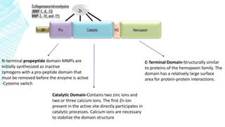
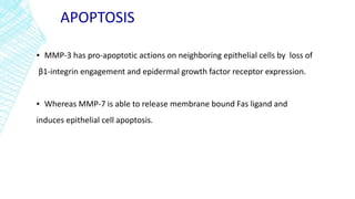
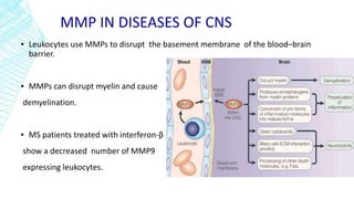
▪ MMPs are synthesized as inactive zymogens and require activation.
▪ The cysteine in the propeptide interacts with a Zn2+ ion in the enzyme’s active site.
▪ This interaction, [Zn2+-cysteine], keeps the proMMP inactive
▪ Activation can occur by physical or chemical means by disturbing the cysteine –zinc
interaction of the cysteine switch .
DEACTIVATION OF MMP
▪ Tissue inhibitors of matrix metalloproteinases (TIMP) are major endogenous
regulators of MMP
▪ The critical points in MMP inhibition by TIMPs are centred on the
disulfidebonds at Cys and Cys.
▪ TIMPs catalysis of disulfide bond formation between cysteines prevents
autocatalytic MMPs activation through the cysteine switch mechanism.
▪ In addition, TIMPs in complexes with other molecules involve in the inhibition
of already active MMP
PHYSIOLOGICAL ROLES
▪ Apoptosis
▪ Ovulation
▪ Inflammation
▪ Angiogenesis
▪ Bone remodeling
▪ Mammary gland development
▪ Embryogenic remodeling
▪ Organ morphogenesis
APOPTOSIS
▪ MMP-3 has pro-apoptotic actions on neighboring epithelial cells by loss of
β1-integrin engagement and epidermal growth factor receptor expression.
▪ Whereas MMP-7 is able to release membrane bound Fas ligand and
induces epithelial cell apoptosis.
OVULATION
▪ The preovulatory LH surge stimulates an increase in ovarian matrix
metalloproteinases (MMPs) and their associated inhibitors, the TIMPs prior to
follicular rupture .
▪ Proteolytic enzymes are locally activated that digest a small part of the dominant
follicle’s wall and extracellular matrix, releasing the oocyte .
OVULATION
Pre Ovulatory phase-IHC-MMP2Early ovulatory phase-TIMPs
INFLAMMATION
▪ MMPs function as inflammatory cytokines during vascular formation or
remodeling.
▪ In matured and quiescent vessels, active MMPs are absent or expressed at low
levels.
▪ But in tissues undergoing abnormal angiogenesis and vascular remodeling, MMPs
are markedly expressed, secreted, and activated.
▪ Macrophages and neutrophils are an important resource of MMPs in vascular
tissue.
MMP IN INFLAMMATION
▪ TNF-α and ILs MMPStimulate
• Degrade ECM to facilitate migration and
recruitment of cells.
• Cleave cell surface receptors and other non-ECM
molecules to mediate adhesion, proliferation, and
apoptosis of cells.
MMP IN WOUND HEALING
▪ Healing of a skin wound requires several processes :
Cell migration
ECM degradation and
Tissue reorganization.
MMP-1 :Keratinocyte migration
MMP-3: Wound contraction
VASCULAR REMODELING
▪ Hemodynamic forces such as shear stress and arterial pressure regulate MMP
expression and participate in vascular remodeling.
▪ Elevated transmural pressure Extracellular signal–regulated kinases (ERKs)
NF-κB
↑MMP 2 and 9
Remodeling due to hemodynamic adaptation of the vein to the arterial condition
leads to saphenous vein graft bypass failure.
BONE REMODELLING
▪ During long bone development, an initial cartilaginous template is formed and
replaced by bone in a coordinated and refined program.
▪ Chondrocyte proliferation and maturation, vascular invasion, recruitment of adult
stem cells and intense remodeling of cartilage and bone matrix.
▪ Osteoclast express –MMP 9 and 13
MAMMARY GLAND DEVELOPMENT
▪ The epithelial ductal network of the mammary gland expands greatly during
puberty in response to both local and systemic signals.
▪ This process requires degradation of the basement membrane and ECM,
restructuring of the endogenous vascular network and large-scale epithelial
morphogenesis
MAMMARY GLAND DEVELOPMENT
▪ MMP 2-Primary invasion
▪ MMP 3-Secondary branching
EMBROGENESIS
▪ Embryonic growth and tissue morphogenesis are fundamental events that require
disruption of ECM barriers to allow cell migration and matrix microenvironment
remodeling.
▪ Trophoblasts express high levels of MMP-9
▪ MMP2 and 9 enable trophoblast cells to penetrate uterine
vasculature during Implantation
▪ Development of the placenta starts with the invasion and migration of trophoblast cells
into the maternal tissue to establish connection with the maternal circulation .
MMPS IN PATHOLOGICAL CONDITIONS
▪ Diseases of CNS
▪ Cardiovascular diseases
▪ Lung fibroblastic disease
▪ Liver fibrosis
▪ Diseases of bone
▪ Tumor development and metastasis
MMP IN DISEASES OF CNS
▪ MMPs are largely absent from the normal CNS and their upregulation has been
reported in several neurological disorders and after injury.
▪ Metalloproteinases in neuroinflammation and Multiple sclerosis: MS is an
immune disorder characterized by demyelination and axonal loss.
▪ The presence of proteinases(MMP 9) in the cerebrospinal fluid (CSF) of patients
with MS has been known for over 20 years.
▪ MS patients show increased MMP9 messenger RNA in leukocytes and elevated
MMP9 levels in serum.
MMP IN DISEASES OF CNS
▪ Leukocytes use MMPs to disrupt the basement membrane of the blood–brain
barrier.
▪ MMPs can disrupt myelin and cause
demyelination.
▪ MS patients treated with interferon-β
show a decreased number of MMP9
expressing leukocytes.
MMP IN DISEASES OF CNS
▪ A role for metalloproteinases in stroke is indicated by the finding that MMP2 and -
9 are rapidly upregulated after focal cerebral ischaemia .
▪ The elevated MMP expression might contribute to the tissue destruction in stroke.
▪ Viral infections of the CNS have been increasingly associated with the production
of MMPs.
▪ Elevated expression of MMP9 has been detected in the CSF of HIV-infected
patients.
MMP IN CARDIOVASCULAR DISEASES
▪ Atherosclerosis:
MMP 9-Adherence of circulating monocytes to the vascular endothelium,
through which they gain entry to the sub intimal tissue.
Plaque ruptures are associated with increased MMP-9 proteolytic activity.
Increased activity of TIMPs-Vascular calcification
MMP IN CARDIOVASCULAR DISEASES
Aneurysm :Consists of destruction and functional loss of elastin in the aortic
medium.
▪ MMP plays a significant role in weakening the ECM components (elastin,
collagen, fibronectin and proteoglycans), thus harming the aortic wall.
Varicose veins: Interstitial collagen III is important for the stretch resistance of the
connective tissue of veins.
▪ MMP-3 may contribute to vein wall weakness and varicose vein formation via
excessive degradation of collagen III.
MMPS IN ACUTE MYOCARDIAL INFARCTION
▪ Myocardial collagens maintain the structural integrity of adjoining myocytes and
cardiac pump function.
▪ Myocardialinfarction (MI) leads to complex architectural alterations involving both the
infarcted and noninfarcted myocardium.
▪ Patients exhibiting extensive infarct expansion after MI are more likely to experience
complications:
Congestive heart failure
Aneurysm formation and
Myocardial rupture.
MMPS IN ACUTE MYOCARDIAL INFARCTION
▪ One of the determinants of left ventricular remodeling is damage to and loss of
the myocardial extracellular matrix (ECM) during the healing process after MI.
▪ Reduction in LV dilatation after MI that can be achieved with MMP inhibitor
treatment.
MMPS IN LUNG FIBROBLASTIC DISEASE
▪ Lung fibrosis is the final result of a large and heterogeneous group of lung
disorders, known as interstitial lung diseases.
▪ Scar formation, the accumulation of excess fibrous
connective tissue leads to thickening of the walls,
and causes reduced oxygen supply in the blood.
▪ MMP1/Collagenase1 :Expressed primarily by epithelial
cells –Cystic spaces (Honey comb pattern)
▪ MMP2- Tissue migration of fibrocytes
MMPS IN EMPHYSEMA
▪ The lung is a sophisticated matrix scaffold on which lung epithelium and
endothelium reside.
▪ When the wall of an alveolus is destroyed, the air sacs coalesce to form larger
ones.
▪ These enlarged airspaces empty more slowly, resulting in airflow obstruction,
the hallmark of COPD.
MMPS IN EMPHYSEMA
▪ Of all the MMPs, the evidence supporting a role for macrophage elastase
(MMP12) in the pathogenesis of emphysema is the strongest.
▪ Mmp12 expression is greatly increased in response to cigarette smoke exposure,
due to a number of overlapping mechanisms.
▪ Activation of the plasmin/thrombin–proteinase activated receptor (PAR-1)
cascade leads to the expression of Mmp12.
▪ Inhibited by A1AT and TGF-β .
MMPS IN LIVER FIBROSIS
▪ Liver fibrosis represents chronic wound repair following diverse insults.
▪ MMPs as bad role: In liver fibrogenesis hepatic stellate cells undergo dramatic
morphological and functional changes –star shaped HSC are converted to
myofibroblastic cells .
▪ Proteolytic degradation of the normal ECM helping inmobilization, migration and
proliferation of cells during repair and regeneneration.
▪ MMPs as good role: The MMP-mediated resolution of tissue fibrosis may act
through ECM degradation as well as by induction of HSC apoptosis.
MMPS IN BONE DISEASES
▪ The irreversible destruction of the cartilage, tendon, and bone that comprise synovial
joints is the hallmark of both rheumatoid arthritis (RA) and osteoarthritis (OA).
▪ Cartilage is made up of proteoglycans and type II collagen
▪ Tendon and bone are composed primarily of type I collagen
▪ RA is an autoimmune disease affecting numerous joints throughout the body
▪ OA develops in a small number of joints, usually resulting from chronic overuse or
injury.
MMPS IN BONE DISEASES
▪ In both RA and OA:
Inflammatory cytokines:IL-1 βand TNF-α MMPs that can degrade all
components of the extracellular matrix.
▪ MMP-1 synovial cells that line the joints
▪ MMP-13 is a product of the chondrocytes that reside in the cartilage
In addition to collagen, MMP-13 also degrades the proteoglycan molecule,
aggrecan, giving it a dual role in matrix destruction.
stimulate
MMPS IN TUMOR GROWTH
MMPs can regulate the growth of tumour cells by the following mechanisms :
▪ Release of cell membrane bound precursors of some growth factors
▪ Modulating the bioavailability of growth factors that are sequestrated by ECM
proteins.
▪ Indirectly regulating proliferative signals through integrins
▪ Because of the shedding of the E cadherin, β catenin translocates to the nucleus
and leads to proliferation
MMPs can inhibit growth by
▪ Activation of TGF-β
▪ Generation of proapoptotic molecules. e.g. Fas ligand or TNF α
REGULATION OF INVASION AND METASTASIS
▪ Tumour invasion is a multistep process in which cell motility is coupled
with proteolysis and involves interaction of cells with ECM .
▪ Cadherin are cell adhesion molecules that mediate cell-cell adhesion in
normal mucosal cells and maintain epithelial integrity and its deregulation is
associated with cancer progression .
REGULATION OF INVASION AND METASTASIS
▪ Decreased expression of E-cadherin causes loss of cell adhesion and
contributes to cell dissociation, increased motility and invasion .
▪ E-cadherin is cleaved by MMP3 and 7
Detachment of cells by loss of intercellular junction
Epithelial to mesenchymal transition(EMT)
Migration
Invasion
SUMMARY
▪ MMPs
▪ Physiological roles Pathological roles
Collagenases
Gelatinases
Stromelysins and
Membrane-type metalloproteinases.
Apoptosis
Ovulation
Inflammation
Angiogenesis
Bone remodeling
Mammary gland development
Embryogenic remodeling
Organ morphogenesis
Diseases of CNS
Cardiovascular diseases
Lung fibroblastic disease
Liver fibrosis
Diseases of bone
Tumor development and metastasis
REFERENCES
▪ Kumar, Vinay., Robbins and Cotran Pathologic Basis of Disease.9th edition. Reed
Elsevier India Private Limited, New Delhi.2015
▪ Brinckerhoff CE, Matrisian LM. Matrix metalloproteinases: a tail of a frog that
became a prince. Nature Rev Mol Cell Biol 2002;3:207–214.
▪ Birkedal-Hansen H, et al. Matrix metalloproteinases: a review. Crit Rev Oral Biol
Med 1993;4:197–250.
▪ Stickens D, et al. Altered endochondral bone development in matrix
metalloproteinase 13-deficient mice. Development 2004;131:5883–5895.
▪ Ducharme A, et al. Targeted deletion of matrix metalloproteinase-9 attenuates
left ventricular enlargement and collagen accumulation after experimental
myocardial infarction. J Clin Invest 2000;106:55–62
▪ Yu Q, Stamenkovic I. Cell surface-localized matrix metalloproteinase-9
proteolytically activates TGF-β and promotes tumor invasion and angiogenesis.
Genes Dev 2000;14:163–176.
THANK YOU

Matrix metalloproteinases

  • 1.
  • 2.
    OVERVIEW ▪ Introduction ▪ Definition ▪Anatomy ▪ Types ▪ Role in maintaining health ▪ Role in diseases and malignancy
  • 3.
    INTRODUCTION ▪ Living tissuesare not just accumulations of tightly packed cells. ▪ Much of a tissue's volume is made up of extracellular space .This void is filled with a complex meshwork called the extracellular matrix. ▪ If cells were seeds then the extracellular matrix (ECM) would be the soil. ▪ In addition to providing structural support for the cells embedded within a tissue, the extracellular matrix guides their division, growth, and development. ▪ Hence ,the extracellular matrix largely determines how a tissue looks and functions.
  • 4.
    ▪ CONTENTS OFECM Collagen Elastin Fibronectin Laminin Growth factors and Metalloproteinases
  • 5.
    CONTENTS OF ECM ▪Collagen-Main structural protein in the extracellular space. Type I: skin, tendon, bone Type II: cartilage Type III: main component of reticular fibers, commonly found alongside type I. Type IV: basement membrane Type V: cell surfaces, hair and placenta ▪ Elastin-Highly elastic protein in connective tissue and allows many tissues in the body to resume their shape after stretching or contracting.
  • 6.
    CONTENTS OF ECM ▪Fibronectin-located within the basement membrane and key role in cell adhesion and wound healing response to injury. ▪ Laminins-are among the first proteins of the ECM to appear in embryos ,role in cell differentiation and migration ▪ Growth Factors-The ECM has been referred to as a ‘reservoir’ for GFs. VEGF, FGF and TGF are tied to the ECM through heparan sulphate. ▪ Matrix Metalloproteinases (MMPs)
  • 7.
    TALE(TAIL) OF MATRIXMETALLOPROTEINASES ▪ MMPs were discovered, in 1962, by Jerome Gross while studying the degradation of triple-helical collagen during the metamorphosis of a tadpole tail. Tad pole tail Area of clearing Collagen gel
  • 8.
    METALLOPROTEINASES ▪ The timelybreakdown of extracellular matrix (ECM) is essential for embryonic development, morphogenesis, reproduction, and tissue resorption and remodeling. ▪ Metalloproteinases /Matrixins are a large group of zinc-dependent proteases responsible for cleaving and rebuilding connective tissue.
  • 9.
    N-terminal propeptide domain-MMPsare initially synthesized as inactive zymogens with a pro-peptide domain that must be removed before the enzyme is active -Cysteine switch C-Terminal Domain-Structurally similar to proteins of the hemopexin family. The domain has a relatively large surface area for protein-protein interactions. Catalytic Domain-Contains two zinc ions and two or three calcium ions. The first Zn ion present in the active site directly participates in catalytic processes. Calcium ions are necessary to stabilize the domain structure
  • 10.
    TYPES ▪ Collagenases ▪ Gelatinases ▪Stromelysins and ▪ Membrane-type metalloproteinases.
  • 11.
    COLLAGENASES ▪ Collagenases areenzymes that break the peptide bonds in collagen. ▪ MMP-1, MMP-8, and MMP-13 are the enzymes with the capacity to cleave the triple helix of fibrillar collagen. ▪ Interstitial collagenase(MMP-1) has preferential activity against type III collagen. ▪ MMP-1 is produced and secreted predominantly by cells prevalent in the post acute stages of wound healing (fibroblasts, endothelial cells)
  • 12.
    COLLAGENASES ▪ Polymorphonuclear collagenase(MMP-8) has an affinity for type I collagen. ▪ MMP-8 is stored in granules within the neutrophils and once activated can be released in seconds. ▪ MMP-13 appears to have the unique ability to cleave type I, type II, and type III collagen.
  • 13.
    GELATINASES ▪ Gelatinase isa proteolytic enzyme that allows a living organism to hydrolyse gelatin into its sub-compounds . ▪ The gelatinases (MMP-2 and MMP-9) cleave collagen types IV, V, VII, and X, elastin, basement membranes and denatured collagen. ▪ The gelatinases may also act synergistically with the collagenase family by further degrading types I, II, and III. ▪ MMP-2 is secreted by fibroblasts, MMP-9 is produced predominantly by leukocytes and keratinocytes.
  • 14.
    STROMELYSINS ▪ Due totheir broad base of substrate specificity, stromelysins (MMP-3, MMP-7, MMP-10, MMP-11, and MMP-12) play a varied role in degradation of the extracellular matrix. ▪ Degrades collagen types II, III, IV, IX, an X, proteoglycans, fibronectin, laminin, and elastin.
  • 15.
    MEMBRANE-TYPE MATRIX METALLOPROTEINASES ▪ Membersof this class of matrix metalloproteinases (MMP-14, MMP-15, MMP- 16, and MMP-17) have certain unique structural characteristics. ▪ They are not secreted into the extracellular matrix. ▪ Rather, they exist on cell membranes and appear to function by binding to other matrix metalloproteinases and activating them.
  • 16.
    ACTIVATION AND DEACTIVATION 1)Genelevel by transcriptional control 2)Molecular level by requiring factors to convert the proenzyme form to the active form. 3)Tissue level -Through Tissue Inhibitors of Metalloproteinases(TIMP)
  • 17.
    TRANSCRIPTIONAL CONTROL ▪ Twoextensively studied factors which influence the MMP transcription are: IL-1β and TGF – β ▪ IL-1 β stimulates, whereas TGF- β inhibits MMP gene expression . ▪ Transcription of many MMPs is promoted by inflammatory cytokines, growth factors, chemokines, oncogenes
  • 18.
    MOLECULAR CONTROL ▪ MMPsare synthesized as inactive zymogens and require activation. ▪ The cysteine in the propeptide interacts with a Zn2+ ion in the enzyme’s active site. ▪ This interaction, [Zn2+-cysteine], keeps the proMMP inactive ▪ Activation can occur by physical or chemical means by disturbing the cysteine –zinc interaction of the cysteine switch .
  • 19.
    DEACTIVATION OF MMP ▪Tissue inhibitors of matrix metalloproteinases (TIMP) are major endogenous regulators of MMP ▪ The critical points in MMP inhibition by TIMPs are centred on the disulfidebonds at Cys and Cys. ▪ TIMPs catalysis of disulfide bond formation between cysteines prevents autocatalytic MMPs activation through the cysteine switch mechanism. ▪ In addition, TIMPs in complexes with other molecules involve in the inhibition of already active MMP
  • 20.
    PHYSIOLOGICAL ROLES ▪ Apoptosis ▪Ovulation ▪ Inflammation ▪ Angiogenesis ▪ Bone remodeling ▪ Mammary gland development ▪ Embryogenic remodeling ▪ Organ morphogenesis
  • 21.
    APOPTOSIS ▪ MMP-3 haspro-apoptotic actions on neighboring epithelial cells by loss of β1-integrin engagement and epidermal growth factor receptor expression. ▪ Whereas MMP-7 is able to release membrane bound Fas ligand and induces epithelial cell apoptosis.
  • 22.
    OVULATION ▪ The preovulatoryLH surge stimulates an increase in ovarian matrix metalloproteinases (MMPs) and their associated inhibitors, the TIMPs prior to follicular rupture . ▪ Proteolytic enzymes are locally activated that digest a small part of the dominant follicle’s wall and extracellular matrix, releasing the oocyte .
  • 23.
  • 24.
    INFLAMMATION ▪ MMPs functionas inflammatory cytokines during vascular formation or remodeling. ▪ In matured and quiescent vessels, active MMPs are absent or expressed at low levels. ▪ But in tissues undergoing abnormal angiogenesis and vascular remodeling, MMPs are markedly expressed, secreted, and activated. ▪ Macrophages and neutrophils are an important resource of MMPs in vascular tissue.
  • 25.
    MMP IN INFLAMMATION ▪TNF-α and ILs MMPStimulate • Degrade ECM to facilitate migration and recruitment of cells. • Cleave cell surface receptors and other non-ECM molecules to mediate adhesion, proliferation, and apoptosis of cells.
  • 26.
    MMP IN WOUNDHEALING ▪ Healing of a skin wound requires several processes : Cell migration ECM degradation and Tissue reorganization. MMP-1 :Keratinocyte migration MMP-3: Wound contraction
  • 27.
    VASCULAR REMODELING ▪ Hemodynamicforces such as shear stress and arterial pressure regulate MMP expression and participate in vascular remodeling. ▪ Elevated transmural pressure Extracellular signal–regulated kinases (ERKs) NF-κB ↑MMP 2 and 9 Remodeling due to hemodynamic adaptation of the vein to the arterial condition leads to saphenous vein graft bypass failure.
  • 28.
    BONE REMODELLING ▪ Duringlong bone development, an initial cartilaginous template is formed and replaced by bone in a coordinated and refined program. ▪ Chondrocyte proliferation and maturation, vascular invasion, recruitment of adult stem cells and intense remodeling of cartilage and bone matrix. ▪ Osteoclast express –MMP 9 and 13
  • 29.
    MAMMARY GLAND DEVELOPMENT ▪The epithelial ductal network of the mammary gland expands greatly during puberty in response to both local and systemic signals. ▪ This process requires degradation of the basement membrane and ECM, restructuring of the endogenous vascular network and large-scale epithelial morphogenesis
  • 30.
    MAMMARY GLAND DEVELOPMENT ▪MMP 2-Primary invasion ▪ MMP 3-Secondary branching
  • 31.
    EMBROGENESIS ▪ Embryonic growthand tissue morphogenesis are fundamental events that require disruption of ECM barriers to allow cell migration and matrix microenvironment remodeling. ▪ Trophoblasts express high levels of MMP-9 ▪ MMP2 and 9 enable trophoblast cells to penetrate uterine vasculature during Implantation ▪ Development of the placenta starts with the invasion and migration of trophoblast cells into the maternal tissue to establish connection with the maternal circulation .
  • 32.
    MMPS IN PATHOLOGICALCONDITIONS ▪ Diseases of CNS ▪ Cardiovascular diseases ▪ Lung fibroblastic disease ▪ Liver fibrosis ▪ Diseases of bone ▪ Tumor development and metastasis
  • 33.
    MMP IN DISEASESOF CNS ▪ MMPs are largely absent from the normal CNS and their upregulation has been reported in several neurological disorders and after injury. ▪ Metalloproteinases in neuroinflammation and Multiple sclerosis: MS is an immune disorder characterized by demyelination and axonal loss. ▪ The presence of proteinases(MMP 9) in the cerebrospinal fluid (CSF) of patients with MS has been known for over 20 years. ▪ MS patients show increased MMP9 messenger RNA in leukocytes and elevated MMP9 levels in serum.
  • 34.
    MMP IN DISEASESOF CNS ▪ Leukocytes use MMPs to disrupt the basement membrane of the blood–brain barrier. ▪ MMPs can disrupt myelin and cause demyelination. ▪ MS patients treated with interferon-β show a decreased number of MMP9 expressing leukocytes.
  • 35.
    MMP IN DISEASESOF CNS ▪ A role for metalloproteinases in stroke is indicated by the finding that MMP2 and - 9 are rapidly upregulated after focal cerebral ischaemia . ▪ The elevated MMP expression might contribute to the tissue destruction in stroke. ▪ Viral infections of the CNS have been increasingly associated with the production of MMPs. ▪ Elevated expression of MMP9 has been detected in the CSF of HIV-infected patients.
  • 36.
    MMP IN CARDIOVASCULARDISEASES ▪ Atherosclerosis: MMP 9-Adherence of circulating monocytes to the vascular endothelium, through which they gain entry to the sub intimal tissue. Plaque ruptures are associated with increased MMP-9 proteolytic activity. Increased activity of TIMPs-Vascular calcification
  • 37.
    MMP IN CARDIOVASCULARDISEASES Aneurysm :Consists of destruction and functional loss of elastin in the aortic medium. ▪ MMP plays a significant role in weakening the ECM components (elastin, collagen, fibronectin and proteoglycans), thus harming the aortic wall. Varicose veins: Interstitial collagen III is important for the stretch resistance of the connective tissue of veins. ▪ MMP-3 may contribute to vein wall weakness and varicose vein formation via excessive degradation of collagen III.
  • 38.
    MMPS IN ACUTEMYOCARDIAL INFARCTION ▪ Myocardial collagens maintain the structural integrity of adjoining myocytes and cardiac pump function. ▪ Myocardialinfarction (MI) leads to complex architectural alterations involving both the infarcted and noninfarcted myocardium. ▪ Patients exhibiting extensive infarct expansion after MI are more likely to experience complications: Congestive heart failure Aneurysm formation and Myocardial rupture.
  • 39.
    MMPS IN ACUTEMYOCARDIAL INFARCTION ▪ One of the determinants of left ventricular remodeling is damage to and loss of the myocardial extracellular matrix (ECM) during the healing process after MI. ▪ Reduction in LV dilatation after MI that can be achieved with MMP inhibitor treatment.
  • 40.
    MMPS IN LUNGFIBROBLASTIC DISEASE ▪ Lung fibrosis is the final result of a large and heterogeneous group of lung disorders, known as interstitial lung diseases. ▪ Scar formation, the accumulation of excess fibrous connective tissue leads to thickening of the walls, and causes reduced oxygen supply in the blood. ▪ MMP1/Collagenase1 :Expressed primarily by epithelial cells –Cystic spaces (Honey comb pattern) ▪ MMP2- Tissue migration of fibrocytes
  • 41.
    MMPS IN EMPHYSEMA ▪The lung is a sophisticated matrix scaffold on which lung epithelium and endothelium reside. ▪ When the wall of an alveolus is destroyed, the air sacs coalesce to form larger ones. ▪ These enlarged airspaces empty more slowly, resulting in airflow obstruction, the hallmark of COPD.
  • 42.
    MMPS IN EMPHYSEMA ▪Of all the MMPs, the evidence supporting a role for macrophage elastase (MMP12) in the pathogenesis of emphysema is the strongest. ▪ Mmp12 expression is greatly increased in response to cigarette smoke exposure, due to a number of overlapping mechanisms. ▪ Activation of the plasmin/thrombin–proteinase activated receptor (PAR-1) cascade leads to the expression of Mmp12. ▪ Inhibited by A1AT and TGF-β .
  • 44.
    MMPS IN LIVERFIBROSIS ▪ Liver fibrosis represents chronic wound repair following diverse insults. ▪ MMPs as bad role: In liver fibrogenesis hepatic stellate cells undergo dramatic morphological and functional changes –star shaped HSC are converted to myofibroblastic cells . ▪ Proteolytic degradation of the normal ECM helping inmobilization, migration and proliferation of cells during repair and regeneneration. ▪ MMPs as good role: The MMP-mediated resolution of tissue fibrosis may act through ECM degradation as well as by induction of HSC apoptosis.
  • 45.
    MMPS IN BONEDISEASES ▪ The irreversible destruction of the cartilage, tendon, and bone that comprise synovial joints is the hallmark of both rheumatoid arthritis (RA) and osteoarthritis (OA). ▪ Cartilage is made up of proteoglycans and type II collagen ▪ Tendon and bone are composed primarily of type I collagen ▪ RA is an autoimmune disease affecting numerous joints throughout the body ▪ OA develops in a small number of joints, usually resulting from chronic overuse or injury.
  • 46.
    MMPS IN BONEDISEASES ▪ In both RA and OA: Inflammatory cytokines:IL-1 βand TNF-α MMPs that can degrade all components of the extracellular matrix. ▪ MMP-1 synovial cells that line the joints ▪ MMP-13 is a product of the chondrocytes that reside in the cartilage In addition to collagen, MMP-13 also degrades the proteoglycan molecule, aggrecan, giving it a dual role in matrix destruction. stimulate
  • 47.
    MMPS IN TUMORGROWTH MMPs can regulate the growth of tumour cells by the following mechanisms : ▪ Release of cell membrane bound precursors of some growth factors ▪ Modulating the bioavailability of growth factors that are sequestrated by ECM proteins. ▪ Indirectly regulating proliferative signals through integrins ▪ Because of the shedding of the E cadherin, β catenin translocates to the nucleus and leads to proliferation MMPs can inhibit growth by ▪ Activation of TGF-β ▪ Generation of proapoptotic molecules. e.g. Fas ligand or TNF α
  • 48.
    REGULATION OF INVASIONAND METASTASIS ▪ Tumour invasion is a multistep process in which cell motility is coupled with proteolysis and involves interaction of cells with ECM . ▪ Cadherin are cell adhesion molecules that mediate cell-cell adhesion in normal mucosal cells and maintain epithelial integrity and its deregulation is associated with cancer progression .
  • 49.
    REGULATION OF INVASIONAND METASTASIS ▪ Decreased expression of E-cadherin causes loss of cell adhesion and contributes to cell dissociation, increased motility and invasion . ▪ E-cadherin is cleaved by MMP3 and 7 Detachment of cells by loss of intercellular junction Epithelial to mesenchymal transition(EMT) Migration Invasion
  • 50.
    SUMMARY ▪ MMPs ▪ Physiologicalroles Pathological roles Collagenases Gelatinases Stromelysins and Membrane-type metalloproteinases. Apoptosis Ovulation Inflammation Angiogenesis Bone remodeling Mammary gland development Embryogenic remodeling Organ morphogenesis Diseases of CNS Cardiovascular diseases Lung fibroblastic disease Liver fibrosis Diseases of bone Tumor development and metastasis
  • 51.
    REFERENCES ▪ Kumar, Vinay.,Robbins and Cotran Pathologic Basis of Disease.9th edition. Reed Elsevier India Private Limited, New Delhi.2015 ▪ Brinckerhoff CE, Matrisian LM. Matrix metalloproteinases: a tail of a frog that became a prince. Nature Rev Mol Cell Biol 2002;3:207–214. ▪ Birkedal-Hansen H, et al. Matrix metalloproteinases: a review. Crit Rev Oral Biol Med 1993;4:197–250. ▪ Stickens D, et al. Altered endochondral bone development in matrix metalloproteinase 13-deficient mice. Development 2004;131:5883–5895. ▪ Ducharme A, et al. Targeted deletion of matrix metalloproteinase-9 attenuates left ventricular enlargement and collagen accumulation after experimental myocardial infarction. J Clin Invest 2000;106:55–62 ▪ Yu Q, Stamenkovic I. Cell surface-localized matrix metalloproteinase-9 proteolytically activates TGF-β and promotes tumor invasion and angiogenesis. Genes Dev 2000;14:163–176.
  • 52.

Editor's Notes

  • #6 Reticular fibres- supporting mesh in soft tissues such as liver, bone marrow, and the tissues and organs of the lymphatic system
  • #7 Vascular endothelial growth factor (VEGF) Fibroblast growth factors, or FGFs Transforming growth factor beta 1 or TGF-β
  • #8 Placing a slice of tadpole tail (centre) on a collagen gel results in a zone of clearing that surrounds the tissue. This indicates collagenase activity.
  • #10 Hemopexin  is a serum glycoprotein that binds haem and transports it to the liver for breakdown and iron recovery, after which the free hemopexin returns to the circulation
  • #22 Degradation of the extracellular matrix by elevated matrix metalloproteinase (MMP) activity following ischemia/reperfusion is implicated in blood–brain barrier disruption and neuronal death. In contrast to their characterized extracellular roles, we previously reported that elevated intranuclear MMP-2 and -9 (gelatinase) activity degrades nuclear DNA repair proteins and promotes accumulation of oxidative DNA damage in neurons in rat brain at 3-h reperfusion after ischemic stroke. 
  • #24 theca interna (TI) and granulosa cells (GC). Basal lamina (BL) is indicated by arrow. (MMP-2) in ovarian tissues. (A) Perifollicular stroma of pre-ovulatory phase with clear staining in layers of the stroma.
  • #26 Therefore, MMPs are inflammatory mediators linking inflammation with angiogenesis and vascular remodeling.
  • #27 At the wound edge, keratinocytes migrate into the denuded area to re-epithelialize the wound. These cells migrate on a collagen substrate. During this process, the provisional matrix consisting of fibrin and plasma proteins is removed. In the subcutaneous tissue, fibroblasts contract the ECM to facilitate wound closure.
  • #28 (ERKs) or classical MAPMitogen-activated protein kinase  are widely expressed protein kinase intracellular signalling molecules Nuclear factor for kappa b-Protein complex involved in cellular responses to stimuli such as stress, cytokines, free radicals,heavy metals, ultraviolet irradiation, 
  • #29 MMP13-osteoclast differentiation and resorption of bone MMP9-Osteoclast migration Matrix metalloproteinase-9 (Mmp9)- and Mmp13-null femurs display greatly expanded hypertrophic cartilage zones (HC; red line) and altered trabecular bone (TB; blue line). Despite this expansion, Mmp9- and Mmp13-null phenotypes eventually resolve, resulting in good bone formation. The Mmp9 Mmp13double mutant has an even greater expansion of hypertrophic cartilage, and significantly and persistently shorter long bones. 
  • #32 MMP9-Degrades basemet membrane and MMP 2 helps trophoblasts to invade endometrium
  • #35 Anoikis is a form of programmed cell death that occurs in anchorage-dependent cells when they detach from the surrounding extracellular matrix (ECM). Fasl-fas ligand
  • #37 MMP 9 helps in breaking the arterial wall which contains collagen 1 and 4
  • #44 T helper cells release il 17 , after this il 17 reacts with receptor of epithelial cell thr will be release of chemocines ccl2 which stimulate macrophage to release mmp12 . This mmp12 wil realse elastototic factors from elastin fibres which are chemotactic for monocytes. Schematic of emphysema pathogenesis. Recruitment of inflammatory cells is depicted on the right. Th17 lymphocytes release IL-17, which interacts with its receptor, IL-17RA, on lung epithelial cells, inducing the release of CC and CXC chemokines. The presence of Th1 immunity drives the release of IFNγ inducible chemokines from CD8+ lymphocytes, such as CXCL10. Interaction with CXCR3 on macrophages leads to the release of MMP12. Destruction of lung matrix is depicted on the left. The release of MMP12 generates EFs from elastic fibers, which are chemotactic for monocytes. Similarly, MMP9 activity nicks collagen fibers enabling prolyl endopeptidase to generate PGP, which is chemotactic for neutrophils. Collectively, the release of NE, MMP1, and MMP12 likely accounts for the majority of elastin and collagen degradation in emphysema.
  • #50 The zinc(II) ion displays suitable properties for catalytic and structural functions within proteins. Amongst these properties are: (a) a great stability towards redox reactions; (b) a d10 electronic configuration where the coordination geometry (4–6) is not dependent on ligand-field stabilization; (c) an intermediate polarizability or borderline hardness allowing coordination of N, S, and O donor atoms; and (d) a Lewis acid useful in activating coordinated substrates, while still maintaining ligand nucleophilicity