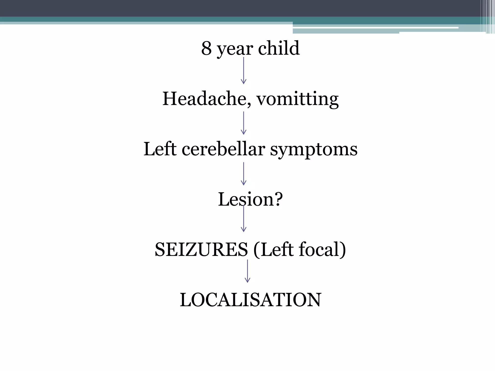
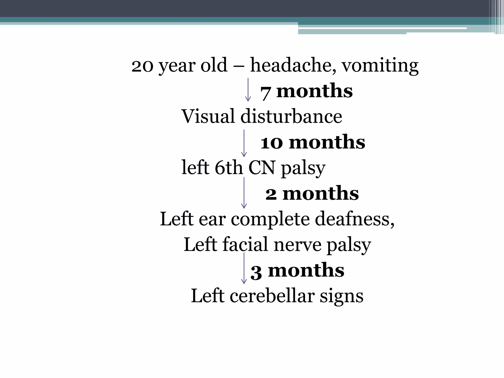
FALSE LOCALISING SIGNS.
Chairperson: Dr.Seetaram.N.K
Student: Dr.Varun.
Neurological signs have been described as ‘false
localizing’ if they reflect dysfunction distant or
remote from the expected anatomical locus of
pathology and hence challenging the traditional
clinicoanatomical correlation paradigm.
Jobin v joseph, False Localising Signs in Neurology, Indian Journal of Clinical Practice, Vol. 23, No. 9
February 2013
William Macewen.
Scottish neurosurgeon.
1876- Frontal lobe abscess.
1879- Left frontal meningioma.
BRAIN
1904
8 year child
Headache, vomitting
Left cerebellar symptoms
Lesion?
SEIZURES (Left focal)
LOCALISATION
• Supratentorial- 13 %
• Infratentorial- 4%
Tumours of the cerebellum rarely fail to
show the signs of cerebellar involvement.
• Paralyses of cranial nerves. 10 %
• Hemianopia.
• Jacksonian epilepsy.
• Bilateral spastic paresis.
• Cerebellar signs.
False localising signs.
Cranial nerve.
1) 6th nerve.
Most common
Seen in both supra and infra tentorial
lesions.
Reason: compression against the petrous
ligament or the ridge of the petrous
temporal bone.
2) Oculomotor nerve : Unilateral fixed dilated
pupil (Hutchinson’s pupil)
May occur with an ipsilateral lesion such as an
intracerebral hemorrhage,
Reason:due to transtentorial herniation of the
brain compressing the oculomotor nerve against
the free edge of the tentorium.
• Because of the fascicular organization of the
fibers within the oculomotor nerve, the
externally placed pupillomotor fibers are most
vulnerable.
• Divisional third nerve palsies may sometimes
occur with more proximal lesions, presumably as
a consequence of the topographic arrangement
of the fascicles within the nerve
• 3) Fourth cranial nerve- Involvement can be
sometimes seen in idiopathic intracranial
hypertension.
• Exact mechanism not clear.
4) Fifth cranial nerve: Seen in posterior fossa
tumour, Idiopathic intracranial hypertension.
Pathophysiology: Brain stem distortion resulting
in traction on cranial nerves.
Vascular compression of the nerve root.
Similar involvement of 7th and 8th cranial nerve is
also noted.
• Concurrent false-localizing involvement of
multiple cranial nerves has been noted on
occasion with a contralateral acoustic neuroma
and trigeminal, glossopharyngeal and vagus
nerves with a contralateral laterally-placed
posterior fossa meningioma.
6) Papilledema- Variable extension of meninges
around the Optic nerve.
Transmission of pressure onto the nerve reduce
axoplasmic flow resulting in papilledema.
MOTOR SYSTEM
• 1)
A supratentorial lesion, such as acute subdural
hematoma, may cause transtentorial herniation
of the temporal lobe, with compression of the
ipsilateral cerebral peduncle against the
tentorial edge; since this is above the pyramidal
decussation, a contralateral hemiparesis results.
Kernohan-Woltman notch phenomenon or
Kernohan’s notch syndrome : hemiparesis may
be ipsilateral to the lesion, and hence false-
localizing; this occurs when the contralateral
cerebral peduncle is compressed by the free edge
of the tentorium.
• 2) Hemidiaphragmatic paralysis with ipsilateral
brainstem (medullary) compression by an
aberrant vertebral artery has been described, in
the absence of pathology localized to the C3-C5
segments of the spinal cord
3) Lesions at the level of the foramen magnum
may produce, false localising signs: paraesthesia
in the hands and lower motor neurone signs in
the upper limbs. The wasting (“remote
atrophy”), weakness, and areflexia may suggest
the involvement of cervical cord segments well
below the level of the foramen magnum lesion.
• Pathophysiology: There is currently no
compelling pathophysiological explanation for
these false localising signs.
• The arterial hypothesis postulates that tumour
in some way compromises descending anterior
spinal artery blood supply to the lower cervical
cord with resultant focal ischaemia.
• Low pressure venous system would seem
inherently more vulnerable to the effects of
compression than the arterial system, with
resultant tissue stasis and hypoxia perhaps
causing the clinically observed neurological
dysfunction.
• Mechanical stresses within the spinal cord,
consequent perhaps on the conjunction of
extrinsic compression with the anchoring of the
spinal cord by the dentate ligaments, have also
been suggested to account for remote signs.
• 4) Lower cervical cord and upper thoracic cord
lesions:
Compressive cervical myelopathy may produce a
false localising thoracic sensory level in addition
to lower limb weakness and hyperreflexia.
• Similarly, lumbar spinal disease may be
simulated by more rostral pathology; for
example, urinary retention, leg weakness, and
lumbar sensory findings may be the presenting
features of high thoracic cord compression, with
clinicoradiological discrepancy of as much as 11
segments.
Cerebellar
• Frontocerebellar pathway damage, for example,
as a result of infarction in the territory of the
anterior cerebral artery, may result in
incoordination of the contralateral limbs,
mimicking cerebellar dysfunction.
Radiculopathy
• False-localising radiculopathy may occur in the
context of IIH and cerebral venous sinus
thrombosis, manifesting as acral paraesthesias,
backache and radicular pain, and less often with
motor deficits.
• Mimic Guillain-Barré syndrome (flaccid-
areflexic quadriplegia).
• The postulated mechanism for such
radiculopathy is mechanical root compression
due to elevated CSF pressure.
False hemineglect
• False-localising neglect has been encountered: in
a patient with a posterior fossa meningioma
causing left pontine compression.
Pseudosyringomyelia
• Pseudosyringomyelia” has been used to describe
a selective loss of pain and temperature
sensation with relative preservation of vibration
and position sense seen in amyloid
polyneuropathy and Tangier disease, (a small
fibre sensory neuropathy), in the absence of any
spinal cord pathology, and hence false localizing.
• Pseudoathetosis.
• Myasthenic nystagmus.
• Subcortical aphasia.
OTHERS
Collier’s Prime findings
• Signs appearing late in the course of intracranial
tumour, where general symptoms and signs
preexisted, are often of false portent.
• Absence of focal neurological deficits during the
early course of illness is in itself a most
important localizing indicator, confining the
disease to the supratentorial compartment.
• As the disease process progress, generalized
symptoms of increased intracranial pressure
may conceal once recognizable true localizing
signs.
• Tumours of the cerebellum rarely fail to show
the signs of cerebellar involvement
8 year child
Headache, vomitting
Left cerebellar symptoms
Lesion?
SEIZURES (Left focal)
LOCALISATION
20 year old – headache, vomiting
7 months
Visual disturbance
10 months
left 6th CN palsy
2 months
Left ear complete deafness,
Left facial nerve palsy
3 months
Left cerebellar signs
• Glioma in left frontal lobe.
References
• James collier, The false localising signs of Intracranial tumour,
Neurology, 1904.
• Companion to clinical neurology, Second edition, William Pryse-
Phillips.
• Clinical neurology, C.David Marsden and Timothy J.Fowler.
• Jobin v joseph, False Localising Signs in Neurology, Indian Journal
of Clinical Practice, Vol. 23, No. 9 February 2013.
• A J Larner, False localising signs, J Neurol Neurosurg Psychiatry
2003;74:415–418.
• Dejong’s, The neurological examination, seventh edition.
• Gassel MM. False localizing signs. A review of the concept and
analysis of the occurrence in 250 cases of intracranial meningioma.
Arch Neurol 1961;4:526–54.
• Davis M, Lucatorto M. The false localizing signs of increased
intracranial pressure. J Neurosci Nursing 1992;24:245–50.
THANK YOU

falselocalisingsigns.pdf

  • 1.
    FALSE LOCALISING SIGNS. Chairperson:Dr.Seetaram.N.K Student: Dr.Varun.
  • 2.
    Neurological signs havebeen described as ‘false localizing’ if they reflect dysfunction distant or remote from the expected anatomical locus of pathology and hence challenging the traditional clinicoanatomical correlation paradigm. Jobin v joseph, False Localising Signs in Neurology, Indian Journal of Clinical Practice, Vol. 23, No. 9 February 2013
  • 3.
    William Macewen. Scottish neurosurgeon. 1876-Frontal lobe abscess. 1879- Left frontal meningioma.
  • 4.
  • 5.
    8 year child Headache,vomitting Left cerebellar symptoms Lesion? SEIZURES (Left focal) LOCALISATION
  • 6.
    • Supratentorial- 13% • Infratentorial- 4% Tumours of the cerebellum rarely fail to show the signs of cerebellar involvement. • Paralyses of cranial nerves. 10 % • Hemianopia. • Jacksonian epilepsy. • Bilateral spastic paresis. • Cerebellar signs.
  • 7.
    False localising signs. Cranialnerve. 1) 6th nerve. Most common Seen in both supra and infra tentorial lesions. Reason: compression against the petrous ligament or the ridge of the petrous temporal bone.
  • 10.
    2) Oculomotor nerve: Unilateral fixed dilated pupil (Hutchinson’s pupil) May occur with an ipsilateral lesion such as an intracerebral hemorrhage, Reason:due to transtentorial herniation of the brain compressing the oculomotor nerve against the free edge of the tentorium.
  • 14.
    • Because ofthe fascicular organization of the fibers within the oculomotor nerve, the externally placed pupillomotor fibers are most vulnerable. • Divisional third nerve palsies may sometimes occur with more proximal lesions, presumably as a consequence of the topographic arrangement of the fascicles within the nerve
  • 15.
    • 3) Fourthcranial nerve- Involvement can be sometimes seen in idiopathic intracranial hypertension. • Exact mechanism not clear.
  • 17.
    4) Fifth cranialnerve: Seen in posterior fossa tumour, Idiopathic intracranial hypertension. Pathophysiology: Brain stem distortion resulting in traction on cranial nerves. Vascular compression of the nerve root. Similar involvement of 7th and 8th cranial nerve is also noted.
  • 18.
    • Concurrent false-localizinginvolvement of multiple cranial nerves has been noted on occasion with a contralateral acoustic neuroma and trigeminal, glossopharyngeal and vagus nerves with a contralateral laterally-placed posterior fossa meningioma.
  • 19.
    6) Papilledema- Variableextension of meninges around the Optic nerve. Transmission of pressure onto the nerve reduce axoplasmic flow resulting in papilledema.
  • 21.
    MOTOR SYSTEM • 1) Asupratentorial lesion, such as acute subdural hematoma, may cause transtentorial herniation of the temporal lobe, with compression of the ipsilateral cerebral peduncle against the tentorial edge; since this is above the pyramidal decussation, a contralateral hemiparesis results.
  • 22.
    Kernohan-Woltman notch phenomenonor Kernohan’s notch syndrome : hemiparesis may be ipsilateral to the lesion, and hence false- localizing; this occurs when the contralateral cerebral peduncle is compressed by the free edge of the tentorium.
  • 24.
    • 2) Hemidiaphragmaticparalysis with ipsilateral brainstem (medullary) compression by an aberrant vertebral artery has been described, in the absence of pathology localized to the C3-C5 segments of the spinal cord
  • 25.
    3) Lesions atthe level of the foramen magnum may produce, false localising signs: paraesthesia in the hands and lower motor neurone signs in the upper limbs. The wasting (“remote atrophy”), weakness, and areflexia may suggest the involvement of cervical cord segments well below the level of the foramen magnum lesion.
  • 26.
    • Pathophysiology: Thereis currently no compelling pathophysiological explanation for these false localising signs. • The arterial hypothesis postulates that tumour in some way compromises descending anterior spinal artery blood supply to the lower cervical cord with resultant focal ischaemia.
  • 27.
    • Low pressurevenous system would seem inherently more vulnerable to the effects of compression than the arterial system, with resultant tissue stasis and hypoxia perhaps causing the clinically observed neurological dysfunction. • Mechanical stresses within the spinal cord, consequent perhaps on the conjunction of extrinsic compression with the anchoring of the spinal cord by the dentate ligaments, have also been suggested to account for remote signs.
  • 28.
    • 4) Lowercervical cord and upper thoracic cord lesions: Compressive cervical myelopathy may produce a false localising thoracic sensory level in addition to lower limb weakness and hyperreflexia.
  • 29.
    • Similarly, lumbarspinal disease may be simulated by more rostral pathology; for example, urinary retention, leg weakness, and lumbar sensory findings may be the presenting features of high thoracic cord compression, with clinicoradiological discrepancy of as much as 11 segments.
  • 30.
    Cerebellar • Frontocerebellar pathwaydamage, for example, as a result of infarction in the territory of the anterior cerebral artery, may result in incoordination of the contralateral limbs, mimicking cerebellar dysfunction.
  • 32.
    Radiculopathy • False-localising radiculopathymay occur in the context of IIH and cerebral venous sinus thrombosis, manifesting as acral paraesthesias, backache and radicular pain, and less often with motor deficits.
  • 33.
    • Mimic Guillain-Barrésyndrome (flaccid- areflexic quadriplegia). • The postulated mechanism for such radiculopathy is mechanical root compression due to elevated CSF pressure.
  • 34.
    False hemineglect • False-localisingneglect has been encountered: in a patient with a posterior fossa meningioma causing left pontine compression.
  • 35.
    Pseudosyringomyelia • Pseudosyringomyelia” hasbeen used to describe a selective loss of pain and temperature sensation with relative preservation of vibration and position sense seen in amyloid polyneuropathy and Tangier disease, (a small fibre sensory neuropathy), in the absence of any spinal cord pathology, and hence false localizing.
  • 36.
    • Pseudoathetosis. • Myasthenicnystagmus. • Subcortical aphasia. OTHERS
  • 37.
    Collier’s Prime findings •Signs appearing late in the course of intracranial tumour, where general symptoms and signs preexisted, are often of false portent.
  • 38.
    • Absence offocal neurological deficits during the early course of illness is in itself a most important localizing indicator, confining the disease to the supratentorial compartment.
  • 39.
    • As thedisease process progress, generalized symptoms of increased intracranial pressure may conceal once recognizable true localizing signs.
  • 40.
    • Tumours ofthe cerebellum rarely fail to show the signs of cerebellar involvement
  • 41.
    8 year child Headache,vomitting Left cerebellar symptoms Lesion? SEIZURES (Left focal) LOCALISATION
  • 42.
    20 year old– headache, vomiting 7 months Visual disturbance 10 months left 6th CN palsy 2 months Left ear complete deafness, Left facial nerve palsy 3 months Left cerebellar signs
  • 43.
    • Glioma inleft frontal lobe.
  • 44.
    References • James collier,The false localising signs of Intracranial tumour, Neurology, 1904. • Companion to clinical neurology, Second edition, William Pryse- Phillips. • Clinical neurology, C.David Marsden and Timothy J.Fowler. • Jobin v joseph, False Localising Signs in Neurology, Indian Journal of Clinical Practice, Vol. 23, No. 9 February 2013. • A J Larner, False localising signs, J Neurol Neurosurg Psychiatry 2003;74:415–418.
  • 45.
    • Dejong’s, Theneurological examination, seventh edition. • Gassel MM. False localizing signs. A review of the concept and analysis of the occurrence in 250 cases of intracranial meningioma. Arch Neurol 1961;4:526–54. • Davis M, Lucatorto M. The false localizing signs of increased intracranial pressure. J Neurosci Nursing 1992;24:245–50.
  • 46.