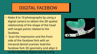
FACEBOW TRANSFER
AND ARTICULATION
DR ABHIDHA TRIPATHI
DEPT OF PROSTHODONTICS
Introduction
1
2
CONTENTS Definition
3
4
5
6
7
Value of Facebow
Recent advances
Facebow Registration and
Transfer
Types of Facebow
Indications
Conclusion
References
8
9
INTRODUCTION
• In 1899, George Snow
introduced the facebow to
help locate the rotation
axis of the lower jaw.
• The procedure of facebow
transfer confirms that the
maxillary arch is oriented in
the same or comparable
distances to the hinges on
the articulator as it is in the
TMJ.
JAW RELATION OR MAXILLO-MANDIBULAR
RELATIONSHIP
• DEFINITION: A registration of any positional relationship of the mandible
relative to the maxillae. These records may be made at any vertical, horizontal
or lateral orientation.
-GPT 9
ORIENTATION JAW RELATION
• DEFINITION: The relations that orient the maxilla to the cranium in such a way
that, when the mandible is kept in its most posterior position, the mandible
can rotate in a sagittal plane around an imaginary transverse axis passing
through or near the condyles.
- GPT 9
• Plane of maxilla may be tilted in some patients - plane of mandible will not be altered
since it articulates with base of the skull.
• Hence , a maxillary tilt will alter the relationship of maxilla to mandible during different
movements, also affects the level of occlusal plane .
ORIENTATION OF MAXILLA IN RELATION TO BASE OF
SKULL
ACCORDING TO GPT 9:
AN INSTRUMENT USED TO RECORD SPATIAL
RELATIONSHIP OF THE MAXILLARY ARCH TO SOME
ANATOMIC REFERENCE POINT OR POINTS AND THEN
TRANSFER THIS RELATIONSHIP TO AN ARTICULATOR; IT
ORIENTS THE DENTAL CAST IN THE SAME SPATIAL
RELATIONSHIP TO THE OPENING AXIS OF THE
ARTICULATOR
DEFINITION
VALUE OF THE FACEBOW
• Failure to use
facebow can
lead to errors
in occlusion of
denture.
• Allows more
accurate arc of
closure on the
articulator.
• An integral
part in
analyzing and
studying the
occlusion.
INDICATIONS
• When interocclusal
check records are used
• When
balanced
occlusion is
desired
• When cusp
form teeth
are used
• In full mouth
rehabilitation when
accurate occlusal
restorations are to be
made
• For diagnostic
mounting and
treatment
planning
• In
gnathological
studies and
treatment
• U SHAPED FRAME
• CONDYLAR RODS
• EAR PIECES
• BITE FORK
• LOCKING DEVICE
• ORBITAL POINTER
PARTS OF
FACEBOW
• Represents the plane of the cranium
• It forms the main frame of the facebow
• All other components are attached to this frame.
• It extends from the region of TMJ on one side to the
other side without contacting the face.
U SHAPED FRAME
• Two small metallic rods on either side of the free end of
the U shaped frame that contact the skin over the TMJ.
• They are used to locate the hinge axis and transfer it to
the articulator.
• Some facebows have ear piece that fit into the external
auditory meatus instead of condylar rods
CONDYLAR RODS
• Represents the plane of the maxilla.
• U shaped plate which is attached to the occlusal rims
while recording the orientation relation
• It is attached to the frame with the help of a rod called
the stem.
• The bite fork should be inserted 3mm below occlusal
surface within occlusal rim
BITE FORK
• Attaches the bite fork to the U shaped frame
• Supports the facebow, occlusal rims and cast during
articulation.
• Consists of transfer rod
• U shaped frame is attached to the vertical transfer rod
LOCKING DEVICE
• Attaches the bite fork to the U shaped frame
• Supports the facebow, occlusal rims and cast during
articulation.
• Consists of transfer rod
• U shaped frame is attached to the vertical transfer rod
THIRD REFERENCE
POINT
• ARBITARY TYPE
• KINEMATIC TYPE
TYPES OF FACEBOW
• EAR PIECE TYPE
• FACIA TYPE
• HANAU FACEBOW
• SLIDEMATIC
• TWIRL BOW
• WHIP MIX
• FACIA TYPE HANAU
FACEBOW ATTACHED TO
HANAU ARTICULATOR
• ARBITARY TYPE
TYPES OF FACEBOW
• KINEMATIC TYPE
Earpiece Facia type
• HANAU
• Denar
Slidematic
• Whipmix
Quick Mount
• HANAU 132-
25 M
• HANAU 132-
2C Model
• TMJ
Instrument
• Pantograph
• Axiograph
• Most commonly used
• In this type the axis is located by using anatomical land
marks. The condylar rods are positioned approximately
13 mm anterior to the auditory meatus on the cantho-
tragal line
ARBITARY TYPE
• This method generally locates the rods within 5mm of
the true hinge axis of the jaws. As this is an arbitary
hinge axis, errors in jaw relation may produce occlusal
discrepancies which should be corrected by minor
occlusal adjustments during insertion.
• Facia type utilizes approximate points on the skin over
the temporomandibular region as the posterior
reference points.
• These points are located by measuring from certain
anatomic landmarks on the face.
• The facebow has a pointer that can be positioned to the
posterior reference point.
FASCIA TYPE
• DISADVANTAGES:
1. As the facebow is placed on the skin which is movable
there is a tendency for the condylar rods to displace.
2.Also requires an assistant to hold the facebow in place.
• It uses external auditory meatus as an arbitary
reference point
• Accurate relationship for most diagnostic and
restorative procedures.
• Posterior reference point is External auditory meatus
and anterior reference point is Orbitale
EARPIECE TYPE
• ADVANTAGES;
1.Simple to use
2. Does not require measurements on face
3.Provides an average anatomic dimension between EAM
and horizontal axis of mandible
• DISADVANTAGES:
1.An error of 0.2mm from the axis can be expected.
• It is an earpiece facebow made of spring steel and
simply springs open and close to various head widths.
• Most commonly used
• This instrument is designed to orient the occlusal plan
to the Frankfort horizontal plane by means of a third
reference point.
• indicated in cases when it is critical to precisely
reproduce the exact opening and closing movement of
the patient to articulator
SPRING BOW (HANAU’S
FACEBOW)
• ADVANTAGES:
1. Easy and efficient to use
2.one piece design of bow eliminates the moving parts
and maintainence problems encountered with other
models
• DISADVANTAGES:
1.Inability to measure inter condylar distance
• It is an earpiece type of facebow
• Allows the maxillary arch to be transferred to the articulator without physically attaching the
facebow to the articulator
• Relates the maxillary arch to FH plane
• A mounting guide is used to mount the transfer rod to the articulator
• Easy to manipulate because the facebow is not needed to mount the maxillary cast in the
articulator
TWIRL BOW
• This facebow has an electronic device, which gives the
reading that can be seen in the anterior region.
• This reading denotes one half of the patient’s inter
condylar distance
• These facebows require specific articulators, which
accept the reading.
• The posterior reference point for this instrument is the
external auditory meatus and the anterior reference
point is 43 mm superior to the incisal edge of the upper
central incisor for dentulous patients.
SLIDEMATIC TYPE (DENAR)
• In an edentulous patient the anterior reference point is
43mm superior to the lower border of the upper lip in a
relaxed state.
• This anterior reference point is also used for Whipmix
articulators.
• The anterior reference point can be marked using a
Denar reference plane locator.
• Tthe plane locator is an instrument which resembles a
facebow.
SLIDEMATIC TYPE (DENAR)
• These facebow have a built in hinge axis locator.
• It automatically locates the hinge axis when the
earpieces are located in the External Auditory Meatus.
• It has a nasion relator assembly with a plastic nose
piece.
• The nasion relator determines the anterior reference
point
WHIPMIX FACEBOW
WHIPMIX FACEBOW
• It is used to determine and locate the exact hinge axis
points.
• Hinge axis of the mandible can be determined can be
determined by a clutch i.e. a segmented impression tray
like device attached onto the mandibular teeth with a
suitable rigid material such as impression plaster.
KINEMATIC FACEBOW
KINEMATIC FACEBOW
• The facebow helps to orient the cast in the patients’s
terminal hinge axis.
• A 12-15degree pure rotational movement of the joint is
possible with maximal incisal separation of 20-25 mm
• This facebow requires a fully adjustable articulator to
accept the true hinge axis.
• Since the patients hinge axis is transferred to the
articulator, the movement of the articulator will
simulate the movements of the joint made at terminal
hinge position.
KINEMATIC FACEBOW
• Quick Mount facebow (with bitefork, nasion relator and Quick
Lock toggle assembly)
• Whip Mix Articulator
• Plaster bowl
• Spatula
• Laboratory Knife with no 25 blade
• Trimmed Maxillary cast
• Mounting Stone
FACEBOW ARMAMENTARIUM
• Two horseshoe wax wafers (Surgident Coprwax Bite
Wafer) are heated in warm tap water until they become
soft and flexible.
• A wafer is adapted to each side of the bite fork so that it is
uniformly covered.
• The wax-covered bite fork is placed between the teeth,
with the bite fork shaft to the patient’s right.
• The fork is centered by aligning the index ring on the fork
with the patient’s midline.
FACEBOW RECORD
TECHNIQUE
• The patient is instructed to bite lightly into the wax to
produce shallow indentations of the cusp tips in the wax.
• The wax is cooled, and the bite fork is removed from the
mouth. Any excess wax is trimmed off the bite fork.
• The maxillary cast is tried in the wax record to ensure that
it will seat without rocking.
• If the cast fails to seat, the occlusal surfaces of the cast are
checked for nodules of stone. If none are evident, there is
a distortion in the registration or the cast
FACEBOW RECORD
TECHNIQUE
• The reference pin is fastened to the underside of the face-
bow by tightening the thumbscrew.
• The bite fork is placed in the mouth, and the patient is
instructed to hold it securely between the maxillary and
mandibular teeth.
• The patient should grip both arms of the facebow to guide
the plastic earpieces into the external auditory meati, in
the same manner as one would place a stethoscope into
the ears
FACEBOW RECORD
TECHNIQUE
• The anterior reference pointer is extended while the
facebow is moved up or down. When the pointer is
properly aligned with the anterior reference point, the
thumbscrew is tightened.
• The facebow should not be allowed to torque or tilt during
the tightening procedure.
FACEBOW RECORD
TECHNIQUE
FACEBOW RECORD
TECHNIQUE
The bite fork is placed against the maxillary teeth
and supported by the dentist as the patient closes
lightly for a shallow impression of the cusp
The patient guides the earpieces as the
dentist places them into the external
auditory meati
FACEBOW RECORD
TECHNIQUE
The dentist places the clamp over the
bitefork shaft while the patient inserts the
earpiece
The thumbscrew on the front of
the facebow is tightened
FACEBOW RECORD
TECHNIQUE
The shaft of the nasion relator is extended,
and the thumbscrew is tightened
The thumbscrew on the top of
the facebow is tightened
FACEBOW RECORD
TECHNIQUE
The Quick Lock Toggle is slipped into the
slot on the bite fork and the thumbscrew is
tightened
Complete facebow record
FACEBOW RECORD
TECHNIQUE
The thumbscrew on the top of the facebow
is loosened
The space between the facebow and the
bite fork is checked for uniformity.
• As the patient opens the mouth, the assembly is removed
from the head. The clamps are rechecked and tightened.
• The bite fork assembly is removed from the underside of
the facebow by loosening the set screw on the clamp by a
quarter turn. Only the bite fork assembly needs to be
used for mounting the maxillary cast and should be
disinfected at this time.
FACEBOW RECORD
TECHNIQUE
• In recent years, the incorporation of CAD/CAM technology
has provided for more efficient protocols by automating
processes and reducing manual labor. Intra-oral scanners
can digitise dental arches and register maxillomandibular
relationships.
• Currently, many CAD/CAM systems include a virtual
articulator simulatory module as a tool to simulate
mandibular movements, which can be adjusted by using
numerical values to represent condylar inclination,
Bennett angle, vertical dimension, etc.
DIGITAL FACEBOW
Solaberrieta E, Garmendia A, Minguez R, Brizuela A, Pradies G. Virtual facebow technique. J Prosthet Dent. 2015 Dec
• Scan the maxillary and mandibular dental arches of the
patient with an intraoral dental scanner to obtain digital
casts
• Place 3 adhesive targets onto the patient’s head. Locate
the first 2 points next to the temporomandibular joints
and the third point onto the infraorbital point.
• Locate scannable elastomeric impression material on a
plastic, colored facebow fork and introduce the facebow
fork into the patient’s mouth
DIGITAL FACEBOW
Solaberrieta E, Garmendia A, Minguez R, Brizuela A, Pradies G. Virtual facebow technique. J Prosthet Dent. 2015 Dec
DIGITAL FACEBOW
Solaberrieta E, Garmendia A, Minguez R, Brizuela A, Pradies G. Virtual facebow technique. J Prosthet Dent. 2015 Dec
DIGITAL FACEBOW
Solaberrieta E, Garmendia A, Minguez R, Brizuela A, Pradies G. Virtual facebow technique. J Prosthet Dent. 2015 Dec
• Make 8 to 10 photographs by using a
digital camera to obtain the 3D spatial
relationship of the shape of the head
with target points related to the
facebow.
• Scan the impression and the front
side of the facebow fork with an
intraoral dental scanner, load the
facebow fork 3D geometry and align it
to the maxillary digital cast by using
DIGITAL FACEBOW
CONCLUSIO
N
Despite lot of controversies in facebow usage,
hinge axis location and establishing plane of
orientation is an important componenet for
recording mandibular movement.
1
Facebow record helps in securing anteroposterior
positioning of the cast in relation to condyles. It
helps in achieving the exact anteroposterior or
vertical positioning of cast irt condyles
2
The virtual facebow is a recent advancement and
can align digital casts directly onto virtual
articulator. this was developed to overcome the
problem of transferring data from patient
simulation to virtual articulators.
3
REFERENCES
• George A Zarb, Hobkirk J, Eckert S, Jacob R. Prosthodontic Treatment for Edentulous Patients: Complete
Denture and Implant Supported Prostheses. South Asia Edition: Elsevier; 2015.
• Shillingburg HT, Hobo S, Whitsett LD, Jacobi R. Fundamentals of fixed prosthodontics. Quintessence
Publishing Company: 1997
• Schuyler CH. The function and importance of incisal guidance in oral rehabilitation. J Prosthet Dent 1963
• Winkler S, editor. Essentials of complete denture prosthodontics. year book medical Pub 1988
• Vivell C, Slavicek G, Slavicek R. Arbitrary versus exact mounting procedure during fabrication of intraoral
splints: An exploratory randomized controlled clinical trial. Int J Stomatol Occl Med. 2009
• Solaberrieta E, Garmendia A, Minguez R, Brizuela A, Pradies G. Virtual facebow technique. J Prosthet
Dent. 2015 Dec;114(6):751-5. doi: 10.1016/j.prosdent.2015.06.012. Epub 2015 Sep 12. PMID: 26372628.

Facebow record, transfer and articulation

  • 1.
    FACEBOW TRANSFER AND ARTICULATION DRABHIDHA TRIPATHI DEPT OF PROSTHODONTICS
  • 2.
    Introduction 1 2 CONTENTS Definition 3 4 5 6 7 Value ofFacebow Recent advances Facebow Registration and Transfer Types of Facebow Indications Conclusion References 8 9
  • 3.
    INTRODUCTION • In 1899,George Snow introduced the facebow to help locate the rotation axis of the lower jaw. • The procedure of facebow transfer confirms that the maxillary arch is oriented in the same or comparable distances to the hinges on the articulator as it is in the TMJ.
  • 4.
    JAW RELATION ORMAXILLO-MANDIBULAR RELATIONSHIP • DEFINITION: A registration of any positional relationship of the mandible relative to the maxillae. These records may be made at any vertical, horizontal or lateral orientation. -GPT 9
  • 5.
    ORIENTATION JAW RELATION •DEFINITION: The relations that orient the maxilla to the cranium in such a way that, when the mandible is kept in its most posterior position, the mandible can rotate in a sagittal plane around an imaginary transverse axis passing through or near the condyles. - GPT 9
  • 6.
    • Plane ofmaxilla may be tilted in some patients - plane of mandible will not be altered since it articulates with base of the skull. • Hence , a maxillary tilt will alter the relationship of maxilla to mandible during different movements, also affects the level of occlusal plane . ORIENTATION OF MAXILLA IN RELATION TO BASE OF SKULL
  • 7.
    ACCORDING TO GPT9: AN INSTRUMENT USED TO RECORD SPATIAL RELATIONSHIP OF THE MAXILLARY ARCH TO SOME ANATOMIC REFERENCE POINT OR POINTS AND THEN TRANSFER THIS RELATIONSHIP TO AN ARTICULATOR; IT ORIENTS THE DENTAL CAST IN THE SAME SPATIAL RELATIONSHIP TO THE OPENING AXIS OF THE ARTICULATOR DEFINITION
  • 8.
    VALUE OF THEFACEBOW • Failure to use facebow can lead to errors in occlusion of denture. • Allows more accurate arc of closure on the articulator. • An integral part in analyzing and studying the occlusion.
  • 9.
    INDICATIONS • When interocclusal checkrecords are used • When balanced occlusion is desired • When cusp form teeth are used • In full mouth rehabilitation when accurate occlusal restorations are to be made • For diagnostic mounting and treatment planning • In gnathological studies and treatment
  • 10.
    • U SHAPEDFRAME • CONDYLAR RODS • EAR PIECES • BITE FORK • LOCKING DEVICE • ORBITAL POINTER PARTS OF FACEBOW
  • 11.
    • Represents theplane of the cranium • It forms the main frame of the facebow • All other components are attached to this frame. • It extends from the region of TMJ on one side to the other side without contacting the face. U SHAPED FRAME
  • 12.
    • Two smallmetallic rods on either side of the free end of the U shaped frame that contact the skin over the TMJ. • They are used to locate the hinge axis and transfer it to the articulator. • Some facebows have ear piece that fit into the external auditory meatus instead of condylar rods CONDYLAR RODS
  • 13.
    • Represents theplane of the maxilla. • U shaped plate which is attached to the occlusal rims while recording the orientation relation • It is attached to the frame with the help of a rod called the stem. • The bite fork should be inserted 3mm below occlusal surface within occlusal rim BITE FORK
  • 14.
    • Attaches thebite fork to the U shaped frame • Supports the facebow, occlusal rims and cast during articulation. • Consists of transfer rod • U shaped frame is attached to the vertical transfer rod LOCKING DEVICE
  • 15.
    • Attaches thebite fork to the U shaped frame • Supports the facebow, occlusal rims and cast during articulation. • Consists of transfer rod • U shaped frame is attached to the vertical transfer rod THIRD REFERENCE POINT
  • 16.
    • ARBITARY TYPE •KINEMATIC TYPE TYPES OF FACEBOW • EAR PIECE TYPE • FACIA TYPE • HANAU FACEBOW • SLIDEMATIC • TWIRL BOW • WHIP MIX • FACIA TYPE HANAU FACEBOW ATTACHED TO HANAU ARTICULATOR
  • 17.
    • ARBITARY TYPE TYPESOF FACEBOW • KINEMATIC TYPE Earpiece Facia type • HANAU • Denar Slidematic • Whipmix Quick Mount • HANAU 132- 25 M • HANAU 132- 2C Model • TMJ Instrument • Pantograph • Axiograph
  • 18.
    • Most commonlyused • In this type the axis is located by using anatomical land marks. The condylar rods are positioned approximately 13 mm anterior to the auditory meatus on the cantho- tragal line ARBITARY TYPE • This method generally locates the rods within 5mm of the true hinge axis of the jaws. As this is an arbitary hinge axis, errors in jaw relation may produce occlusal discrepancies which should be corrected by minor occlusal adjustments during insertion.
  • 19.
    • Facia typeutilizes approximate points on the skin over the temporomandibular region as the posterior reference points. • These points are located by measuring from certain anatomic landmarks on the face. • The facebow has a pointer that can be positioned to the posterior reference point. FASCIA TYPE • DISADVANTAGES: 1. As the facebow is placed on the skin which is movable there is a tendency for the condylar rods to displace. 2.Also requires an assistant to hold the facebow in place.
  • 20.
    • It usesexternal auditory meatus as an arbitary reference point • Accurate relationship for most diagnostic and restorative procedures. • Posterior reference point is External auditory meatus and anterior reference point is Orbitale EARPIECE TYPE • ADVANTAGES; 1.Simple to use 2. Does not require measurements on face 3.Provides an average anatomic dimension between EAM and horizontal axis of mandible • DISADVANTAGES: 1.An error of 0.2mm from the axis can be expected.
  • 21.
    • It isan earpiece facebow made of spring steel and simply springs open and close to various head widths. • Most commonly used • This instrument is designed to orient the occlusal plan to the Frankfort horizontal plane by means of a third reference point. • indicated in cases when it is critical to precisely reproduce the exact opening and closing movement of the patient to articulator SPRING BOW (HANAU’S FACEBOW) • ADVANTAGES: 1. Easy and efficient to use 2.one piece design of bow eliminates the moving parts and maintainence problems encountered with other models • DISADVANTAGES: 1.Inability to measure inter condylar distance
  • 22.
    • It isan earpiece type of facebow • Allows the maxillary arch to be transferred to the articulator without physically attaching the facebow to the articulator • Relates the maxillary arch to FH plane • A mounting guide is used to mount the transfer rod to the articulator • Easy to manipulate because the facebow is not needed to mount the maxillary cast in the articulator TWIRL BOW
  • 23.
    • This facebowhas an electronic device, which gives the reading that can be seen in the anterior region. • This reading denotes one half of the patient’s inter condylar distance • These facebows require specific articulators, which accept the reading. • The posterior reference point for this instrument is the external auditory meatus and the anterior reference point is 43 mm superior to the incisal edge of the upper central incisor for dentulous patients. SLIDEMATIC TYPE (DENAR)
  • 24.
    • In anedentulous patient the anterior reference point is 43mm superior to the lower border of the upper lip in a relaxed state. • This anterior reference point is also used for Whipmix articulators. • The anterior reference point can be marked using a Denar reference plane locator. • Tthe plane locator is an instrument which resembles a facebow. SLIDEMATIC TYPE (DENAR)
  • 25.
  • 26.
  • 27.
    • These facebowhave a built in hinge axis locator. • It automatically locates the hinge axis when the earpieces are located in the External Auditory Meatus. • It has a nasion relator assembly with a plastic nose piece. • The nasion relator determines the anterior reference point WHIPMIX FACEBOW
  • 28.
  • 29.
    • It isused to determine and locate the exact hinge axis points. • Hinge axis of the mandible can be determined can be determined by a clutch i.e. a segmented impression tray like device attached onto the mandibular teeth with a suitable rigid material such as impression plaster. KINEMATIC FACEBOW
  • 30.
  • 31.
    • The facebowhelps to orient the cast in the patients’s terminal hinge axis. • A 12-15degree pure rotational movement of the joint is possible with maximal incisal separation of 20-25 mm • This facebow requires a fully adjustable articulator to accept the true hinge axis. • Since the patients hinge axis is transferred to the articulator, the movement of the articulator will simulate the movements of the joint made at terminal hinge position. KINEMATIC FACEBOW
  • 32.
    • Quick Mountfacebow (with bitefork, nasion relator and Quick Lock toggle assembly) • Whip Mix Articulator • Plaster bowl • Spatula • Laboratory Knife with no 25 blade • Trimmed Maxillary cast • Mounting Stone FACEBOW ARMAMENTARIUM
  • 33.
    • Two horseshoewax wafers (Surgident Coprwax Bite Wafer) are heated in warm tap water until they become soft and flexible. • A wafer is adapted to each side of the bite fork so that it is uniformly covered. • The wax-covered bite fork is placed between the teeth, with the bite fork shaft to the patient’s right. • The fork is centered by aligning the index ring on the fork with the patient’s midline. FACEBOW RECORD TECHNIQUE
  • 34.
    • The patientis instructed to bite lightly into the wax to produce shallow indentations of the cusp tips in the wax. • The wax is cooled, and the bite fork is removed from the mouth. Any excess wax is trimmed off the bite fork. • The maxillary cast is tried in the wax record to ensure that it will seat without rocking. • If the cast fails to seat, the occlusal surfaces of the cast are checked for nodules of stone. If none are evident, there is a distortion in the registration or the cast FACEBOW RECORD TECHNIQUE
  • 35.
    • The referencepin is fastened to the underside of the face- bow by tightening the thumbscrew. • The bite fork is placed in the mouth, and the patient is instructed to hold it securely between the maxillary and mandibular teeth. • The patient should grip both arms of the facebow to guide the plastic earpieces into the external auditory meati, in the same manner as one would place a stethoscope into the ears FACEBOW RECORD TECHNIQUE
  • 36.
    • The anteriorreference pointer is extended while the facebow is moved up or down. When the pointer is properly aligned with the anterior reference point, the thumbscrew is tightened. • The facebow should not be allowed to torque or tilt during the tightening procedure. FACEBOW RECORD TECHNIQUE
  • 37.
    FACEBOW RECORD TECHNIQUE The bitefork is placed against the maxillary teeth and supported by the dentist as the patient closes lightly for a shallow impression of the cusp The patient guides the earpieces as the dentist places them into the external auditory meati
  • 38.
    FACEBOW RECORD TECHNIQUE The dentistplaces the clamp over the bitefork shaft while the patient inserts the earpiece The thumbscrew on the front of the facebow is tightened
  • 39.
    FACEBOW RECORD TECHNIQUE The shaftof the nasion relator is extended, and the thumbscrew is tightened The thumbscrew on the top of the facebow is tightened
  • 40.
    FACEBOW RECORD TECHNIQUE The QuickLock Toggle is slipped into the slot on the bite fork and the thumbscrew is tightened Complete facebow record
  • 41.
    FACEBOW RECORD TECHNIQUE The thumbscrewon the top of the facebow is loosened The space between the facebow and the bite fork is checked for uniformity.
  • 42.
    • As thepatient opens the mouth, the assembly is removed from the head. The clamps are rechecked and tightened. • The bite fork assembly is removed from the underside of the facebow by loosening the set screw on the clamp by a quarter turn. Only the bite fork assembly needs to be used for mounting the maxillary cast and should be disinfected at this time. FACEBOW RECORD TECHNIQUE
  • 43.
    • In recentyears, the incorporation of CAD/CAM technology has provided for more efficient protocols by automating processes and reducing manual labor. Intra-oral scanners can digitise dental arches and register maxillomandibular relationships. • Currently, many CAD/CAM systems include a virtual articulator simulatory module as a tool to simulate mandibular movements, which can be adjusted by using numerical values to represent condylar inclination, Bennett angle, vertical dimension, etc. DIGITAL FACEBOW Solaberrieta E, Garmendia A, Minguez R, Brizuela A, Pradies G. Virtual facebow technique. J Prosthet Dent. 2015 Dec
  • 44.
    • Scan themaxillary and mandibular dental arches of the patient with an intraoral dental scanner to obtain digital casts • Place 3 adhesive targets onto the patient’s head. Locate the first 2 points next to the temporomandibular joints and the third point onto the infraorbital point. • Locate scannable elastomeric impression material on a plastic, colored facebow fork and introduce the facebow fork into the patient’s mouth DIGITAL FACEBOW Solaberrieta E, Garmendia A, Minguez R, Brizuela A, Pradies G. Virtual facebow technique. J Prosthet Dent. 2015 Dec
  • 45.
    DIGITAL FACEBOW Solaberrieta E,Garmendia A, Minguez R, Brizuela A, Pradies G. Virtual facebow technique. J Prosthet Dent. 2015 Dec
  • 46.
    DIGITAL FACEBOW Solaberrieta E,Garmendia A, Minguez R, Brizuela A, Pradies G. Virtual facebow technique. J Prosthet Dent. 2015 Dec
  • 47.
    • Make 8to 10 photographs by using a digital camera to obtain the 3D spatial relationship of the shape of the head with target points related to the facebow. • Scan the impression and the front side of the facebow fork with an intraoral dental scanner, load the facebow fork 3D geometry and align it to the maxillary digital cast by using DIGITAL FACEBOW
  • 48.
    CONCLUSIO N Despite lot ofcontroversies in facebow usage, hinge axis location and establishing plane of orientation is an important componenet for recording mandibular movement. 1 Facebow record helps in securing anteroposterior positioning of the cast in relation to condyles. It helps in achieving the exact anteroposterior or vertical positioning of cast irt condyles 2 The virtual facebow is a recent advancement and can align digital casts directly onto virtual articulator. this was developed to overcome the problem of transferring data from patient simulation to virtual articulators. 3
  • 49.
    REFERENCES • George AZarb, Hobkirk J, Eckert S, Jacob R. Prosthodontic Treatment for Edentulous Patients: Complete Denture and Implant Supported Prostheses. South Asia Edition: Elsevier; 2015. • Shillingburg HT, Hobo S, Whitsett LD, Jacobi R. Fundamentals of fixed prosthodontics. Quintessence Publishing Company: 1997 • Schuyler CH. The function and importance of incisal guidance in oral rehabilitation. J Prosthet Dent 1963 • Winkler S, editor. Essentials of complete denture prosthodontics. year book medical Pub 1988 • Vivell C, Slavicek G, Slavicek R. Arbitrary versus exact mounting procedure during fabrication of intraoral splints: An exploratory randomized controlled clinical trial. Int J Stomatol Occl Med. 2009 • Solaberrieta E, Garmendia A, Minguez R, Brizuela A, Pradies G. Virtual facebow technique. J Prosthet Dent. 2015 Dec;114(6):751-5. doi: 10.1016/j.prosdent.2015.06.012. Epub 2015 Sep 12. PMID: 26372628.