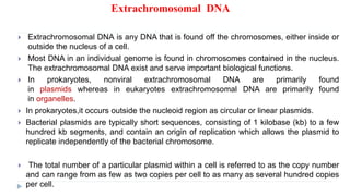
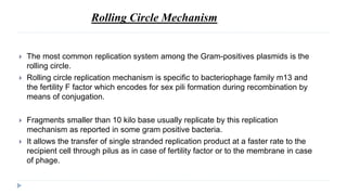
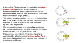
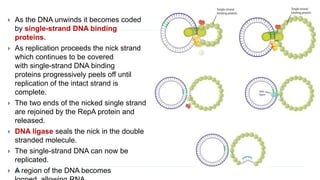
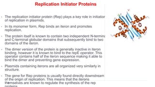
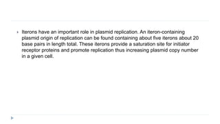
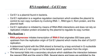
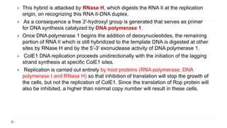
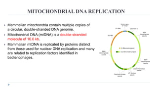
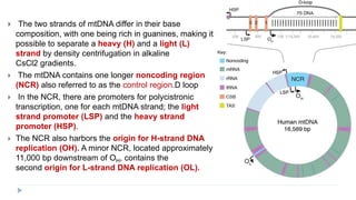
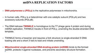
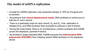
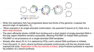
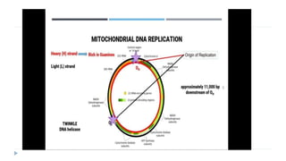
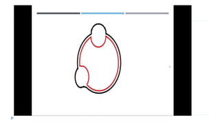
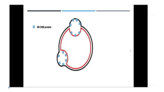
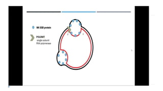
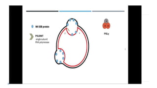
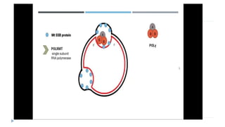
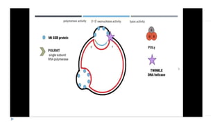
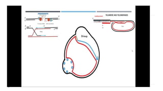
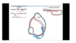
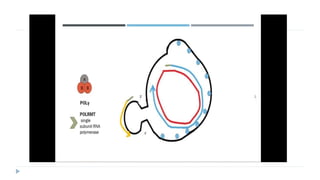
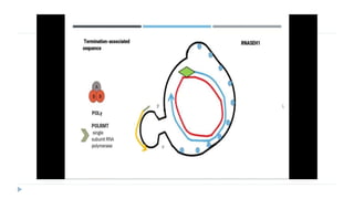
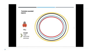
2. contents
Extrachromosomal DNA
Prokaryotic extrachromosomal DNA
Plasmid
Replication mechanisms
Eukaryotic extrachomosomal DNA
Mitochondria
Replication mechanism
Chloroplast
Replication mechanism
3. Extrachromosomal DNA
Extrachromosomal DNA is any DNA that is found off the chromosomes, either inside or
outside the nucleus of a cell.
Most DNA in an individual genome is found in chromosomes contained in the nucleus.
The extrachromosomal DNA exist and serve important biological functions.
In prokaryotes, nonviral extrachromosomal DNA are primarily found
in plasmids whereas in eukaryotes extrachromosomal DNA are primarily found
in organelles.
In prokaryotes,it occurs outside the nucleoid region as circular or linear plasmids.
Bacterial plasmids are typically short sequences, consisting of 1 kilobase (kb) to a few
hundred kb segments, and contain an origin of replication which allows the plasmid to
replicate independently of the bacterial chromosome.
The total number of a particular plasmid within a cell is referred to as the copy number
and can range from as few as two copies per cell to as many as several hundred copies
per cell.
4. Extrachromosomal DNA in the cytoplasm have been found to be structurally different
from nuclear DNA. Cytoplasmic DNA are less methylated than DNA found within the
nucleus.
In addition to DNA found outside the nucleus in cells, infection of viral genomes also
provides an example of extrachromosomal DNA.
Extrachromosomal DNA are often used in research of replication because they are easy
to identify and isolate.
5. PLASMID
Plasmids are small, circular, double stranded, few kilo base self-replicating extra
DNA fragments commonly recognized in different gram negative and positive
bacterial strains as well as in some fungi including unicellular yeasts
They are capable of self replication independent of the host genome.
Though plasmids are not required for survival of a living organism it encodes
essential genetic determinants that enable an organism to adapt and resist
unfavourable conditions for better survival.
Most of them are covalently closed circular double-stranded DNA molecules,
recently linear plasmids have been isolated from different bacteria.
Their existence was initially revealed as the "F factor" in Escherichia coli even
before the double-helix structure of DNA was elucidated by Watson and Crick.
6.
7. REPLICATION
Several host- and plasmid-encoded factors are required for plasmid replication.
Plasmid replicons consists of one or more origin of replication (ori) and
few regulatory elements such as Rep proteins, localized in the 4 kilo base region
of the DNA fragment.
In addition, most plasmid replicons harbor a gene encoding either a protein or an
RNA molecule that functions as a primer for DNA replication.
It was reported that many plasmid origins follow a molecular mechanism similar
to oriC, the origin of replication of the E. coli chromosome. However, the major
difference is that plasmids require an origin-specific plasmid-encoded protein for the
initiation step, generally called Rep proteins.
Rolling circle, Col E1 type and iteron-containing replicons are the
common modes through which plasmid replicates, each mechanism with
unique significance to the organism.
8. Mainly three mechanism;
1. Rolling circle mechanism
2.Iteron regulated mechanism
3.RNA regulated-Col E1 mechanism
9. Rolling Circle Mechanism
The most common replication system among the Gram-positives plasmids is the
rolling circle.
Rolling circle replication mechanism is specific to bacteriophage family m13 and
the fertility F factor which encodes for sex pili formation during recombination by
means of conjugation.
Fragments smaller than 10 kilo base usually replicate by this replication
mechanism as reported in some gram positive bacteria.
It allows the transfer of single stranded replication product at a faster rate to the
recipient cell through pilus as in case of fertility factor or to the membrane in case
of phage.
10. Rolling circle DNA replication is initiated by an initiator
protein (RepA) encoded by the plasmid or
bacteriophage DNA, which nicks one strand of the
double-stranded, circular DNA molecule at a site called
the double-strand origin, or DSO.
The initiator protein remains bound to the 5' phosphate
end of the nicked strand, and the free 3' hydroxyl end is
released to serve as a primer for DNA synthesis
by host DNA polymerase III.
Using the unnicked strand as a template, replication
proceeds around the circular DNA molecule, displacing
the nicked strand as single-stranded DNA.
Displacement (unwinding) of the nicked strand is
carried out by a host-encoded helicase called PcrA
(the abbreviation standing for plasmid copy reduced) in
the presence of RepA.
11. As the DNA unwinds it becomes coded
by single-strand DNA binding
proteins.
As replication proceeds the nick strand
which continues to be covered
with single-strand DNA binding
proteins progressively peels off until
replication of the intact strand is
complete.
The two ends of the nicked single strand
are rejoined by the RepA protein and
released.
DNA ligase seals the nick in the double
stranded molecule.
The single-strand DNA can now be
replicated.
A region of the DNA becomes
12. Host DNA polymerases use the primer
as a starting point for the synthesis of
DNA.
RNA polymerase and DNA
polymerase III then replicate the
single-stranded origin (SSO) DNA to
make another double-stranded circle.
DNA polymerase I removes the primer,
replacing it with DNA, and DNA
ligase joins the ends to make another
molecule of double-stranded circular
DNA.
Each of these plasmids can
undergo replication again by the same
method.
13. Iteron-regulated plasmids
Iterons are directly repeated DNA sequences which play an important role in regulation
of plasmid copy number in bacterial cells. Iterons complex with cognate replication
(Rep) initiator proteins to achieve the required regulatory effect.
This replicon consists of :
a gene that encodes Rep protein ( replication initiator protein ) for plasmid
replication initiation
Origin of replication (ori)
set of direct repeat sequences called iteron (located within the ori )
adjacent AT-rich region
DnaA boxes which is a protein required for bacterial chromosome replication
initiation.
Iterons are the primary DNA binding sites for Rep protein and these sequences
arranged in tandem, direct repeats. (DR)
The conjugative plasmid R6K, a naturally occurring extrachromosomal element that
codes for resistance to the antibiotics ampicillin and streptomycin belongs to the
group of iteron-regulated replicons. It is about 38 kb in size and has a copy number of
13 to 40 per cell.
14. Mechanism:
Iteron contain replication begins with the
binding of Rep proteins to the iteron
being organized in the same orientation
of the DNA helix.
Then this binds to the DnaA boxes in the
replicon, the Rep-DnaA-DNA assembly
promotes melting of the strand at the
nearby AT-rich region to which host
replication factors subsequently gain
access and promote leading and lagging
strand synthesis in a manner analogous
to initiation of replication at the
chromosomal origin, oriC.
15.
16. Replication Initiator Proteins
• The replication initiator protein (Rep) plays a key role in initiation
of replication in plasmids.
• In its monomer form, Rep binds an iteron and promotes
replication.
• The protein itself is known to contain two independent N-terminal
and C-terminal globular domains that subsequently bind to two
domains of the iteron.
• The dimer version of the protein is generally inactive in iteron
binding, however it is known to bind to the repE operator. This
operator contains half of the iteron sequence making it able to
bind the dimer and preventing gene expression.
• Plasmids containing iterons are all organized very similarly in
structure.
• The gene for Rep proteins is usually found directly downstream
of the origin of replication. This means that the iterons
themselves are known to regulate the synthesis of the rep
17. Iterons have an important role in plasmid replication. An iteron-containing
plasmid origin of replication can be found containing about five iterons about 20
base pairs in length total. These iterons provide a saturation site for initiator
receptor proteins and promote replication thus increasing plasmid copy number
in a given cell.
18. RNA regulated – Col E1 type
Col E1 is a plasmid found in bacteria.
Col E1 replication is a negative regulation mechanism which enables the plasmid to
control its own copy numbers by involving RNA I , RNA type II, Rom protein, and the
plasmid itself.
Col E1 replication is initiated by means of RNA-RNA interactions and does not rely on
replication initiation protein encoded by the plasmid to regulate its copy number.
Mechanism :
RNA polymerase initiates transcription of RNA II that originates 555 base pairs
upstream from the replication origin of Col E1 plasmid which marks the start of Col E1
replication.
A determined hybrid with the DNA strand is formed by a loop enriched in G nucleotide
of RNAII and a C-rich region on the template strand upstream from the origin.
The transcript folds into a secondary structure which stabilises the interaction between
the nascent RNA and the origin's DNA. Several stems and loops are exhibited by the
19. This hybrid is attacked by RNase H, which digests the RNA II at the replication
origin, on recognizing this RNA II-DNA duplex.
As a consequence a free 3'-hydroxyl group is generated that serves as primer
for DNA synthesis catalyzed by DNA polymerase 1.
Once DNA polymerase 1 begins the addition of deoxynucleotides, the remaining
portion of RNA II which is still hybridized to the template DNA is digested at other
sites by RNase H and by the 5'-3' exonuclease activity of DNA polymerase 1.
ColE1 DNA replication proceeds unidirectionally with the initiation of the lagging
strand synthesis at specific ColE1 sites.
Replication is carried out entirely by host proteins (RNA polymerase, DNA
polymerase I and RNase H) so that inhibition of translation will stop the growth of
the cells, but not the replication of ColE1. Since the translation of Rop protein will
also be inhibited, a higher than normal copy number will result in these cells.
20. RNAI is a counter-transcript to a
section of RNAII and so binds to its 5'
end.
This alters the folding of RNAII so
that the DNA-RNA hybrid is not
stabilized and cleavage does
not occur.
This ensures that at high copy
numbers, replication is slowed down
due to increased RNAI concentration.
Rop is a secondary replication
repressor, it stabilizes the RNAI-
RNAII hybrid. Rop may be especially
important at preventing runaway
replication, at slow growth rates.
21. COPY NUMBER AND PARTITIONING
Copy number refers to the average or expected number of copies per host cell.
Plasmids are either low, medium or high copy number. Plasmids vary widely in
copy number depending on three main factors:
1) The ori and its constituents – (e.g. ColE1 RNA I and RNA II).
2) The size of the plasmid and its associated insert (bigger inserts and plasmids
may be replicated at a lower number as they represent a great metabolic burden
for the cell).
3) Culture conditions (i.e. factors that influence the metabolic burden on the host).
22. Partition is generally the most important determinant of the stability of low-copy-
number plasmids, which are common in bacteria. In contrast, high-copy-
number plasmids typically do not encode partition systems because random
segregation is sufficient for stability.
Partition is a dynamic process; plasmids are moved and positioned inside the cell
so that cell division separates at least one copy into each daughter cell.
Random segregation of low-copy-number plasmids (only 2 to 4 copies per cell)
would likely mean that, following cell division, one of the daughter cells would not
receive a plasmid. The plasmid would eventually be diluted from the population.
Consequently, regulated partitioning mechanisms are essential for these
plasmids.
23. MITOCHONDRIAL DNA REPLICATION
Mammalian mitochondria contain multiple copies of
a circular, double-stranded DNA genome.
Mitochondrial DNA (mtDNA) is a double-stranded
molecule of 16.6 kb.
Mammalian mtDNA is replicated by proteins distinct
from those used for nuclear DNA replication and many
are related to replication factors identified in
bacteriophages.
24. The two strands of mtDNA differ in their base
composition, with one being rich in guanines, making it
possible to separate a heavy (H) and a light (L)
strand by density centrifugation in alkaline
CsCl2 gradients.
The mtDNA contains one longer noncoding region
(NCR) also referred to as the control region.D loop
In the NCR, there are promoters for polycistronic
transcription, one for each mtDNA strand; the light
strand promoter (LSP) and the heavy strand
promoter (HSP).
The NCR also harbors the origin for H-strand DNA
replication (OH). A minor NCR, located approximately
11,000 bp downstream of OH, contains the
second origin for L-strand DNA replication (OL).
25. mtDNA REPLICATION FACTORS
DNA polymerase γ (POLγ) is the replicative polymerase in mitochondria.
In human cells, POLγ is a heterotrimer with one catalytic subunit (POLγA) and two
accessory subunits (POLγB).
The DNA helicase TWINKLE is homologous to the T7 phage gene 4 protein and during
mtDNA replication, TWINKLE travels in front of POLγ, unwinding the double-stranded DNA
template.
TWINKLE forms a hexamer and requires a fork structure (a single-stranded 5′-DNA
loading site and a short 3′-tail) to load and initiate unwinding.
Mitochondrial single-stranded DNA-binding protein (mtSSB) binds to the formed
ssDNA, protects it against nucleases, and prevents secondary structure formation.
26. The model of mtDNA replication
A model for mtDNA replication was presented already in 1972 by Vinograd and
co-workers.
According to their strand displacement model, DNA synthesis is continuous on
both the H- and L-strand.
There is a dedicated origin for each strand, OH and OL. First, replication is
initiated at OH and DNA synthesis then proceeds to produce a new H-strand.
During the initial phase, there is no simultaneous L-strand synthesis and mtSSB
covers the displaced, parental H-strand.
By binding to single-stranded DNA, mtSSB prevents the mitochondrial RNA
polymerase (POLRMT) from initiating random RNA synthesis on the displaced
strand.
27. When the replication fork has progressed about two-thirds of the genome, it passes the
second origin of replication, OL.
When exposed in its single-stranded conformation, the parental H-strand at OL folds into a
stem–loop structure.
The stem efficiently blocks mtSSB from binding and a short stretch of single-stranded DNA in
the loop region therefore remains accessible, allowing POLRMT to initiate RNA synthesis.
POLRMT is not processive on a single-stranded DNA templates.
Already after about 25 nt, it is replaced by POLγ and L-strand DNA synthesis is initiated.
From this point, H- and L-strand synthesis proceeds continuously until the two strands have
reached full circle. Replication of the two strands is linked, since H-strand synthesis is required
for initiation of L-strand synthesis.
28. When POLγ has completed synthesis, the newly formed DNA strands are ligated by
DNA ligase III.
To allow for efficient ligation, the RNA primers used to initiate mtDNA synthesis must
first be removed. A likely candidate for primer removal is RNASEH1.
After completing a full circle-replication, POLγ encounters the 5′-end of the nascent full-
length mtDNA strand it has just produced.
At this point, POLγ initiates successive cycles of polymerization and 3′–5′ exonuclease
degradation at the nick. This process, idling, is required for proper ligation..
29. SEPARATION mtDNA
During DNA replication, the parental molecule remains intact, which poses a
steric problem for the moving replication machinery.
Topoisomerases belonging to the type 1 family can relieve torsional strain
formed in this way, by allowing one of the strands to pass through the other. In
mammalian mitochondria, TOP1MT a type IB enzyme can act as a DNA
“swivel”.
Replication of intact, circular DNA generates daughter molecules linked together
as catenanes, i.e. mechanically interlocked, but not yet completely finished
DNA circles.
Therefore, replication of circular genomes requires decatenation to generate
complete daughter molecules separation.
.
30. The existence of catenanes in mitochondria was reported already in
1967 by Vinograd and co-workers, who identified mtDNA molecules
linked together during completion of mtDNA replication. Recently, it was
demonstrated that these structures are hemicatenanes, i.e. double-
stranded DNA molecules linked together via a single-stranded linkage
A mitochondrial isoform of Topoisomerase 3α (Top3α) is required to
resolve the hemicatenane structure
31.
32.
33.
34.
35.
36.
37.
38.
39.
40.
41.
42.
43.
44. CHLOROPLASTS
Chloroplasts contain multiple copies of a DNA molecule(the plastome) that
encodes many of the gene products required to perform photosynthesis.
The plastome is replicated by nuclear-encoded proteins and its copy number
seems to be highly regulated by the cell in a tissue-specific and developmental
manner.
The chloroplast genome (plastome) exists as a covalently closed, double
stranded circular ranging in size from 120 kilobase pairs (kbp) in some species to
over 200 kbp in others.
45. ctDNA REPLICATION
The currently prevailing model of DNA replication in chloroplasts is based on electron
microscopic examination of replication intermediates in isolated
pea and maize ctDNA, and was put forth by Kolodner and Tewari.
Replication begins at two displacement (D-) loop initiation sites located on opposite
strands approximately 7 kbp apart.
The D-loops expand unidirectionally toward each other until the advancing forks pass
the D-loop initiation site on the opposite strand, at which
point discontinuous replication begins, resulting in two Cairns-
type, bidirectional forks moving away from each other.
Presumably the forks meet at some point approximately 180 degrees from
the starting point and give rise to two daughter molecules.
46. A. Unidirectional elongation of
nascent strand initiated from
both origins.
B. Unidirectional fork movement
toward each other.
C. Fusion of D-loops.
D. Bidirectional, semi-
discontinuous replication.
E. Resulting daughter
molecules.
47. Replication enzymes:
These include origin recognition protein(s), DNA unwinding activity (helicase),
single stranded DNA binding protein topoisomerase I and II, single stranded DNA
binding protein DNA polymerase(s),
Results obtained with diverse plant types and with plastomes of
different morphologies do not yet allow us to present a generalized picture
of ctDNA replication. What can be stated with confidence is that in higher plants
the ctDNA replication machinery seems to be encoded in the nucleus, which
would place regulation of plastome synthesis under cellular control.