This was a research paper I wrote for my Integrated Laboratory Techniques in Biological Sciences II course.
Evaluation of the lacZ gene in Escherichia coli mutagenesis using pBluescript and pTn5: Km vectors
Organization and Regulation of Mitochondrial Protein SynthesisHeena36363
PRESENTATION OUTLINE
Basics about mitochondria-size structure and functions
Origin of Mitochondria
Mitochondrial Genetic System
Structure of mt-DNA
Translational requirements of mitochondria
Mitoribosomes
Adaptation Of translation machinery To operate within mitochondria
Translation cycle in mitochondria
Regulation Of mitochondrial translation
Culture of Renal Proximal Tubule Epithelial Cell Line SA7K Using Extracellula...mdmitc
MilliporeSigma's Toni Steiner recently presented a poster at the 2016 AAPS/ITC Transporter Workshop demonstrating how culture conditions can influence drug transporter expression and activity in renal proximal tubule epithelial cells.
Generation of MRP2 Efflux Transporter Knock-Out in HepaRG Cell Linemdmitc
MilliporeSigma's Jennifer Pratt recently presented a poster at the 2016 AAPS/ITC Transporter Workshop demonstrating the utility of HepaRG MRP2 Knockout cells for investigating drug-transporter interactions in the liver involving MRP2.
A ribozyme is a ribonucleic acid (RNA) enzyme that catalyses specific reactions in a similar way to that of protein enzymes; it also known as catalytic RNA, ribozymes are found in the ribosome for protein formation and play a role in other vital mechanisms such as RNA splicing, transfer RNA biosynthesis, and viral replication. Discovery of catalytic RNA contributed to the hypothesis of prebiotic RNA world i.e. how life may have originated from an “RNA World” inhabited by self-replicating ribozymes. The ribosome is indeed a ribozyme underlines the relevance of RNA catalysis in today’s protein-dominated world.
The recent discoveries of RNA interference and micro-RNA associated mechanisms of gene regulation further emphasize the central importance of RNA to understanding gene regulation and leads to design new RNA-based technologies for gene manipulation and silencing.
The discovery that riboswitches and in some cases ribozymes, including a variant of the hammerhead ribozyme are also involved in regulating gene expression explains how intimately RNA structure, function, and catalysis are involved in many aspects of biological control.
This presentation explains the fundamentals of Genetic Code, Protein synthesis mechanism and Antibiotics that inhibits at various stages of Translation.
definition of Mitochondrial gene expression
structure of mitochondrial dna
requirment for transcriptional activity
transcription elongation and termination
post transcriptional modification
translation of mitochondrial transcripts
ONLY THE LAST QUESTION IS THE POINT OF POST. THE OTHER PAGES ARE B.pdfamzonknr
ONLY THE LAST QUESTION IS THE POINT OF POST. THE OTHER PAGES ARE
BACKGROUND CONTEXT Lab: Differential Expression Differential gene expression provides
the ability for a cell or organism to respond to a constantly changing external environment. The
specific constellation of proteins required for optimal function and growth varies with cellular
age and environmental context. Thus, protein production is carefully regulated by multiple
mechanisms that modulate both transcriptional and translational pathways. Control of
transcription initiation by RNA polymerase is a predominant mechanism for regulating
expression of specific proteins, presumably because it provides maximal conservation of energy
for the cell. We can often observe the consequence of differential transcription due to the
presence or absence of particular proteins or the growth in particular environments. Control can
also occur at translation; the mRNA is synthesized, but only in certain circumstances is it
translated. Control can also occur at the level of protein function; the protein is inactive, or
activity is not observed due to the lack of the substrate. In this lab we will observe differential
expression of two different genes encoded on plasmids. We will analyze transcriptional activity,
translational activity, and protein function. Plasmids are extra-chromosomal DNA. Bacteria often
have plasmids and will replicate the plasmid and pass it to daughter cells (vertical transmission)
and to neighboring cells (horizontal). Plasmids are a mechanism of gene diversity. In order to
stably retain the plasmid, there needs to be some type of metabolic reason for the bacteria to
maintain the plasmid. In other words, the plasmid confers an advantage. Plasmids contain: 1. Ori:
the plasmid may present is low or high copy number. 2. Lab generated plasmids typically also
contain a selectable marker (antibiotic resistance), 3. Additional gene for ease of visual screening
4. Multiple cloning site
pUC19 is one of a series of plasmid cloning vectors created by Joachim Messing and co-workers.
The designation "pUC" is derived from the classical "p" prefix (denoting "plasmid") and the
abbreviation for the University of California, where early work on the plasmid series had been
conducted. It is a circular double stranded DNA and has 2686 base pairs. pUC19 is one of the
most widely used vector molecules as the recombinants, or the cells into which foreign DNA has
been introduced, can be easily distinguished from the non-recombinants based on color
differences of colonies on growth media. pUC18 is similar to pUC19, but the MCS region is
reversed. - pUC 19 has an origin of replication and is maintained at a high copy number. -
pUC19 encodes for an ampicillin resistance gene (amopR), via a -lactamase enzyme that
functions by degrading ampicillin and reducing its toxicity to the host. - It has an N-terminal
fragment of -galactosidase (lacZ) gene of E. coli which allows for visual screening of
recombinant.
ONLY THE LAST QUESTION IS THE POINT OF POST. THE OTHER PAGES ARE BAC.pdfamzonknr
ONLY THE LAST QUESTION IS THE POINT OF POST. THE OTHER PAGES ARE
BACKGROUND CONTEXT Lab: Differential Expression Differential gene expression provides
the ability for a cell or organism to respond to a constantly changing external environment. The
specific constellation of proteins required for optimal function and growth varies with cellular
age and environmental context. Thus, protein production is carefully regulated by multiple
mechanisms that modulate both transcriptional and translational pathways. Control of
transcription initiation by RNA polymerase is a predominant mechanism for regulating
expression of specific proteins, presumably because it provides maximal conservation of energy
for the cell. We can often observe the consequence of differential transcription due to the
presence or absence of particular proteins or the growth in particular environments. Control can
also occur at translation; the mRNA is synthesized, but only in certain circumstances is it
translated. Control can also occur at the level of protein function; the protein is inactive, or
activity is not observed due to the lack of the substrate. In this lab we will observe differential
expression of two different genes encoded on plasmids. We will analyze transcriptional activity,
translational activity, and protein function. Plasmids are extra-chromosomal DNA. Bacteria often
have plasmids and will replicate the plasmid and pass it to daughter cells (vertical transmission)
and to neighboring cells (horizontal). Plasmids are a mechanism of gene diversity. In order to
stably retain the plasmid, there needs to be some type of metabolic reason for the bacteria to
maintain the plasmid. In other words, the plasmid confers an advantage. Plasmids contain: 1. Ori:
the plasmid may present is low or high copy number. 2. Lab generated plasmids typically also
contain a selectable marker (antibiotic resistance), 3. Additional gene for ease of visual screening
4. Multiple cloning site
pUC19 is one of a series of plasmid cloning vectors created by Joachim Messing and co-workers.
The designation "pUC" is derived from the classical "p" prefix (denoting "plasmid") and the
abbreviation for the University of California, where early work on the plasmid series had been
conducted. It is a circular double stranded DNA and has 2686 base pairs. pUC19 is one of the
most widely used vector molecules as the recombinants, or the cells into which foreign DNA has
been introduced, can be easily distinguished from the non-recombinants based on color
differences of colonies on growth media. pUC18 is similar to pUC19, but the MCS region is
reversed. - pUC 19 has an origin of replication and is maintained at a high copy number. -
pUC19 encodes for an ampicillin resistance gene (amopR), via a -lactamase enzyme that
functions by degrading ampicillin and reducing its toxicity to the host. - It has an N-terminal
fragment of -galactosidase (lacZ) gene of E. coli which allows for visual screening of
recombinant.
Lab: Differential Expression Differential gene expression provides the ability for a cell or
organism to respond to a constantly changing external environment. The specific constellation of
proteins required for optimal function and growth varies with cellular age and environmental
context. Thus, protein production is carefully regulated by multiple mechanisms that modulate
both transcriptional and translational pathways. Control of transcription initiation by RNA
polymerase is a predominant mechanism for regulating expression of specific proteins,
presumably because it provides maximal conservation of energy for the cell. We can often
observe the consequence of differential transcription due to the presence or absence of particular
proteins or the growth in particular environments. Control can also occur at translation; the
mRNA is synthesized, but only in certain circumstances is it translated. Control can also occur at
the level of protein function; the protein is inactive, or activity is not observed due to the lack of
the substrate. In this lab we will observe differential expression of two different genes encoded
on plasmids. We will analyze transcriptional activity, translational activity, and protein function.
Plasmids are extra-chromosomal DNA. Bacteria often have plasmids and will replicate the
plasmid and pass it to daughter cells (vertical transmission) and to neighboring cells (horizontal).
Plasmids are a mechanism of gene diversity. In order to stably retain the plasmid, there needs to
be some type of metabolic reason for the bacteria to maintain the plasmid. In other words, the
plasmid confers an advantage. Plasmids contain: 1. Ori: the plasmid may present is low or high
copy number. 2. Lab generated plasmids typically also contain a selectable marker (antibiotic
resistance), 3. Additional gene for ease of visual screening 4. Multiple cloning site
pUC19 is one of a series of plasmid cloning vectors created by Joachim Messing and co-workers.
The designation "pUC" is derived from the classical "p" prefix (denoting "plasmid") and the
abbreviation for the University of California, where early work on the plasmid series had been
conducted. It is a circular double stranded DNA and has 2686 base pairs. pUC19 is one of the
most widely used vector molecules as the recombinants, or the cells into which foreign DNA has
been introduced, can be easily distinguished from the non-recombinants based on color
differences of colonies on growth media. pUC18 is similar to pUC19, but the MCS region is
reversed. - pUC 19 has an origin of replication and is maintained at a high copy number. -
pUC19 encodes for an ampicillin resistance gene (amopR), via a -lactamase enzyme that
functions by degrading ampicillin and reducing its toxicity to the host. - It has an N-terminal
fragment of -galactosidase (lacZ) gene of E. coli which allows for visual screening of
recombinant plasmids. The transformed cells containing the plasmid with the gene of interest ca.
DNA damage repair Neil3 gene Knockout in MOLT-4iosrjce
RNAi is superannuated cellular mechanism that protect organism against viruses that replicate
through double- stranded RNA. RNAi can be used to diminish gene expression from plasmid expressing and
inserted sequence repeat. A stable harpin would be expressed after the vector was integrated into the genome.
In this paper a shiRNA expressing vector for Neil3 was designed and developed which is capable of replication
in MOLT-4. This shiRNA vector had the ability to arose the RNAi pathway, and reduce the gene expression of
Neil3. This was assessed by using pSilence 4.1CMV as a vector, and Gapdh as positive control.
This (final exam) is part of the requirement for Southeast Asian Studies, a course I took at Mahidol University International College. There are five different responses, which all discuss social issues related to Southeast Asia.
This is a presentation I created for my ICBI 436 Industrial Enzymology, a Biotechnology course I took at Mahidol University International College (MUIC)
This is a presentation I presented during my pathobiology course in college. The presentation discusses vitamin B3 deficiency. in terms of background, nutrition, pathogenesis, and signs & symptoms.
Serum Ischemia-Modified Albumin in Preterm Babies with Respiratory Distress S...Emilio Solomon
This is a presentation from my seminar in biological sciences course. The presentations discusses serum ischemia-modified albumin in preterm babies with respiratory distress syndrome.
This was an evaluation of a study I presented in my seminar class for my major.
Daily sesame oil supplement attenuates joint pain by inhibiting muscular oxidative stress in osteoarthritis rat model
“Immobilization of urease using glycidyl methacrylate grafted nylon-6-membranes”Emilio Solomon
This was a paper I did for Industrial Enzymology; a Biotechnology elective course in my college.
“Immobilization of urease using glycidyl methacrylate grafted nylon-6-membranes”
Evaluation of antioxidant properties in different colors of asian rice (oryza...Emilio Solomon
Senior project for my major (Biological Sciences: Biomedical Science concentration)
Evaluation of antioxidant properties in different colors of asian rice (oryza sativa) against insecticide carbosulfan using mealworm (Tenebrio molitor) assay
This is a pdf file showing how I study for college Human Anatomy (Introduction). I used a surface pro 3 for these notes. Any aspiring doctors and nurses can take a look at this document
This is an essay I wrote during my sophomore year of college. It's for my Introduction to Philosophy class. It's a redo assignment, which discusses Hume's and Descartes' skeptical views.
This is my senior thesis. My project focuses on the evaluation of antioxidant properties of different colors of asian rice against carbosulfan insecticide using mealworms.
THE IMPORTANCE OF MARTIAN ATMOSPHERE SAMPLE RETURN.Sérgio Sacani
The return of a sample of near-surface atmosphere from Mars would facilitate answers to several first-order science questions surrounding the formation and evolution of the planet. One of the important aspects of terrestrial planet formation in general is the role that primary atmospheres played in influencing the chemistry and structure of the planets and their antecedents. Studies of the martian atmosphere can be used to investigate the role of a primary atmosphere in its history. Atmosphere samples would also inform our understanding of the near-surface chemistry of the planet, and ultimately the prospects for life. High-precision isotopic analyses of constituent gases are needed to address these questions, requiring that the analyses are made on returned samples rather than in situ.
A brief information about the SCOP protein database used in bioinformatics.
The Structural Classification of Proteins (SCOP) database is a comprehensive and authoritative resource for the structural and evolutionary relationships of proteins. It provides a detailed and curated classification of protein structures, grouping them into families, superfamilies, and folds based on their structural and sequence similarities.
Richard's entangled aventures in wonderlandRichard Gill
Since the loophole-free Bell experiments of 2020 and the Nobel prizes in physics of 2022, critics of Bell's work have retreated to the fortress of super-determinism. Now, super-determinism is a derogatory word - it just means "determinism". Palmer, Hance and Hossenfelder argue that quantum mechanics and determinism are not incompatible, using a sophisticated mathematical construction based on a subtle thinning of allowed states and measurements in quantum mechanics, such that what is left appears to make Bell's argument fail, without altering the empirical predictions of quantum mechanics. I think however that it is a smoke screen, and the slogan "lost in math" comes to my mind. I will discuss some other recent disproofs of Bell's theorem using the language of causality based on causal graphs. Causal thinking is also central to law and justice. I will mention surprising connections to my work on serial killer nurse cases, in particular the Dutch case of Lucia de Berk and the current UK case of Lucy Letby.
Introduction:
RNA interference (RNAi) or Post-Transcriptional Gene Silencing (PTGS) is an important biological process for modulating eukaryotic gene expression.
It is highly conserved process of posttranscriptional gene silencing by which double stranded RNA (dsRNA) causes sequence-specific degradation of mRNA sequences.
dsRNA-induced gene silencing (RNAi) is reported in a wide range of eukaryotes ranging from worms, insects, mammals and plants.
This process mediates resistance to both endogenous parasitic and exogenous pathogenic nucleic acids, and regulates the expression of protein-coding genes.
What are small ncRNAs?
micro RNA (miRNA)
short interfering RNA (siRNA)
Properties of small non-coding RNA:
Involved in silencing mRNA transcripts.
Called “small” because they are usually only about 21-24 nucleotides long.
Synthesized by first cutting up longer precursor sequences (like the 61nt one that Lee discovered).
Silence an mRNA by base pairing with some sequence on the mRNA.
Discovery of siRNA?
The first small RNA:
In 1993 Rosalind Lee (Victor Ambros lab) was studying a non- coding gene in C. elegans, lin-4, that was involved in silencing of another gene, lin-14, at the appropriate time in the
development of the worm C. elegans.
Two small transcripts of lin-4 (22nt and 61nt) were found to be complementary to a sequence in the 3' UTR of lin-14.
Because lin-4 encoded no protein, she deduced that it must be these transcripts that are causing the silencing by RNA-RNA interactions.
Types of RNAi ( non coding RNA)
MiRNA
Length (23-25 nt)
Trans acting
Binds with target MRNA in mismatch
Translation inhibition
Si RNA
Length 21 nt.
Cis acting
Bind with target Mrna in perfect complementary sequence
Piwi-RNA
Length ; 25 to 36 nt.
Expressed in Germ Cells
Regulates trnasposomes activity
MECHANISM OF RNAI:
First the double-stranded RNA teams up with a protein complex named Dicer, which cuts the long RNA into short pieces.
Then another protein complex called RISC (RNA-induced silencing complex) discards one of the two RNA strands.
The RISC-docked, single-stranded RNA then pairs with the homologous mRNA and destroys it.
THE RISC COMPLEX:
RISC is large(>500kD) RNA multi- protein Binding complex which triggers MRNA degradation in response to MRNA
Unwinding of double stranded Si RNA by ATP independent Helicase
Active component of RISC is Ago proteins( ENDONUCLEASE) which cleave target MRNA.
DICER: endonuclease (RNase Family III)
Argonaute: Central Component of the RNA-Induced Silencing Complex (RISC)
One strand of the dsRNA produced by Dicer is retained in the RISC complex in association with Argonaute
ARGONAUTE PROTEIN :
1.PAZ(PIWI/Argonaute/ Zwille)- Recognition of target MRNA
2.PIWI (p-element induced wimpy Testis)- breaks Phosphodiester bond of mRNA.)RNAse H activity.
MiRNA:
The Double-stranded RNAs are naturally produced in eukaryotic cells during development, and they have a key role in regulating gene expression .
Nutraceutical market, scope and growth: Herbal drug technologyLokesh Patil
As consumer awareness of health and wellness rises, the nutraceutical market—which includes goods like functional meals, drinks, and dietary supplements that provide health advantages beyond basic nutrition—is growing significantly. As healthcare expenses rise, the population ages, and people want natural and preventative health solutions more and more, this industry is increasing quickly. Further driving market expansion are product formulation innovations and the use of cutting-edge technology for customized nutrition. With its worldwide reach, the nutraceutical industry is expected to keep growing and provide significant chances for research and investment in a number of categories, including vitamins, minerals, probiotics, and herbal supplements.
Cancer cell metabolism: special Reference to Lactate PathwayAADYARAJPANDEY1
Normal Cell Metabolism:
Cellular respiration describes the series of steps that cells use to break down sugar and other chemicals to get the energy we need to function.
Energy is stored in the bonds of glucose and when glucose is broken down, much of that energy is released.
Cell utilize energy in the form of ATP.
The first step of respiration is called glycolysis. In a series of steps, glycolysis breaks glucose into two smaller molecules - a chemical called pyruvate. A small amount of ATP is formed during this process.
Most healthy cells continue the breakdown in a second process, called the Kreb's cycle. The Kreb's cycle allows cells to “burn” the pyruvates made in glycolysis to get more ATP.
The last step in the breakdown of glucose is called oxidative phosphorylation (Ox-Phos).
It takes place in specialized cell structures called mitochondria. This process produces a large amount of ATP. Importantly, cells need oxygen to complete oxidative phosphorylation.
If a cell completes only glycolysis, only 2 molecules of ATP are made per glucose. However, if the cell completes the entire respiration process (glycolysis - Kreb's - oxidative phosphorylation), about 36 molecules of ATP are created, giving it much more energy to use.
IN CANCER CELL:
Unlike healthy cells that "burn" the entire molecule of sugar to capture a large amount of energy as ATP, cancer cells are wasteful.
Cancer cells only partially break down sugar molecules. They overuse the first step of respiration, glycolysis. They frequently do not complete the second step, oxidative phosphorylation.
This results in only 2 molecules of ATP per each glucose molecule instead of the 36 or so ATPs healthy cells gain. As a result, cancer cells need to use a lot more sugar molecules to get enough energy to survive.
introduction to WARBERG PHENOMENA:
WARBURG EFFECT Usually, cancer cells are highly glycolytic (glucose addiction) and take up more glucose than do normal cells from outside.
Otto Heinrich Warburg (; 8 October 1883 – 1 August 1970) In 1931 was awarded the Nobel Prize in Physiology for his "discovery of the nature and mode of action of the respiratory enzyme.
WARNBURG EFFECT : cancer cells under aerobic (well-oxygenated) conditions to metabolize glucose to lactate (aerobic glycolysis) is known as the Warburg effect. Warburg made the observation that tumor slices consume glucose and secrete lactate at a higher rate than normal tissues.
Slide 1: Title Slide
Extrachromosomal Inheritance
Slide 2: Introduction to Extrachromosomal Inheritance
Definition: Extrachromosomal inheritance refers to the transmission of genetic material that is not found within the nucleus.
Key Components: Involves genes located in mitochondria, chloroplasts, and plasmids.
Slide 3: Mitochondrial Inheritance
Mitochondria: Organelles responsible for energy production.
Mitochondrial DNA (mtDNA): Circular DNA molecule found in mitochondria.
Inheritance Pattern: Maternally inherited, meaning it is passed from mothers to all their offspring.
Diseases: Examples include Leber’s hereditary optic neuropathy (LHON) and mitochondrial myopathy.
Slide 4: Chloroplast Inheritance
Chloroplasts: Organelles responsible for photosynthesis in plants.
Chloroplast DNA (cpDNA): Circular DNA molecule found in chloroplasts.
Inheritance Pattern: Often maternally inherited in most plants, but can vary in some species.
Examples: Variegation in plants, where leaf color patterns are determined by chloroplast DNA.
Slide 5: Plasmid Inheritance
Plasmids: Small, circular DNA molecules found in bacteria and some eukaryotes.
Features: Can carry antibiotic resistance genes and can be transferred between cells through processes like conjugation.
Significance: Important in biotechnology for gene cloning and genetic engineering.
Slide 6: Mechanisms of Extrachromosomal Inheritance
Non-Mendelian Patterns: Do not follow Mendel’s laws of inheritance.
Cytoplasmic Segregation: During cell division, organelles like mitochondria and chloroplasts are randomly distributed to daughter cells.
Heteroplasmy: Presence of more than one type of organellar genome within a cell, leading to variation in expression.
Slide 7: Examples of Extrachromosomal Inheritance
Four O’clock Plant (Mirabilis jalapa): Shows variegated leaves due to different cpDNA in leaf cells.
Petite Mutants in Yeast: Result from mutations in mitochondrial DNA affecting respiration.
Slide 8: Importance of Extrachromosomal Inheritance
Evolution: Provides insight into the evolution of eukaryotic cells.
Medicine: Understanding mitochondrial inheritance helps in diagnosing and treating mitochondrial diseases.
Agriculture: Chloroplast inheritance can be used in plant breeding and genetic modification.
Slide 9: Recent Research and Advances
Gene Editing: Techniques like CRISPR-Cas9 are being used to edit mitochondrial and chloroplast DNA.
Therapies: Development of mitochondrial replacement therapy (MRT) for preventing mitochondrial diseases.
Slide 10: Conclusion
Summary: Extrachromosomal inheritance involves the transmission of genetic material outside the nucleus and plays a crucial role in genetics, medicine, and biotechnology.
Future Directions: Continued research and technological advancements hold promise for new treatments and applications.
Slide 11: Questions and Discussion
Invite Audience: Open the floor for any questions or further discussion on the topic.
Observation of Io’s Resurfacing via Plume Deposition Using Ground-based Adapt...Sérgio Sacani
Since volcanic activity was first discovered on Io from Voyager images in 1979, changes
on Io’s surface have been monitored from both spacecraft and ground-based telescopes.
Here, we present the highest spatial resolution images of Io ever obtained from a groundbased telescope. These images, acquired by the SHARK-VIS instrument on the Large
Binocular Telescope, show evidence of a major resurfacing event on Io’s trailing hemisphere. When compared to the most recent spacecraft images, the SHARK-VIS images
show that a plume deposit from a powerful eruption at Pillan Patera has covered part
of the long-lived Pele plume deposit. Although this type of resurfacing event may be common on Io, few have been detected due to the rarity of spacecraft visits and the previously low spatial resolution available from Earth-based telescopes. The SHARK-VIS instrument ushers in a new era of high resolution imaging of Io’s surface using adaptive
optics at visible wavelengths.
Evaluation of the lacZ gene in Escherichia coli mutagenesis using pBluescript and pTn5: Km vectors
1. 1
Evaluation of the lacZ gene in Escherichia coli mutagenesis using pBluescript and pTn5:
Km vectors
Emilio Solomon
5580132
Abstract
The lacZ gene in Escherichia coli was used for mutagenesis using pBluescript and pTn5:
Km vectors to evaluate its functionality and the activity of β-D-galactosidase, its encoded
enzyme. Microbiological and molecular lab protocols were performed, including medium
preparation, competent cell preparation, transposition, transformation, screening, alkaline lysis,
restriction endonuclease digestion, and gel electrophoresis. Medium preparation, competent cell
preparation, transposition, transformation, and screening were conducted to evaluate the lacZ
gene’s functionality and the activity of β-D-galactosidase. Total loss of lacZ gene function and a
non-functional β-D-galactosidase were observed. Alkaline lysis, restriction endonuclease
digestion, and gel electrophoresis were used to determine the size of the DNA fragments in
recombinant plasmid; pBluescript and pTn5: Km and its non-recombinant counterpart;
pBluescript, as well as to map restriction sites in both recombinant and non-recombinant
plasmids. Proposed restriction maps corresponded with the DNA fragment sizes from gel
electrophoresis and were validated by the concentration of agarose used.
1. Introduction
Transposons are transposable elements that are capable of undergoing translocation
within chromosomal, phage, or plasmid DNA. There are three classes of transposons, including
Class I elements, Class II elements, and Helitrons. Class I elements are known as
retrotransposons. These transposons transpose via a copy and paste mechanism. In this copy and
paste mechanism, the mRNA transcribed from RNA polymerase II is converted into cDNA by
reverse transcription and then integrated at a new position in the genome. Class I elements can
further be sub-divided into long terminal repeats (LTR) and non-LTR elements. Both differ in
the mechanism of integration. Long terminal repeats encode all of the necessary proteins for
2. 2
transposition. Non-LTR elements require enzymes, encoded by LTR elements. Class II elements
transpose via a cut and paste mechanism. In the cut and paste mechanism, the element excised
from the chromosome is reintegrated at a new location. This process involves a transposase
enzyme encoded by the transposon. Helitrons are transposons that transpose via a rolling circle
mechanism. This process involves nicking at the Helitron terminus, followed by strand invasion,
DNA synthesis, strand displacement, and the resolution of a heteroduplex by DNA replication.
Its rolling circle mechanism is regulated through flanking. Flanking occurs when DNA synthesis
and strand displacement proceeds farther than the end of the Helitron.
The lacZ gene is a gene that encodes for the enzyme, β-D-galactosidase. This enzyme is
responsible for hydrolyzing lactose into galactose and glucose. The lacZ gene is regulated in the
lac operon. For example, when the lac operon is turned on, the lacZ gene will facilitate the
hydrolysis of lactose into galactose and glucose. When the lac operon is turned off, the lacZ gene
will not be activated. This will only occur when glucose is present in high concentrations in cells.
The lacZ gene is used for a variety of purposes, according to various journals. According to a
journal from FEMS Microbiology Letters, the lacZ gene can be used for the characterization and
expression of genes from Yersinia pestis and Escherichia coli (Bobrov & Perry, 2006). The lacZ
gene can also be used for cloning PCR-amplified gene promoters on antibiotic resistant plasmids
in the lac operon (Datsenko & Wanner, 2000). According to a journal from Gene Expression
Patterns, the lacZ gene can be used for investigating the functions of Dapper antagonist of
catenin-1 (Dact1) in Wnt-mediated organogenesis and tissue homeostasis in mice (Suzuki, Leu,
Brice, & Senoo, 2014). In humans, the lacZ gene can be used to study Tem1 ontogeny (Huang et
al., 2011). Researchers from Mutation Research/Genetic Toxicology and Environmental
Mutagenesis have used the lacZ gene in order to detect mutagens and clastogens in mice
(Mahabir et al., 2008).
X-gal, or 5-bromo-4-chloro-3-indolyl-β-D-galactopyranoside is an analogue of lactose. β-
D-galactosidase is also capable of digesting X-gal, however the product after hydrolysis is
different. X-gal is digested into galactose and 5-bromo-4-chloro-3-hydroxyindole. The
byproducts can further be dimerized and oxidized into 5, 5’ –dibromo- 4, 4’-dichloroindigo. 5,
5’-dibromo- 4, 4’- dichloroindigo would appear as an insoluble, blue product. Escherichia coli
(E. coli) is a gram-negative, facultative anaerobic bacterium commonly found in the intestine of
3. 3
warm-blooded organisms, including humans. It is capable of growing rapidly and is able to grow
with or without the presence of oxygen. Its genome is well understood by scientists and is often
used as a host for cell culturing and molecular cloning. E. coli contains genes, including the lacZ
gene in the form of plasmid DNA. Plasmid DNA in E. coli contains restriction sites, where
restriction enzyme digestion can occur. Restriction enzyme digestion is facilitated through
restriction enzymes, restriction endonucleases. Many restriction endonucleases, including EcoRI,
HinDIII, and BamHI are capable of digesting the plasmid DNA at various restriction sites. These
restriction sites contain specific segments of nucleotide bases.
This research investigation aims to evaluate the lacZ gene in Escherichia coli
mutagenesis using vectors pBluescript (pGEM), containing the lacZ gene and pTn5: Km
(pMOD). Parameters such as the functionality of the lacZ gene and β-D-galactosidase activity
will be investigated. The research investigation also aims to determine the restriction sites in
pBluescript with or without pTn5: Km. The experiment will involve the use of Luria-Bertani
(LB), a medium present in complex and chemically-defined forms. The complex medium
appears as a liquid, composing of Peptone, yeast extract, and sodium chloride, NaCl. Peptone
acts as a protein source, which will provide amino acids and peptides to E. coli. Yeast extract
contains vitamins, minerals, and other nutrients, acting as a carbon source. NaCl in LB helps in
providing sodium and chlorine ions. The chemically-defined medium appears as a solid. This
medium is composed of the broth, with the addition of agar. The agar helps in solidifying the
medium. Both complex (broth) and chemically defined (agar) media will be prepared. The
prepared agar and broth will then be used for making competent cells. Competent cells will be
made using techniques such as cell suspension, centrifugation, and incubation. Cell suspension
will help increase cell competence. Centrifugation will help separate the pellet (protein) from the
supernatant (DNA) in the competent cells. Incubation will help maintain the competent cells
under optimal conditions. Once the competent cells are made, the cells will be transferred into
agar plates containing ampicillin only. These competent cells will be spread through the agar to
distribute cells evenly. The spreading of competent cells will be done aseptically to avoid
contamination. Ampicillin agar plates, containing competent E. coli cells will be treated with X-
gal (5-bromo-4-chloro-3-indolyl-β-D-galactopyranoside) and reaction mixture containing
reaction buffer, vectors pBluescript (pGEM) and pTn5: Km (pMOD), enzyme transposase, and
sterile water for the transposition reaction, followed by transformation. Transformation will
4. 4
result in either the formation of blue colonies, white colonies, or both. Blue colonies will
demonstrate β-D-galactosidase activity, while white colonies will not show any β-D-
galactosidase activity. After transformation, only the white colonies will be selected for
screening. The screening process will help determine the functionality of the lacZ gene.
Ampicillin and ampicillin/kanamycin agar plates will be used for screening. Blue and white
colonies will be observed following the screening process. Blue colonies will indicate a
functional lacZ gene whereas white colonies will indicate a non-functional lacZ gene. The
frequency of transposition, which is defined as the number of colonies after transformation over
the number of colonies before transformation will help determine the extent of lacZ gene
functionality. Techniques such as alkaline lysis and restriction enzyme digestion, will be
employed subsequently in order to extract and purify the plasmid DNA of E. coli and cut the
plasmid DNA/determine the site of insertion of pTn5: Km vector, respectively. Alkaline lysis
will be performed using suspension, lysis, neutralizing, and alcohol solutions. Finally, the
plasmid DNA will be separated through gel electrophoresis using 1% agarose, Tris-base EDTA
buffer, purple tracking dye, and fluorescein staining solution. Tris-base EDTA buffer will help
maintain the ionization of the DNA. Purple tracking-dye will help make the DNA denser. This
will help in visualizing the separated DNA. Fluorescein staining solution will help in visualizing
the DNA under UV light. The sizes of the DNA fragments will be determined and used to map
restriction sites in pBluescript, with or without pTn5: Km insert.
2. Materials and Methods
2.1 Chemicals/solutions
0.2 M NaOH
1% (w/v) SDS
100x BSA
10x buffer
15% sucrose
70% ethanol
Acetic acid
Agar
Agarose powder
Ampicillin
Calcium chloride
Distilled water
EDTA
Ethanol
EZ- Tn5 reaction buffer
Fluorescein solution
5. 5
Glycerol
Ice
Isopropanol
Kanamycin
Manganese (II) chloride
Milli-Q water
pBluescript
Peptone
Potassium acetate
pTn5: Km
Restriction enzyme (EcoRI,
BamHI)
Sodium acetate
Sodium chloride (NaCl)
Stop solution
SYBR®
Transposase
Tris-base EDTA (TBE)
buffer
Tris-HCl
X-gal (5-bromo-4-chloro-3-
indolyl-β-D-galacto-pyranoside)
Yeast extract
2.2 Devices/machines and miscellaneous materials
Aliquot
Autoclave
Beaker
Burner
Centrifuge
Eppendorf tube
Erlenmeyer flask
Horizontal electrophoresis
chamber
Incubator
Petri dish
Pipettes
Refrigerator
Spreader
Tip
Toothpick
UV trans illuminator
2.3.1 Identification of an unknown sequence
Using the partial DNA sequence and partial amino acid sequence of an unknown gene, a
BLAST search was performed. Through the BLAST search, the unknown gene of the partial
DNA sequence and partial amino acid sequence was identified. Information on the name of the
unknown gene, host organism, protein encoded by the gene, metabolic reaction, and the use of
the gene in molecular cloning were obtained.
6. 6
2.3.2 Medium preparation
Luria-Bertani (LB) broth and agar were prepared. 100 ml of LB broth was prepared using
1 gm of Peptone, 0.5 gm of Yeast extract, 0.5 gm of sodium chloride (NaCl), and 100 ml of
distilled water. 30 ml and 3 ml of LB broth were then aliquoted into a flask and plastic tubes,
respectively. The medium was then autoclaved. LB agar was made using LB broth and agar and
transferred to bottles. Antibiotics, ampicillin and kanamycin were added into the bottles,
containing LB agar. Ampicillin was added to one bottle to a final concentration of 100 μg/ml.
Both ampicillin and kanamycin were added to another bottle. Ampicillin was added to a
concentration of 100 μg/ml, followed by the addition of kanamycin to a final concentration of 50
μg/ml. LB agar with Amp and LB agar with Amp/Kan were then poured into their respective agar plates
and were left to solidify.
2.3.3 Competent cell preparation
Starter cultures were grown in 5 ml of LB broth until the stationary phase was achieved.
0.3 ml of starter cultures were then transferred into pre-warmed (37° C) 30-ml LB broths. Broths
were then grown at 37° C, followed by shaking for 2.5 hrs. Shaking was done until OD600 of 0.3-
0.4 was achieved. After the growth and shaking of culture, culture was poured carefully into a 50
ml centrifuge tube. The tube was chilled on ice for 15 minutes and cells were centrifuged for 10
min at 7520 ×g and 4° C for harvest. The supernatant was discarded into a special disposal bin
and the pellet was resuspended into culture medium containing 10 mM of sodium acetate,
CH3COONa (pH= 5.6), 50 mM of manganese (II) chloride MnCl2, and 5 mM of sodium
chloride, NaCl. The mixture was then incubated on ice for 20 min and centrifuged at 7520 ×g, 4°
C for 10 min. The supernatant was discarded and the pellet was resuspended in culture medium
containing 10 mM of CH3COONa (pH= 5.0), 70 mM of CaCl2, 5 mM of MnCl2, and 5%
glycerol. The mixture was then incubated on ice for at least 30 minutes, but no more than 1 hour.
Meanwhile, five Eppendorf tubes were prepared and labelled. Once cold incubation was done,
100 μl of the mixture, containing the competent cells were transferred into 1.5 ml Eppendorf
tubes (with labels). This was done aseptically. The mixture was then stored at -80° C.
7. 7
2.3.4 Transposition
10 µl of reaction mixtures were prepared for the transposition reaction. One reaction
mixture using 1 μl of 10x EZ- Tn5 reaction buffer, 1 μl of pBluescript (pGEM), 1 µl of pTn5:
Km (pMOD), 1 μl of transposase, and 6 µl of distilled water. One reaction mixture (without
pMOD) containing 1 μl of 10x EZ- Tn5 reaction buffer, 1 μl of pGEM, 1 μl of transposase, and
7 μl of distilled water. One reaction mixture (without transposase) containing 1 μl of 10x EZ-
Tn5 reaction buffer, 1 μl of pGEM, 1 μl of pMOD, and 7 µl of distilled water. Reaction mixtures
were then incubated at 37° C for 2 hrs. Stop solution was then added into each mixture and
further incubated at 70° C for 10 min. Reaction mixtures were then transferred into the
Eppendorf tubes (containing competent cells) and incubated on ice for 30 min. Tubes containing
competent cells and transposition reaction mixture were transferred into new, empty Eppendorf
tubes. Tubes were then placed in a water bath at 42° C and incubated for exactly 45 seconds.
Tubes were transferred and placed in a cold bath, for 30 min. After incubation, 1 ml of LB broth
was added into each tube and resuspended. 300 µl, 100 µl, 50 µl, and 5 µl of suspension were
added into 4 ampicillin-LB agar plates. 300 µl of reaction mixture without pMOD and 300 µl of
reaction mixture without transposase were added into their respective ampicillin-LB agar plates.
All agar plates were incubated for 24 hrs for the transposition reaction to occur. Transposition
was repeated using two reaction mixtures; one with 1 µl of reaction buffer, 1 µl of pGEM, 1 µl
of pMOD, 1 µl of transposase, and 6 µl of distilled water and another one with 1 µl of reaction
buffer, 1 µl of pMOD, 1 µl of transposase, and 7 µl of distilled water, if no growth was observed
at all during the transformation process. For the protocol repeat, only two amp agar plates with
100 µl of reaction mixture were used.
2.3.5 Transformation/Screening
After transposition, white colonies were selected for transformation. Prior to
transformation, ampicillin agar plates were gridded into 50 regions. Selected white colonies were
inoculated/transferred to one region in the ampicillin-LB agar plates (repeat protocol) one by one
using toothpicks aseptically. Ampicillin-LB agar plates containing the colonies were then,
incubated for 24 hrs for growth. Following incubation, only white colonies were selected and
transferred to two gridded LB agar plates (1 ampicillin-LB, 1 ampicillin/kanamycin- LB) using
8. 8
toothpicks aseptically. Both agar plates were incubated for 24 hrs for growth. Clones from only
amp/kan agar plates were then inoculated in 3 ml LB broth using a pipette aseptically. The tip
was left inside the tube with LB broth containing ampicillin and kanamycin and incubated for 24
hrs for growth. Cells were then, harvested by centrifugation for alkaline lysis.
2.3.6 Alkaline lysis
The supernatant was discarded and the pellet was resuspended in 250 µl of suspension
solution containing 25 mM Tris-HCl (pH= 8.0), 10 mM EDTA (pH= 8.0), and 15% to create
pores in the cell membrane, as well as maintain the cell’s osmolarity. 250 µl of lysis solution
containing 0.2 M sodium hydroxide (NaOH) and 1% (w/v) sodium dodecyl sulfate (SDS) was
then applied. The tube was then inverted for cell lysis. This helped in separating the DNA from
protein. Once the cells have been lysed, 350 µl of neutralizing solution containing 2 M potassium
acetate (CH3COOK) and 1 M acetic acid (CH3COOH) was added to prevent further lysis. This
was followed by tube inverting and centrifugation to reanneal the lysed DNA. The supernatant
was then transferred into a new tube and added with 500 µl of isopropanol. Tube was inverted
and centrifuged for 5 minutes. The supernatant was discarded and the pellet was resuspended in
500 µl of 70% ethanol and centrifuged for 2 minutes. The addition of isopropanol and 70%
ethanol helped in precipitating the DNA. The supernatant was discarded and the pellet was air
dried over paper towels at room temperature for approximately 30 min. The pellet was then
resuspended in 50 µl of sterile Milli-Q water for purification.
2.3.7 Restriction enzyme digestion
10 to 15 µl of purified plasmid DNA was mixed with 5 µl of 10x buffer, 0.5 µl of 100x
BSA, 1 µl of restriction enzyme in a new, empty Eppendorf tube. Milli-Q water was added to a
total volume of 50 µl. Mixture was then incubated for 24 hrs for restriction enzyme digestion to
occur.
9. 9
2.3.8 Gel electrophoresis
5 µl of digested DNA was aliquoted into 5 new, empty Eppendorf tubes each. 1 µl of 6x
purple loading dye was added into all 5 tubes. Meanwhile, 1% agarose with Tris-base EDTA
(TBE) buffer was prepared, using 0.2 gm of agarose powder and 10 ml of TBE buffer. The
mixture was then poured into a well to solidify into a gel-like form. Once solidified, the gel was
transferred to a horizontal electrophoresis chamber containing TBE buffer. The horizontal
electrophoresis chamber was then powered with electricity and left for 30 min for the DNA to
migrate/separate. Gel electrophoresis results were analyzed and restriction maps were proposed.
Proposed restriction maps were checked for validation using the concentration of agarose used.
3. Results
3.1 Unknown sequence
After the BLAST search was performed using the given partial DNA and partial amino
acid sequences, the name of the unknown gene, as well as information about its host organism,
protein encoded, metabolic reaction, and uses in molecular cloning were obtained. The name of
the unknown gene is the lacZ gene. This gene encodes for the enzyme, β-D-galactosidase, an
enzyme that catalyzes the hydrolysis of lactose into galactose and glucose (Formula 3.1.1). The
lacZ gene is found in a variety of warm-blooded organisms, including humans. The gene is
commonly used in the molecular cloning of E. coli.
Formula 3.1.1 Enzymatic reaction
lactose
β-D-galactosidase
→ galactose + glucose
3.2 Transformation/screening
White colonies were transformed using Amp agar and screened using both Amp and
Amp/Kan agar to determine the functionality of the lacZ gene.
10. 10
Table 3.2.1 Number of colonies in transposition reaction mixtures
Colony
1 2 3
300 µl 100 µl 50 µl 5
µl
300
µl
300
µl
White 0 0 0 0 0 0
Blue 0 0 0 0 0 0
Table 3.2.1 presents the number of colonies observed after transposition. Colonies,
including white and blue colonies were not observed in any of the agar plates, including 1 (EZ-
Tn5 reaction buffer, pGEM, pMOD: Km, transposase) and controls 2 (EZ- Tn5 reaction buffer,
pGEM, transposase), and 3 (EZ- Tn5 reaction buffer, pGEM, pMOD: Km). Results
demonstrated that transformation did not occur with, nor without transposition. This indicated
poor execution of the transposition reaction protocol. The protocol was repeated using two amp
agar plates (Table 3.2.2).
Table 3.2.2 Number of white and blue colonies in ampicillin agar plates
Agar (Amp) Number of white
colonies
Number of blue
colonies
1 807 0
2 807 0
Table 3.2.2 presents the number of white and blue colonies, observed in ampicillin agar
plates. Only white colonies were observed in both samples, with 807 colonies present in both.
Blue colonies were not observed. The sole presence of white colonies in both ampicillin agar
plates denoted the total loss of function of the lacZ gene, following transformation. The sole
presence of white colonies also indicated a non-functional β-D-galactosidase.
11. 11
Table 3.2.3 Number of colonies in Amp and Amp/Kan agar plates (n= 200)
Agar Plate Number of white
colonies
Number of
blue colonies
Amp 1 50 0
2 50 0
Amp/Kan 1 0 0
2 0 0
Table 3.2.3 shows the number of colonies in amp and amp/kan agar plates after
screening. Only white colonies were observed in amp plates. There were 100 white colonies in
both ampicillin plates, with 50 colonies in both. Blue colonies were not observed in both
ampicillin plates, however. In other words, the 50 white colonies observed in both amp plates
were able to grow in the presence of ampicillin only (ampicillin resistance), but demonstrated
indigestion of X-gal. On the other hand, both white and blue colonies were not observed in
amp/kan plates. Both plates had 0 white colonies and 0 blue colonies. This indicated that
colonies were not able to grow in the presence of amp/kan, making the digestion of X-gal non-
existent.
Table 3.2.4 Frequency of transposition between Amp and Amp/Kan agars
Agar (Amp + Amp/Kan) Frequency of Transposition
1 0
2 0
Using the results from Table 3.2.4, the frequency of transposition was calculated for both
sets of amp and amp/kan agar plates using Formula 3.2.1. Both sets had a frequency of
transposition of 0, indicating that all of the colonies grown in the presence of ampicillin were not
able to grow in the presence of ampicillin with kanamycin. In other words, transformation did
not occur.
Formula 3.2.1 Frequency of transposition
12. 12
Frequency of transposition =
# of colonies on Amp, Kan agar
# of colonies on Amp
3.3 Gel electrophoresis
Gel electrophoresis was employed in order to separate the DNA into fragments for size
determination. Prior to gel electrophoresis, DNA was extracted/purified and cut during alkaline
lysis and restriction enzyme digestion protocols, respectively.
Figure 3.3.1 DNA fragments after gel electrophoresis
Figure 3.3.2 1 kb DNA ladder (GeneRuler™)
M 1 2 3 4 5 6 7 8 9 10 11 12 13 14 15 16
M= DNA size marker
Well
Size of
marker
(bp)
4000
2000
1500
1000
13. 13
Figure 3.3.1 displays the DNA fragments after gel electrophoresis. The sizes of the DNA
fragments (bp) in wells 14 (pBluescript), 15 (pBluescript + pTn5: Km), and 16 (control) (Figure
3.3.1) were determined using the sizes from the DNA marker (bp); 1 kb DNA ladder (Figure
3.3.2), the distance of migration (cm), and the logarithm of fragment size from DNA marker
(Table 3.3.1). The distance of migration was plotted with the log of the DNA marker fragment
size and the linear equation was obtained from the line of best fit of the graph (Figure 3.3.2).
The linear equation was then used to estimate the size of the DNA fragments, observed after gel
electrophoresis (Table 3.3.2).
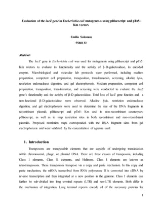
Figure 3.3.3 Migration distance vs Log (fragment size)
f(x) = -0.2824x + 3.9404
R² = 0.9039
2.50
2.70
2.90
3.10
3.30
3.50
3.70
1.00 1.50 2.00 2.50 3.00 3.50 4.00
Log(fragmentsize)
Distance(cm)
Migration distance vs Log(fragmentsize)
14. 14
Table 3.3.1 Estimated sizes of the DNA fragments (kb)
DNA
ladder
fragment
sizes (bp)
Distance of
migration
(cm)
Log(DNA
ladder fragment
sizes)
Size of
fragments (bp)
Estimated size
of fragments
(kb)
4000
1.50
3.60
3287
3.0
2000
2.00
3.30
2374
2.0
1500
2.50
3.18
1716
2.0
1000
3.50
3.00
895
1.0
Table 3.3.2 Estimated sizes of plasmids
Plasmid Size (bp)
Estimated size
(kb)
pBluescript +
pTn5: Km 5661
5.0
pBluescript 2611 3.0
The sizes of fragments (Table 3.3.3) were used to determine the size of plasmids
pBluescript with and without pTn5: Km insert (Table 3.3.4). Using the sizes of pBluescript with
or without insert, restriction maps were deduced.
1.0 kb
pBluescript + pTn5:
Km (5.0 kb)
BamHI
EcoRI
EcoRI
pTn5: Km (2.0 kb)
pBluescript (3.0 kb)
pBluescript (3.0 kb)
BamHIEcoRI
2.0 kb
15. 15
Figure 3.3.4 Restriction maps of pBluescript + pTn5: Km and pBluescript
Figure 3.3.4 shows the restriction maps of pBluescript with insert (pBluescript + pTn5:
Km) and without insert (pBluescript). In first restriction map, pBluescript and pTn5: Km were
present. It is known that the size of the recombinant plasmid is approximately 5.0 kb (Table
3.3.2). It is also known that the size of pBluescript is approximately 3.0 kb. This means that the
size of pTn5: Km is approximately 2.0 kb. Since the restriction map included pBluescript and
pTn5: Km, this suggested that two restriction enzymes were involved in digesting the plasmid.
Those enzymes included EcoRI and BamHI. EcoRI digested the plasmid at two restriction sites,
whereas BamHI digested the plasmid at only one restriction site. In the second restriction map,
pBluescript was present only. The plasmid without insert is approximately 3.0 kb, suggesting
that EcoRI and BamHI cut at one restriction site only. The restriction maps in Figure 3.3.4
corresponded with the results from Tables 3.3.1 and 3.3.2, as well as Figure 3.3.1.
Figure 3.3.5 Recognition sites of various restriction enzymes in E. coli
Figure 3.3.5 shows the recognition sites of various restriction enzymes capable of
digesting E. coli. Since only EcoRI and BamHI were used, the recognition sites of these
restriction enzymes were sought. According to Figure 3.3.5, EcoRI digested between A and G
(5’ to 3’) and BamHI digested between G and G (5’ to 3’) in E. coli, resulting in a sticky end
cleavage patterns.
16. 16
Table 3.3.3 Percent agarose (w/v) and their DNA resolution size
Percent agarose (w/v) DNA resolution size (1 kb = 1000 bp)
0.5 1 – 30 kb
0.7 800 bp – 12 kb
1.0 500 bp – 10 kb
1.2 400 bp – 7 kb
1.5 200 bp – 3 kb
2.0 50 bp – 2 kb
Table 3.3.3 displays the concentration of agarose used and their uses in specific DNA
sizes. Since 1.0% agarose was used, the DNA should have ranged from 500 bp- 10 kb. The
estimated sizes of both plasmids corresponded with the DNA resolution size of 1.0% agarose.
5.0 kb (pBluescript + pTn5: Km) and 3.0 kb (pBluescript) lie within the range, indicating a
successful gel electrophoresis.
4. Discussion
It was initially hypothesized that the function of the lacZ gene would be lost following
the mutagenesis of Escherichia coli using vectors pBluescript and pTn5: Km. Mutagenesis
involved techniques, including transposition, transformation, and screening, alkaline lysis,
restriction endonuclease digestion, and gel electrophoresis. Transposition, transformation, and
screening were used for evaluate the functionality of the lacZ gene and the activity of β-D-
galactosidase. Alkaline lysis, restriction endonuclease digestion, and gel electrophoresis were
used to determine the restriction sites in pBluescript with or without pTn5: Km insert. After all
processes, it was confirmed that the function lacZ gene was lost.
The loss of function in the lacZ gene was expected following transposition,
transformation, and screening. However, during the transposition, transformation, and screening
processes, the lacZ gene demonstrated a total loss of gene function. According to Table 3.2.2,
white colonies were observed in ampicillin agar plates only, with a total of 807 colonies in both.
17. 17
Blue colonies however, were not observed in the agar plates. Results from the screening process
(Tables 3.2.3 and 3.2.4) helped confirm the total loss of lacZ gene function observed during the
transformation process. In Table 3.2.3, white colonies were observed in ampicillin agar plates
following the screening process only. Both ampicillin agar plates had 50 white colonies. Blue
colonies were not observed in the ampicillin agar plates. On the other hand, both white and blue
colonies were not present in the ampicillin/kanamycin agar plates. There were 0 white colonies
and 0 blue colonies in the ampicillin/kanamycin agar plates. The sole presence of white colonies
in ampicillin plates following the screening process, indicated that colonies were able to grow in
the presence of ampicillin only, but were not able to grow in the presence of ampicillin with
kanamycin. The total absence of white and blue colonies in both ampicillin/kanamycin agar
plates demonstrated that growth did not occur, making the digestion of X-gal non-existent. In
Table 3.2.4, both sets of ampicillin and ampicillin/kanamycin agar plates had a frequency of
transposition of 0. These frequencies supported the results from Table 3.2.3. Results from the
transformation and screening process did not support the literatures. According to Analytical
Biochemistry and the Journal of Virological Methods, blue and white colonies were expected to
be present (Wessels et al., 2015), (Winnard Jr, Challa, Bhujwalla, & Raman, 2014). The total
loss of function of the lacZ gene may have been caused by poor execution of the lab protocols,
including media preparation, competent cell preparation, and transposition. Perhaps, not enough
LB broth nutrients such as Peptone, yeast extract, and sodium chloride or antibiotics such as
ampicillin and kanamycin were supplied when preparing the LB broth and agar. Also, the
competent cells may have been incubated for too long during competent cell preparation, causing
the cells to reach the death phase. Perhaps, the competent cells were not distributed evenly
enough in the agar plates or that the alcohol spreader was excessively hot during transposition.
The total loss of function of the lacZ gene may have also been caused by non-technical errors,
such as the cell’s innate competence.
Following alkaline lysis, restriction endonuclease digestion, and gel electrophoresis, the
sizes of the DNA fragments and the size of plasmids were determined and the restriction sites in
pBluescript with or without pTn5: Km were mapped. Using the sizes of the DNA fragments (bp)
in wells 14, 15, and 16, the sizes from the DNA marker (bp); 1 kb DNA ladder (Figure 3.3.2),
the distance of migration (cm), and the logarithm of fragment size from DNA marker (Table
3.3.3) four DNA fragments with sizes 1.0, 2.0, 3.0, and 5.0 kb were observed. The sizes of the
18. 18
DNA fragments and prior knowledge of vector sizes helped map the restriction sites in
pBluescript with or without pTn5: Km insert (Figure 3.3.4). According to Figure 3.3.4, the
recombinant plasmid (pBluescript + pTn5: Km) has three restriction sites, including two EcoRI
restriction sites and one BamHI restriction site. The non-recombinant plasmid (pBluescript) has
two restriction sites, including one EcoRI and one BamHI restriction site. In other words, EcoRI
is capable of digesting the recombinant plasmid twice and once in the non-recombinant plasmid,
whereas BamHI is capable of digesting in both the recombinant and non-recombinant plasmids
once. EcoRI and BamHI restriction/recognition sites were specifically determined in E. coli
(Figure 3.3.5), with EcoRI digesting between A and G (5’ to 3’) and BamHI digesting between G
and G (5’ to 3’). Overall, the restriction maps in Figure 3.3.4 corresponded with results from
Figures 3.3.1, 3.3.3, 3.3.4 and Tables 3.3.1 and 3.3.2. Proposed restriction maps were validated
by the concentration of agarose used (Table 3.3.3). 1.0% agarose was used for the separation of
DNA, with sizes ranging from 500 bp to 10 kb. Sizes of plasmids ranged from 500 bp to 10 kb
indicating a successful gel electrophoresis.
5. Conclusion
This research investigation primarily aimed to determine the restriction sites in plasmid
vector pBluescript with or without pTn5: Km. The investigation also aimed to evaluate the
functionality of the lacZ gene, as well as the activity of β-D-galactosidase, the enzyme encoded
by the lacZ gene. Escherichia coli mutagenesis was performed in order to evaluate such
parameters. The mutagenesis of E. coli involved the use standard microbiological lab procedures
such as transformation and screening and molecular lab procedures such as transposition,
alkaline lysis, restriction endonuclease digestion, and gel electrophoresis. Transposition,
transformation, and screening helped determine the functionality of the lacZ gene, as well as
evaluate the activity of its encoded enzyme, β-D-galactosidase. Following these three processes,
both the lacZ gene and β-D-galactosidase appeared to be non-functional at all, indicating a total
loss of function of the lacZ gene. White colonies were present in ampicillin plates during the
transformation and screening processes only. Blue colonies were absent in both ampicillin and
ampicillin/kanamycin agar plates during these processes. This was shown in Tables 3.2.1. 3.2.2.
3.2.3 and 3.2.4. Alkaline lysis, restriction endonuclease digestion, and gel electrophoresis on the
19. 19
other hand, helped determine the sizes of the DNA fragments, as well as helped map the
restriction sites of EcoRI and BamHI in pBluescript with or without pTn5: Km insert. The
proposed restriction maps (Figure 3.3.4) corresponded with the results obtained from gel
electrophoresis (Figures 3.3.1-3.3.4, Tables 3.3.1-3.3.2) and were validated by the concentration
of agarose used. For further research, other genes involved in the lac operon, such as lacY and
lacA should be used to determine their functionalities and their encoded enzymes’ properties and
activities. Using lacY and lacA, the activities and properties of galactoside permease and
galactoside transacetylase can be observed. Sufficient knowledge on these genes can help
researchers understand the lac operon better in terms of its regulation and roles in various
organisms.
References
Bobrov, A. G., & Perry, R. D. (2006). Yersinia pestis lacZ expresses a β-galactosidase with low
enzymatic activity. FEMS Microbiology Letters, 255(1), 43-51. doi: 10.1111/j.1574-
6968.2005.00039.x
Datsenko, K. A., & Wanner, B. L. (2000). One-step inactivation of chromosomal genes in
Escherichia coli K-12 using PCR products. Proceedings of the National Academy of
Sciences of the United States of America, 97(12), 6640-6645.
Huang, H.-P., Hong, C.-L., Kao, C.-Y., Lin, S.-W., Lin, S.-R., Wu, H.-L., . . . Yu, I. S. (2011).
Gene targeting and expression analysis of mouse Tem1/endosialin using a lacZ reporter.
Gene Expression Patterns, 11(5–6), 316-326. doi:
http://dx.doi.org/10.1016/j.gep.2011.03.001
Mahabir, A. G., van Benthem, J., Korsten, H., Lynch, A. M., Bailey, L., de Vries, A., . . . van
Steeg, H. (2008). Detecting genotoxic effects of potential clastogens: An in vivo study
using the transgenic lacZ plasmid and the Muta™Mouse model. Mutation
Research/Genetic Toxicology and Environmental Mutagenesis, 652(2), 151-157. doi:
http://dx.doi.org/10.1016/j.mrgentox.2008.01.007
Suzuki, D., Leu, N. A., Brice, A. K., & Senoo, M. (2014). Expression analysis of Dact1 in mice
using a LacZ reporter. Gene Expression Patterns, 15(1), 21-30. doi:
http://dx.doi.org/10.1016/j.gep.2014.03.002
20. 20
Wessels, U., Stech, O., Abdelwhab, E.-S. M., Judel, A., Mettenleiter, T. C., & Stech, J. (2015).
Improved universal cloning of influenza A virus genes by LacZα-mediated blue/white
selection. Journal of Virological Methods, 225, 87-89. doi:
http://dx.doi.org/10.1016/j.jviromet.2015.09.009
Winnard Jr, P. T., Challa, R., Bhujwalla, Z. M., & Raman, V. (2014). Direct facile screening of
recombinant DNA vector constructs. Analytical Biochemistry, 450, 1-3. doi:
http://dx.doi.org/10.1016/j.ab.2013.12.034