Establishment of Body Axis in Humans
•
4 likes•3,917 views

A note on the development of the body axes in human embryos.

Recommended
More Related Content
What's hot
What's hot (20)
Similar to Establishment of Body Axis in Humans
Similar to Establishment of Body Axis in Humans (20)
More from Syed Muhammad Khan
More from Syed Muhammad Khan (20)
Recently uploaded
Recently uploaded (20)
Establishment of Body Axis in Humans
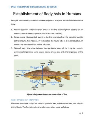
- 1. SYED MUHAMMAD KHAN (BS HONS. ZOOLOGY) pg. 1 Establishment of Body Axis in Humans Embryos must develop three crucial axes (singular – axis) that are the foundation of the body: 1. Anterior-posterior (anteroposterior) axis: it is the line extending from head to tail (or mouth to anus in those organisms that lack a head and tail). 2. Dorsal-ventral (dorsoventral) axis: it is the line extending from the back (dorsum) to belly (ventrum). For instance, in vertebrates, the neural tube is a dorsal structure. In insects, the neural cord is a ventral structure. 3. Right-left axis: it is a line between the two lateral sides of the body, i.e. even in symmetrical organisms, some organs belong on one side and other organs go on the other. Figure: Body axes drawn over the surface of fish. Axis Formation in Mammals Mammals have three body axes: anterior-posterior axis, dorsal-ventral axis, and lateral / left-right axis. The formation of mammalian axes takes place as follows:
- 2. SYED MUHAMMAD KHAN (BS HONS. ZOOLOGY) pg. 2 1. Anterior-Posterior Axis Formation: Node & AVE: The mammalian embryo appears to have two signaling centers: one in the node and one in the anterior visceral endoderm (AVE). The node is responsible for the creation of all of the body, and it works with AVE to form the anterior region of the embryo. The notochord forms by the dorsal in-folding of the cells of the node. The AVE originates from the visceral endoderm, it secretes two antagonists (that work opposite) of the Nodal protein, Lefty-1 and Cerberus. Nodal proteins in the epiblast activate the expression of posterior genes that are required for mesoderm formation. AVE creates an anterior region where Nodal cannot act. The AVE also begins expressing anterior markers such as Wnt-inhibitor Dickkopf. AVE promotes anterior specification by suppressing the formation of the primitive streak, a posterior structure, by Nodal and Wnt proteins. However, the AVE alone cannot induce neural tissue, but the node can. Once the node is formed (due to AVE), it will initiate head development, with the help of the notochord. AVE functions in the epiblast to restrict Nodal activity, thereby it promotes the head- forming genes to be expressed in the anterior portion of the epiblast (with the help of the mes-endoderm). FGF Gradient The head region of the mammalian embryo is devoid of FGFs. The posterior region is characterized by FGFs and retinoic acid (along with other proteins). There appears to be a gradient of FGF proteins that is highest in the posterior region and lower in the anterior region. The Fgf8 gradient is created by the decay of its mRNA. Fgf8 is expressed at the growing posterior tip of the embryo, but the Fgf8 message is slowly degraded in the newly formed tissues. Thus there is a gradient of Fgf8 mRNA across the posterior of the embryo,
- 3. SYED MUHAMMAD KHAN (BS HONS. ZOOLOGY) pg. 3 which is then converted into an Fgf8 protein gradient. The FGF gradient patterns the posterior portion of the embryo. Retinoic Acid Gradient Retinoic acid (RA) is a vitamin A derivative that is important in specifying the anterior- posterior axis and in forming the jaws and heart of the mammalian embryo. The late gastrula has a gradient of retinoic acid. RA levels are high in the posterior regions and low in the anterior portions of the embryo. This is due to RA-synthesizing enzymes in the embryo's posterior and RA-degrading enzymes in the anterior parts of the embryo. 2. Dorsal-Ventral Axis Formation: In mice and humans, the hypoblast (ventral) forms on the side of the inner cell mass that is exposed to the blastocyst fluid, while the dorsal axis forms from those inner cell mass cells that are in contact with the trophoblast and amnionic cavity. Thus, the dorsal-ventral axis of the embryo is defined, in part, by the embryonic-abembryonic axis of the blastocyst. The embryonic region contains the inner cell mass, while the abembryonic region is that part of the blastocyst opposite to the inner cell mass. The first dorsal-ventral polarity is seen at the blastocyst stage, and as development proceeds, the primitive streak maintains this polarity by causing migration ventrally from the dorsal surface of the embryo. 3. Lateral / Left-Right Axis Formation: The mammalian body is not symmetrical. Although the human heart begins its formation at the midline of the embryo, it moves to the left side of the chest cavity and loops to the right. The spleen is found solely on the left side of the abdomen, the major lobe of the liver forms on the right side of the abdomen, the large intestine loops right to left as it traverses the abdominal cavity, and the right lung has one more lobe than the left lung. Thus, a lateral / left-right axis exists in mammals.