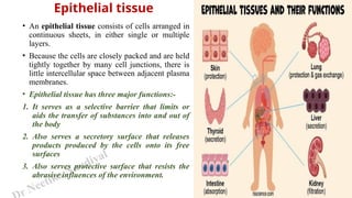
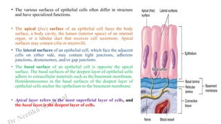
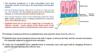
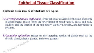
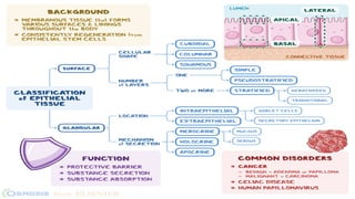
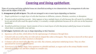
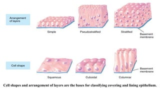
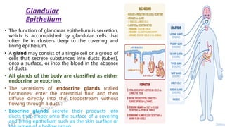
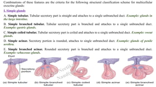
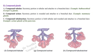
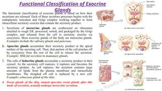
Epithelial Tissue
Dr. Neetika Naudiyal
Assistant Professor,
Department of Life Sciences,
Kristu Jayanti College (Autonomous),
Bengaluru
r Neetika Naudiyal
• An epithelial tissue consists of cells arranged in
continuous sheets, in either single or multiple
layers.
• Because the cells are closely packed and are held
tightly together by many cell junctions, there is
little intercellular space between adjacent plasma
membranes.
• Epithelial tissue has three major functions:-
1. It serves as a selective barrier that limits or
aids the transfer of substances into and out of
the body
2. Also serves a secretory surface that releases
products produced by the cells onto its free
surfaces
3. Also serves protective surface that resists the
abrasive influences of the environment.
Epithelial tissue
r Neetika Naudiyal
• The various surfaces of epithelial cells often differ in structure
and have specialized functions.
• The apical (free) surface of an epithelial cell faces the body
surface, a body cavity, the lumen (interior space) of an internal
organ, or a tubular duct that receives cell secretions. Apical
surfaces may contain cilia or microvilli.
• The lateral surfaces of an epithelial cell, which face the adjacent
cells on either side, may contain tight junctions, adherens
junctions, desmosomes, and/or gap junctions.
• The basal surface of an epithelial cell is opposite the apical
surface. The basal surfaces of the deepest layer of epithelial cells
adhere to extracellular materials such as the basement membrane.
Hemidesmosomes in the basal surfaces of the deepest layer of
epithelial cells anchor the epithelium to the basement membrane.
• Apical layer refers to the most superficial layer of cells, and
the basal layer is the deepest layer of cells.
r Neetika Naudiyal
• The basement membrane is a thin extracellular layer that
commonly consists of two layers, the basal lamina and reticular
lamina.
• The basal lamina (lamina thin layer) is closer to—and secreted
by—the epithelial cells. It contains proteins such as laminin and
collagen, as well as glycoproteins and proteoglycans.
• The reticular lamina is closer to the underlying connective tissue
and contains proteins such as collagen produced by connective
tissue cells called fibroblasts.
• Epithelial tissue has its own nerve supply but, is avascular (a-
without; -vascular vessel), relying on the blood vessels of the
adjacent connective tissue to bring nutrients and remove wastes.
 Exchange of substances between an epithelial tissue and connective tissue occurs by diffusion.
 Epithelial tissue forms boundaries between the body’s organs, or between the body and the external environment,
it is repeatedly subjected to physical stress and injury.
 A high rate of cell division allows epithelial tissue to constantly renew and repair itself by sloughing off dead or
injured cells and replacing them with new ones.
r Neetika Naudiyal
Epithelial Tissue Classification
Epithelial tissue may be divided into two types:-
A.Covering and lining epithelium forms the outer covering of the skin and some
internal organs. It also forms the inner lining of blood vessels, ducts, and body
cavities, and the interior of the respiratory, digestive, urinary, and reproductive
systems.
B.Glandular epithelium makes up the secreting portion of glands such as the
thyroid gland, adrenal glands, and sweat glands.
r Neetika Naudiyal
Types of covering and lining epithelial tissue are classified according to two characteristics: the arrangement of cells into
layers and the shapes of the cells.
1. Arrangement of cells in layers: The cells are arranged in one or more layers depending on function:
i. Simple epithelium is a single layer of cells that functions in diffusion, osmosis, filtration, secretion, or absorption.
ii. Pseudostratified epithelium (pseudo- false) appears to have multiple layers of cells because the cell nuclei lie at different
levels and not all cells reach the apical surface; it is actually a simple epithelium because all its cells rest on the basement
membrane.
iii. Stratified epithelium (stratum layer) consists of two or more layers of cells that protect underlying tissues in locations
where there is considerable wear and tear.
2. Cell shapes: Epithelial cells vary in shape depending on their function:
iv. Squamous cells are thin, which allows for the rapid passage of substances through them.
v. Cuboidal cells are as tall as they are wide and are shaped like cubes or hexagons. They may have microvilli at their apical
surface and function in either secretion or absorption.
vi. Columnar cells are much taller than they are wide, like columns, and protect underlying tissues. They are often
specialized for secretion and absorption.
vii. Transitional cells change shape, from squamous to cuboidal and back, as organs such as the urinary bladder stretch
(distend) to a larger size and then collapse to a smaller size.
Covering and lining epithelium
Cell shapes and arrangement of layers are the bases for classifying covering and lining epithelium.
r Neetika Naudiyal
On combining the two characteristics (arrangements of layers and cell shapes), the following types of epithelial tissues
are there:
I. Simple epithelium
A. Simple squamous epithelium
B. Simple cuboidal epithelium
C. Simple columnar epithelium (non-ciliated and ciliated)
D. Pseudostratified columnar epithelium (non-ciliated and ciliated)
II. Stratified epithelium
A. Stratified squamous epithelium (keratinized, when surface cells are dead and become hardened, and nonkeratinized,
when surface cells remain alive)
B. Stratified cuboidal epithelium
C. Stratified columnar epithelium
D. Transitional epithelium
r Neetika Naudiyal
Name Location Function
Simple Squamous
Epithelium
• Lines the cardiovascular and lymphatic system where it is
known as endothelium
• Epithelial layer of serous membranes, where it is called
mesothelium
• Air sacs of lungs
• Glomerular capsule of kidneys
• Inner surface of eardrum
• Blood filtration in kidneys
• Diffusion of oxygen into blood vessels of
lungs
• Secretion in serous membranes
Simple Cuboidal
Epithelium
• Ovary surface layer
• Anterior surface of lens capsule of the eye
• Lines kidney tubules
• Secreting portion of glands like thyroid gland and ducts of
some glands like pancreas
• Secretion
• Absorption
Non-ciliated Simple
Columnar Epithelium
• Gastrointestinal tract (from stomach to anus)
• Ducts of many glands
• Gallbladder
• Secretion and absorption
• Lubrication of linings of different tracts
• Prevent destruction of stomach lining by
acidic gastric juice secreted by stomach
Ciliated Simple
Columnar Epithelium
• Lines some bronchioles of respiratory tract, uterine
(fallopian) tubes
• Central canal of spinal cord
• Brain ventricles
• Removal of mucus and foreign particles
• oocytes expulsion from ovaries through
uterine (fallopian) tubes into uterus
r Neetika Naudiyal
Name Location Function
Pseudostratified Columnar
Epithelium
• Ciliated variety lines airways of most of
upper respiratory tract
• Non-ciliated variety lines larger ducts of
many glands, epididymis, and part of male
urethra.
• Ciliated variety secretes mucus that
traps foreign particles, and cilia sweep
away mucus for elimination from body
• Non-ciliated variety functions in
absorption and protection
Stratified Squamous Epithelium • Keratinized variety forms superficial layer
of skin
• Non-keratinized variety lines wet surfaces
• Covers tongue
• Protection against abrasion, water loss,
UV, and foreign invasion
• First line of defense against microbes
Stratified Cuboidal Epithelium • Ducts of adult sweat glands and esophageal
glands
• Part of male urethra
• Protection
• Limited secretion and absorption
Stratified Columnar Epithelium • Lines part of urethra; large excretory ducts
of some glands
• Small areas in anal mucous membrane
• Part of conjunctiva of eye
• Protection
• Secretion
Transitional Epithelium • Lines urinary bladder
• Portions of ureters and urethra
• Allows urinary organs to stretch and
maintain protective lining while
holding variable amounts of fluid
without rupturing
r Neetika Naudiyal
• The function of glandular epithelium is secretion,
which is accomplished by glandular cells that
often lie in clusters deep to the covering and
lining epithelium.
• A gland may consist of a single cell or a group of
cells that secrete substances into ducts (tubes),
onto a surface, or into the blood in the absence
of ducts.
• All glands of the body are classified as either
endocrine or exocrine.
• The secretions of endocrine glands (called
hormones, enter the interstitial fluid and then
diffuse directly into the bloodstream without
flowing through a duct.
• Exocrine glands secrete their products into
ducts that empty onto the surface of a covering
and lining epithelium such as the skin surface or
Glandular
Epithelium
Name Description Location Function
Endocrine Glands Secretions (hormones) enter interstitial
fluid and diffuse directly into
bloodstream without flowing through a
duct.
• Pituitary gland
• Pineal gland
• Thyroid and parathyroid
glands
• Adrenal glands
• Pancreas near stomach,
Ovaries
• Testes
• Thymus
Regulation of metabolic and
physiological activities to
maintain homeostasis.
Exocrine Glands Secretory products released into ducts
that empty onto surface of a covering
and lining epithelium.
• Sweat, oil, and earwax
glands of skin
• Digestive glands
Produce substances such as
sweat to help lower body
temperature, oil, earwax,
saliva, or digestive enzymes.
r Neetika Naudiyal
• Exocrine glands are classified as unicellular or multicellular.
• Unicellular glands are single-celled glands. Goblet cells are important unicellular exocrine glands that
secrete mucus directly onto the apical surface of a lining epithelium.
• Most exocrine glands are multicellular glands, composed of many cells that form a distinctive microscopic
structure or macroscopic organ. Examples include sudoriferous (sweat), sebaceous (oil), and salivary glands.
• Multicellular glands are categorized according to two criteria:
(1) whether their ducts are branched or unbranched and
(2) the shape of the secretory portions of the gland
• If the duct of the gland does not branch, it is a simple gland and if the duct branches, it is a compound
gland.
• Glands with tubular secretory parts are tubular glands; those with rounded secretory portions are acinar
glands, also called alveolar glands. Tubuloacinar glands have both tubular and more rounded secretory
parts.
Structural Classification of Exocrine
Glands
Dr Neetika
Naudiyal
r Neetika Naudiyal
Combinations of these features are the criteria for the following structural classification scheme for multicellular
exocrine glands:
I. Simple glands
A. Simple tubular. Tubular secretory part is straight and attaches to a single unbranched duct. Example: glands in
the large intestine.
B. Simple branched tubular. Tubular secretory part is branched and attaches to a single unbranched duct.
Example: gastric glands.
C. Simple coiled tubular. Tubular secretory part is coiled and attaches to a single unbranched duct. Example: sweat
glands.
D. Simple acinar. Secretory portion is rounded, attaches to single unbranched duct. Example: glands of penile
urethra.
E. Simple branched acinar. Rounded secretory part is branched and attaches to a single unbranched duct.
Example: sebaceous glands.
Dr Neetika
Naudiyal
r Neetika Naudiyal
II. Compound glands
A. Compound tubular. Secretory portion is tubular and attaches to a branched duct. Example: bulbourethral
(Cowper’s) glands.
B. Compound acinar. Secretory portion is rounded and attaches to a branched duct. Example: mammary
glands.
C. Compound tubuloacinar. Secretory portion is both tubular and rounded and attaches to a branched duct.
Example: acinar glands of the pancreas.
Dr Neetika
audiyal
The functional classification of exocrine glands is based on how their
secretions are released. Each of these secretory processes begins with the
endoplasmic reticulum and Golgi complex working together to form
intracellular secretory vesicles that contain the secretory product.
1. Secretions of merocrine glands are synthesized on ribosomes
attached to rough ER; processed, sorted, and packaged by the Golgi
complex; and released from the cell in secretory vesicles via
exocytosis. Most exocrine glands of the body are merocrine glands.
Examples include the salivary glands and pancreas.
2. Apocrine glands accumulate their secretory product at the apical
surface of the secreting cell. Then, that portion of the cell pinches off
by exocytosis from the rest of the cell to release the secretion.
Example: Milk fat secretion in mammary glands.
3. The cells of holocrine glands accumulate a secretory product in their
cytosol. As the secretory cell matures, it ruptures and becomes the
secretory product. As cell ruptures, the secretion contains large
amounts of lipids from the plasma membrane and intracellular
membranes. The sloughed off cell is replaced by a new cell.
Example: sebaceous gland of the skin.
4. Sweat glands of the skin, named apocrine sweat glands after this
mode of secretion, actually undergo merocrine secretion.
Functional Classification of Exocrine
Glands
Naudiyal

Epithelial tissue and their classification

  • 1.
    Epithelial Tissue Dr. NeetikaNaudiyal Assistant Professor, Department of Life Sciences, Kristu Jayanti College (Autonomous), Bengaluru
  • 2.
    r Neetika Naudiyal •An epithelial tissue consists of cells arranged in continuous sheets, in either single or multiple layers. • Because the cells are closely packed and are held tightly together by many cell junctions, there is little intercellular space between adjacent plasma membranes. • Epithelial tissue has three major functions:- 1. It serves as a selective barrier that limits or aids the transfer of substances into and out of the body 2. Also serves a secretory surface that releases products produced by the cells onto its free surfaces 3. Also serves protective surface that resists the abrasive influences of the environment. Epithelial tissue
  • 3.
    r Neetika Naudiyal •The various surfaces of epithelial cells often differ in structure and have specialized functions. • The apical (free) surface of an epithelial cell faces the body surface, a body cavity, the lumen (interior space) of an internal organ, or a tubular duct that receives cell secretions. Apical surfaces may contain cilia or microvilli. • The lateral surfaces of an epithelial cell, which face the adjacent cells on either side, may contain tight junctions, adherens junctions, desmosomes, and/or gap junctions. • The basal surface of an epithelial cell is opposite the apical surface. The basal surfaces of the deepest layer of epithelial cells adhere to extracellular materials such as the basement membrane. Hemidesmosomes in the basal surfaces of the deepest layer of epithelial cells anchor the epithelium to the basement membrane. • Apical layer refers to the most superficial layer of cells, and the basal layer is the deepest layer of cells.
  • 4.
    r Neetika Naudiyal •The basement membrane is a thin extracellular layer that commonly consists of two layers, the basal lamina and reticular lamina. • The basal lamina (lamina thin layer) is closer to—and secreted by—the epithelial cells. It contains proteins such as laminin and collagen, as well as glycoproteins and proteoglycans. • The reticular lamina is closer to the underlying connective tissue and contains proteins such as collagen produced by connective tissue cells called fibroblasts. • Epithelial tissue has its own nerve supply but, is avascular (a- without; -vascular vessel), relying on the blood vessels of the adjacent connective tissue to bring nutrients and remove wastes.  Exchange of substances between an epithelial tissue and connective tissue occurs by diffusion.  Epithelial tissue forms boundaries between the body’s organs, or between the body and the external environment, it is repeatedly subjected to physical stress and injury.  A high rate of cell division allows epithelial tissue to constantly renew and repair itself by sloughing off dead or injured cells and replacing them with new ones.
  • 5.
    r Neetika Naudiyal EpithelialTissue Classification Epithelial tissue may be divided into two types:- A.Covering and lining epithelium forms the outer covering of the skin and some internal organs. It also forms the inner lining of blood vessels, ducts, and body cavities, and the interior of the respiratory, digestive, urinary, and reproductive systems. B.Glandular epithelium makes up the secreting portion of glands such as the thyroid gland, adrenal glands, and sweat glands.
  • 7.
    r Neetika Naudiyal Typesof covering and lining epithelial tissue are classified according to two characteristics: the arrangement of cells into layers and the shapes of the cells. 1. Arrangement of cells in layers: The cells are arranged in one or more layers depending on function: i. Simple epithelium is a single layer of cells that functions in diffusion, osmosis, filtration, secretion, or absorption. ii. Pseudostratified epithelium (pseudo- false) appears to have multiple layers of cells because the cell nuclei lie at different levels and not all cells reach the apical surface; it is actually a simple epithelium because all its cells rest on the basement membrane. iii. Stratified epithelium (stratum layer) consists of two or more layers of cells that protect underlying tissues in locations where there is considerable wear and tear. 2. Cell shapes: Epithelial cells vary in shape depending on their function: iv. Squamous cells are thin, which allows for the rapid passage of substances through them. v. Cuboidal cells are as tall as they are wide and are shaped like cubes or hexagons. They may have microvilli at their apical surface and function in either secretion or absorption. vi. Columnar cells are much taller than they are wide, like columns, and protect underlying tissues. They are often specialized for secretion and absorption. vii. Transitional cells change shape, from squamous to cuboidal and back, as organs such as the urinary bladder stretch (distend) to a larger size and then collapse to a smaller size. Covering and lining epithelium
  • 8.
    Cell shapes andarrangement of layers are the bases for classifying covering and lining epithelium.
  • 9.
    r Neetika Naudiyal Oncombining the two characteristics (arrangements of layers and cell shapes), the following types of epithelial tissues are there: I. Simple epithelium A. Simple squamous epithelium B. Simple cuboidal epithelium C. Simple columnar epithelium (non-ciliated and ciliated) D. Pseudostratified columnar epithelium (non-ciliated and ciliated) II. Stratified epithelium A. Stratified squamous epithelium (keratinized, when surface cells are dead and become hardened, and nonkeratinized, when surface cells remain alive) B. Stratified cuboidal epithelium C. Stratified columnar epithelium D. Transitional epithelium
  • 10.
    r Neetika Naudiyal NameLocation Function Simple Squamous Epithelium • Lines the cardiovascular and lymphatic system where it is known as endothelium • Epithelial layer of serous membranes, where it is called mesothelium • Air sacs of lungs • Glomerular capsule of kidneys • Inner surface of eardrum • Blood filtration in kidneys • Diffusion of oxygen into blood vessels of lungs • Secretion in serous membranes Simple Cuboidal Epithelium • Ovary surface layer • Anterior surface of lens capsule of the eye • Lines kidney tubules • Secreting portion of glands like thyroid gland and ducts of some glands like pancreas • Secretion • Absorption Non-ciliated Simple Columnar Epithelium • Gastrointestinal tract (from stomach to anus) • Ducts of many glands • Gallbladder • Secretion and absorption • Lubrication of linings of different tracts • Prevent destruction of stomach lining by acidic gastric juice secreted by stomach Ciliated Simple Columnar Epithelium • Lines some bronchioles of respiratory tract, uterine (fallopian) tubes • Central canal of spinal cord • Brain ventricles • Removal of mucus and foreign particles • oocytes expulsion from ovaries through uterine (fallopian) tubes into uterus
  • 11.
    r Neetika Naudiyal NameLocation Function Pseudostratified Columnar Epithelium • Ciliated variety lines airways of most of upper respiratory tract • Non-ciliated variety lines larger ducts of many glands, epididymis, and part of male urethra. • Ciliated variety secretes mucus that traps foreign particles, and cilia sweep away mucus for elimination from body • Non-ciliated variety functions in absorption and protection Stratified Squamous Epithelium • Keratinized variety forms superficial layer of skin • Non-keratinized variety lines wet surfaces • Covers tongue • Protection against abrasion, water loss, UV, and foreign invasion • First line of defense against microbes Stratified Cuboidal Epithelium • Ducts of adult sweat glands and esophageal glands • Part of male urethra • Protection • Limited secretion and absorption Stratified Columnar Epithelium • Lines part of urethra; large excretory ducts of some glands • Small areas in anal mucous membrane • Part of conjunctiva of eye • Protection • Secretion Transitional Epithelium • Lines urinary bladder • Portions of ureters and urethra • Allows urinary organs to stretch and maintain protective lining while holding variable amounts of fluid without rupturing
  • 12.
    r Neetika Naudiyal •The function of glandular epithelium is secretion, which is accomplished by glandular cells that often lie in clusters deep to the covering and lining epithelium. • A gland may consist of a single cell or a group of cells that secrete substances into ducts (tubes), onto a surface, or into the blood in the absence of ducts. • All glands of the body are classified as either endocrine or exocrine. • The secretions of endocrine glands (called hormones, enter the interstitial fluid and then diffuse directly into the bloodstream without flowing through a duct. • Exocrine glands secrete their products into ducts that empty onto the surface of a covering and lining epithelium such as the skin surface or Glandular Epithelium
  • 13.
    Name Description LocationFunction Endocrine Glands Secretions (hormones) enter interstitial fluid and diffuse directly into bloodstream without flowing through a duct. • Pituitary gland • Pineal gland • Thyroid and parathyroid glands • Adrenal glands • Pancreas near stomach, Ovaries • Testes • Thymus Regulation of metabolic and physiological activities to maintain homeostasis. Exocrine Glands Secretory products released into ducts that empty onto surface of a covering and lining epithelium. • Sweat, oil, and earwax glands of skin • Digestive glands Produce substances such as sweat to help lower body temperature, oil, earwax, saliva, or digestive enzymes.
  • 14.
    r Neetika Naudiyal •Exocrine glands are classified as unicellular or multicellular. • Unicellular glands are single-celled glands. Goblet cells are important unicellular exocrine glands that secrete mucus directly onto the apical surface of a lining epithelium. • Most exocrine glands are multicellular glands, composed of many cells that form a distinctive microscopic structure or macroscopic organ. Examples include sudoriferous (sweat), sebaceous (oil), and salivary glands. • Multicellular glands are categorized according to two criteria: (1) whether their ducts are branched or unbranched and (2) the shape of the secretory portions of the gland • If the duct of the gland does not branch, it is a simple gland and if the duct branches, it is a compound gland. • Glands with tubular secretory parts are tubular glands; those with rounded secretory portions are acinar glands, also called alveolar glands. Tubuloacinar glands have both tubular and more rounded secretory parts. Structural Classification of Exocrine Glands
  • 15.
  • 16.
    r Neetika Naudiyal Combinationsof these features are the criteria for the following structural classification scheme for multicellular exocrine glands: I. Simple glands A. Simple tubular. Tubular secretory part is straight and attaches to a single unbranched duct. Example: glands in the large intestine. B. Simple branched tubular. Tubular secretory part is branched and attaches to a single unbranched duct. Example: gastric glands. C. Simple coiled tubular. Tubular secretory part is coiled and attaches to a single unbranched duct. Example: sweat glands. D. Simple acinar. Secretory portion is rounded, attaches to single unbranched duct. Example: glands of penile urethra. E. Simple branched acinar. Rounded secretory part is branched and attaches to a single unbranched duct. Example: sebaceous glands. Dr Neetika Naudiyal
  • 17.
    r Neetika Naudiyal II.Compound glands A. Compound tubular. Secretory portion is tubular and attaches to a branched duct. Example: bulbourethral (Cowper’s) glands. B. Compound acinar. Secretory portion is rounded and attaches to a branched duct. Example: mammary glands. C. Compound tubuloacinar. Secretory portion is both tubular and rounded and attaches to a branched duct. Example: acinar glands of the pancreas. Dr Neetika audiyal
  • 18.
    The functional classificationof exocrine glands is based on how their secretions are released. Each of these secretory processes begins with the endoplasmic reticulum and Golgi complex working together to form intracellular secretory vesicles that contain the secretory product. 1. Secretions of merocrine glands are synthesized on ribosomes attached to rough ER; processed, sorted, and packaged by the Golgi complex; and released from the cell in secretory vesicles via exocytosis. Most exocrine glands of the body are merocrine glands. Examples include the salivary glands and pancreas. 2. Apocrine glands accumulate their secretory product at the apical surface of the secreting cell. Then, that portion of the cell pinches off by exocytosis from the rest of the cell to release the secretion. Example: Milk fat secretion in mammary glands. 3. The cells of holocrine glands accumulate a secretory product in their cytosol. As the secretory cell matures, it ruptures and becomes the secretory product. As cell ruptures, the secretion contains large amounts of lipids from the plasma membrane and intracellular membranes. The sloughed off cell is replaced by a new cell. Example: sebaceous gland of the skin. 4. Sweat glands of the skin, named apocrine sweat glands after this mode of secretion, actually undergo merocrine secretion. Functional Classification of Exocrine Glands Naudiyal