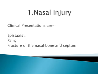
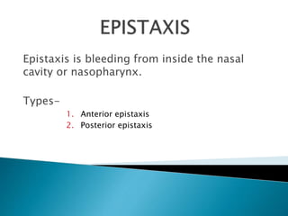
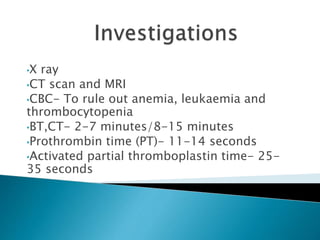
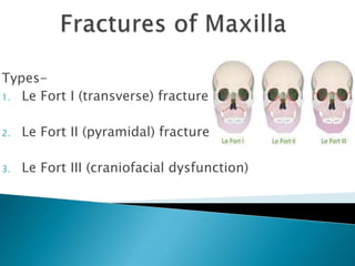
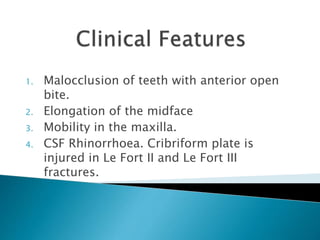
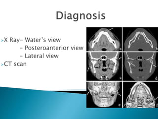
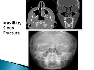
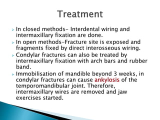
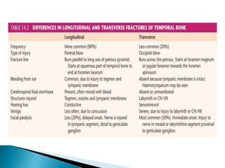
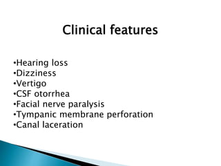
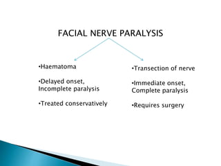
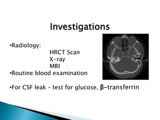
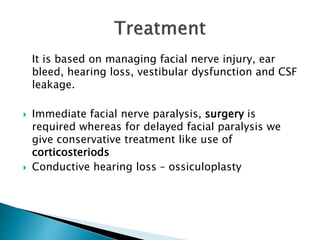
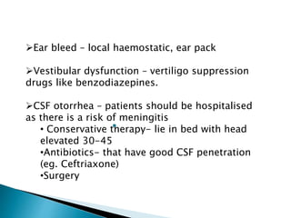
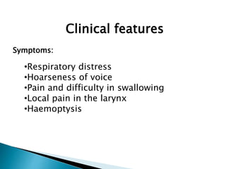
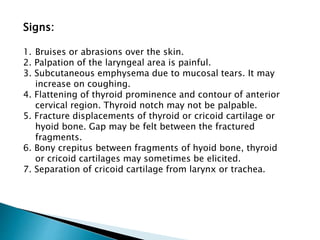
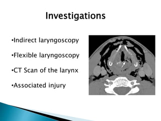
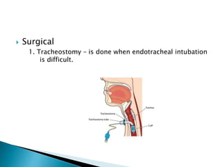
Presenters-
Prerana Malakar, Roll No.-109
Phibadonbok Nongtraw, Roll no.-110
Moderator- Proff. Dr. Arup Kakati Sir
Some of the common ENT injuries in
polytrauma are-
1. Nasal injury
2. Facio- maxillary injury
3. Injury in the temporal region
4. Injury over the neck region
Clinical Presentations are-
Epistaxis ,
Pain,
Fracture of the nasal bone and septum
Epistaxis is bleeding from inside the nasal
cavity or nasopharynx.
Types-
1. Anterior epistaxis
2. Posterior epistaxis
•X ray
•CT scan and MRI
•CBC- To rule out anemia, leukaemia and
thrombocytopenia
•BT,CT- 2-7 minutes/8-15 minutes
•Prothrombin time (PT)- 11-14 seconds
•Activated partial thromboplastin time- 25-
35 seconds
•First aid-If bleeding occurs through the little’s
area ,it is controlled by pinching the nose with
thumb and index finger for about 5 mins.
•Trotter’s method
•Cauterization
•Anterior nasal packing
•Posterior nasal packing
•Ligation of Vessels
•Monitor the BP,pulse and respiration
•Antibiotics can be given to prevent sinusitis
Most common fracture.
Types-
1. Depressed-Due to frontal blow. A severe
frontal blow will cause ‘open-book fracture’
in which nasal septum is collapsed and nasal
bones splayed out.
2. Angulated-A lateral blow may cause
unilateral depression of nasal bone on the
same side or may fracture both the nasal
bones and the septum with deviation of the
nasal bridge.
Clinical features
•Periorbital ecchymosis
•Tenderness
•Nasal deformity
•Crepitus
•Epistaxis
•Lacerations of the nasal skin with exposure
of the nasal bones and cartilage.
•Physical examination
•X Ray- Water’s view, right and left
lateral views and occlusal view.
•CT scan
The best time to reduce a fracture is
before the appearance of oedema, or
after it has subsided , which is usually
5-7 days.
1. Closed Reduction- Impacted
fragments sometimes require
disimpaction with Walsham or
Asch’s forceps before alignment.
Unstable fractures require intranasal
packing.
2. Open Reduction- indicated when
closed methods fail.
Clinical Presentations are-
•Swelling
•Peri-orbital oedema
•Tenderness
•Limitation in mouth opening
•Lacerations
Types-
1. Le Fort I (transverse) fracture
2. Le Fort II (pyramidal) fracture
3. Le Fort III (craniofacial dysfunction)
1. Malocclusion of teeth with anterior open
bite.
2. Elongation of the midface
3. Mobility in the maxilla.
4. CSF Rhinorrhoea. Cribriform plate is
injured in Le Fort II and Le Fort III
fractures.
X Ray- Water’s view
- Posteroanterior view
- Lateral view
CT scan
Maxillary
Sinus
Fracture
•Antibiotics
•Steroids for inflammation
•Analgesics like Diclofenac for pain
•In case of maxillary fracture, immediate attention is paid
to restore the airway and stop severe haemorrhage from
maxillary artery or its branches.
•Fixation of maxillary fractures can be achieved by-
1. Interdental wiring
2. Intermaxillary wiring using arch bars
3. Open reduction and interosseous wiring
4. Wire slings from frontal bone, zygoma or infraorbital
rim to the teeth or arch bars.
 Fractures of mandible are classified according
to their location (Dingman classification)
 Condylar fractures are the most common.
 They are followed by fractures of the angle,
body and sympysis.
• If fragments are not displaced, pain and
trismus are the main features.
• If fragments are displaced, there is, in
addition, malocclusion of teeth and deviation
of jaw to the opposite side on opening the
mouth.
 X –rays-Posteroanterior view ( for condyle)
-Right and left oblique views of mandible
-Panorex view
 In closed methods- Interdental wiring and
intermaxillary fixation are done.
 In open methods-Fracture site is exposed and
fragments fixed by direct interosseous wiring.
 Condylar fractures can also be treated by
intermaxillary fixation with arch bars and rubber
band.
 Immobilisation of mandible beyond 3 weeks, in
condylar fractures can cause ankylosis of the
temporomandibular joint. Therefore,
intermaxillary wires are removed and jaw
exercises started.
• Temporal bone
fracture
• Longitudinal
• Transverse
• Mixed
Clinical features
•Hearing loss
•Dizziness
•Vertigo
•CSF otorrhea
•Facial nerve paralysis
•Tympanic membrane perforation
•Canal laceration
FACIAL NERVE PARALYSIS
•Haematoma
•Delayed onset,
Incomplete paralysis
•Treated conservatively
•Transection of nerve
•Immediate onset,
Complete paralysis
•Requires surgery
Investigations
•Radiology:
HRCT Scan
X-ray
MRI
•Routine blood examination
•For CSF leak – test for glucose, β-transferrin
It is based on managing facial nerve injury, ear
bleed, hearing loss, vestibular dysfunction and CSF
leakage.
 Immediate facial nerve paralysis, surgery is
required whereas for delayed facial paralysis we
give conservative treatment like use of
corticosteriods
 Conductive hearing loss – ossiculoplasty
Ear bleed – local haemostatic, ear pack
Vestibular dysfunction – vertiligo suppression
drugs like benzodiazepines.
CSF otorrhea – patients should be hospitalised
as there is a risk of meningitis
• Conservative therapy- lie in bed with head
elevated 30-45
•Antibiotics- that have good CSF penetration
(eg. Ceftriaxone)
•Surgery
• Laryngotracheal
Trauma
Clinical features
Symptoms:
•Respiratory distress
•Hoarseness of voice
•Pain and difficulty in swallowing
•Local pain in the larynx
•Haemoptysis
Signs:
1. Bruises or abrasions over the skin.
2. Palpation of the laryngeal area is painful.
3. Subcutaneous emphysema due to mucosal tears. It may
increase on coughing.
4. Flattening of thyroid prominence and contour of anterior
cervical region. Thyroid notch may not be palpable.
5. Fracture displacements of thyroid or cricoid cartilage or
hyoid bone. Gap may be felt between the fractured
fragments.
6. Bony crepitus between fragments of hyoid bone, thyroid
or cricoid cartilages may sometimes be elicited.
7. Separation of cricoid cartilage from larynx or trachea.
Investigations
•Indirect laryngoscopy
•Flexible laryngoscopy
•CT Scan of the larynx
•Associated injury
 CONSERVATIVE
1. Patient should be hospitalized and observed for
respiratory distress.
2. Voice rest is essential.
3. Humidification of inspired air is essential.
4. Steroid therapy should be started immediately and
in full dose. It helps to resolve oedema and
haematoma and prevent scarring and stenosis.
5. Antibiotics are given to prevent perichondritis and
cartilage necrosis.
 Surgical
1. Tracheostomy – is done when endotracheal intubation
is difficult.
2. Open reduction and internal fixation (ORIF)
a) Fractues of hyoid bone and cartilages- can be
wired/suture and replaced in their anatomical
position. Miniplates of titanium can be used for
immobilization of cartilaginous fragments.
b) Mucosal lacerations are repaired with catgut
c) Epiglottis is anchored in its normal position and if
already avulsed, may be excised.
d) Arytenoid cartilages can be repositioned or
removed completely.
e) End-to-end anastomosis, in laryngotracheal
separation
f) Lateral stenting if internal splintage is required.

ENT injuries in polytrauma.pptx

  • 1.
    Presenters- Prerana Malakar, RollNo.-109 Phibadonbok Nongtraw, Roll no.-110 Moderator- Proff. Dr. Arup Kakati Sir
  • 2.
    Some of thecommon ENT injuries in polytrauma are- 1. Nasal injury 2. Facio- maxillary injury 3. Injury in the temporal region 4. Injury over the neck region
  • 3.
    Clinical Presentations are- Epistaxis, Pain, Fracture of the nasal bone and septum
  • 4.
    Epistaxis is bleedingfrom inside the nasal cavity or nasopharynx. Types- 1. Anterior epistaxis 2. Posterior epistaxis
  • 5.
    •X ray •CT scanand MRI •CBC- To rule out anemia, leukaemia and thrombocytopenia •BT,CT- 2-7 minutes/8-15 minutes •Prothrombin time (PT)- 11-14 seconds •Activated partial thromboplastin time- 25- 35 seconds
  • 6.
    •First aid-If bleedingoccurs through the little’s area ,it is controlled by pinching the nose with thumb and index finger for about 5 mins. •Trotter’s method •Cauterization •Anterior nasal packing •Posterior nasal packing •Ligation of Vessels •Monitor the BP,pulse and respiration •Antibiotics can be given to prevent sinusitis
  • 8.
    Most common fracture. Types- 1.Depressed-Due to frontal blow. A severe frontal blow will cause ‘open-book fracture’ in which nasal septum is collapsed and nasal bones splayed out. 2. Angulated-A lateral blow may cause unilateral depression of nasal bone on the same side or may fracture both the nasal bones and the septum with deviation of the nasal bridge.
  • 10.
    Clinical features •Periorbital ecchymosis •Tenderness •Nasaldeformity •Crepitus •Epistaxis •Lacerations of the nasal skin with exposure of the nasal bones and cartilage.
  • 11.
    •Physical examination •X Ray-Water’s view, right and left lateral views and occlusal view. •CT scan
  • 12.
    The best timeto reduce a fracture is before the appearance of oedema, or after it has subsided , which is usually 5-7 days. 1. Closed Reduction- Impacted fragments sometimes require disimpaction with Walsham or Asch’s forceps before alignment. Unstable fractures require intranasal packing. 2. Open Reduction- indicated when closed methods fail.
  • 13.
    Clinical Presentations are- •Swelling •Peri-orbitaloedema •Tenderness •Limitation in mouth opening •Lacerations
  • 14.
    Types- 1. Le FortI (transverse) fracture 2. Le Fort II (pyramidal) fracture 3. Le Fort III (craniofacial dysfunction)
  • 15.
    1. Malocclusion ofteeth with anterior open bite. 2. Elongation of the midface 3. Mobility in the maxilla. 4. CSF Rhinorrhoea. Cribriform plate is injured in Le Fort II and Le Fort III fractures.
  • 16.
    X Ray- Water’sview - Posteroanterior view - Lateral view CT scan
  • 17.
  • 18.
    •Antibiotics •Steroids for inflammation •Analgesicslike Diclofenac for pain •In case of maxillary fracture, immediate attention is paid to restore the airway and stop severe haemorrhage from maxillary artery or its branches. •Fixation of maxillary fractures can be achieved by- 1. Interdental wiring 2. Intermaxillary wiring using arch bars 3. Open reduction and interosseous wiring 4. Wire slings from frontal bone, zygoma or infraorbital rim to the teeth or arch bars.
  • 19.
     Fractures ofmandible are classified according to their location (Dingman classification)  Condylar fractures are the most common.  They are followed by fractures of the angle, body and sympysis.
  • 20.
    • If fragmentsare not displaced, pain and trismus are the main features. • If fragments are displaced, there is, in addition, malocclusion of teeth and deviation of jaw to the opposite side on opening the mouth.
  • 21.
     X –rays-Posteroanteriorview ( for condyle) -Right and left oblique views of mandible -Panorex view
  • 22.
     In closedmethods- Interdental wiring and intermaxillary fixation are done.  In open methods-Fracture site is exposed and fragments fixed by direct interosseous wiring.  Condylar fractures can also be treated by intermaxillary fixation with arch bars and rubber band.  Immobilisation of mandible beyond 3 weeks, in condylar fractures can cause ankylosis of the temporomandibular joint. Therefore, intermaxillary wires are removed and jaw exercises started.
  • 23.
  • 24.
  • 27.
    Clinical features •Hearing loss •Dizziness •Vertigo •CSFotorrhea •Facial nerve paralysis •Tympanic membrane perforation •Canal laceration
  • 28.
    FACIAL NERVE PARALYSIS •Haematoma •Delayedonset, Incomplete paralysis •Treated conservatively •Transection of nerve •Immediate onset, Complete paralysis •Requires surgery
  • 29.
    Investigations •Radiology: HRCT Scan X-ray MRI •Routine bloodexamination •For CSF leak – test for glucose, β-transferrin
  • 30.
    It is basedon managing facial nerve injury, ear bleed, hearing loss, vestibular dysfunction and CSF leakage.  Immediate facial nerve paralysis, surgery is required whereas for delayed facial paralysis we give conservative treatment like use of corticosteriods  Conductive hearing loss – ossiculoplasty
  • 31.
    Ear bleed –local haemostatic, ear pack Vestibular dysfunction – vertiligo suppression drugs like benzodiazepines. CSF otorrhea – patients should be hospitalised as there is a risk of meningitis • Conservative therapy- lie in bed with head elevated 30-45 •Antibiotics- that have good CSF penetration (eg. Ceftriaxone) •Surgery
  • 32.
  • 33.
    Clinical features Symptoms: •Respiratory distress •Hoarsenessof voice •Pain and difficulty in swallowing •Local pain in the larynx •Haemoptysis
  • 34.
    Signs: 1. Bruises orabrasions over the skin. 2. Palpation of the laryngeal area is painful. 3. Subcutaneous emphysema due to mucosal tears. It may increase on coughing. 4. Flattening of thyroid prominence and contour of anterior cervical region. Thyroid notch may not be palpable. 5. Fracture displacements of thyroid or cricoid cartilage or hyoid bone. Gap may be felt between the fractured fragments. 6. Bony crepitus between fragments of hyoid bone, thyroid or cricoid cartilages may sometimes be elicited. 7. Separation of cricoid cartilage from larynx or trachea.
  • 35.
  • 36.
     CONSERVATIVE 1. Patientshould be hospitalized and observed for respiratory distress. 2. Voice rest is essential. 3. Humidification of inspired air is essential. 4. Steroid therapy should be started immediately and in full dose. It helps to resolve oedema and haematoma and prevent scarring and stenosis. 5. Antibiotics are given to prevent perichondritis and cartilage necrosis.
  • 37.
     Surgical 1. Tracheostomy– is done when endotracheal intubation is difficult.
  • 38.
    2. Open reductionand internal fixation (ORIF) a) Fractues of hyoid bone and cartilages- can be wired/suture and replaced in their anatomical position. Miniplates of titanium can be used for immobilization of cartilaginous fragments. b) Mucosal lacerations are repaired with catgut c) Epiglottis is anchored in its normal position and if already avulsed, may be excised. d) Arytenoid cartilages can be repositioned or removed completely. e) End-to-end anastomosis, in laryngotracheal separation f) Lateral stenting if internal splintage is required.