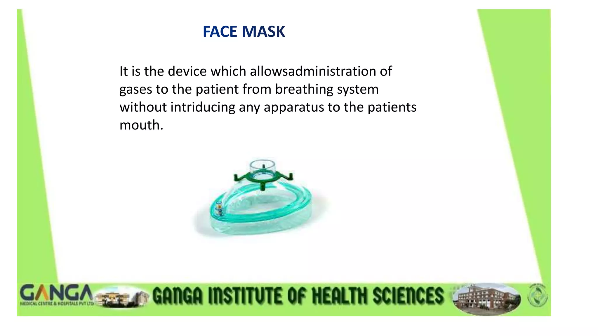
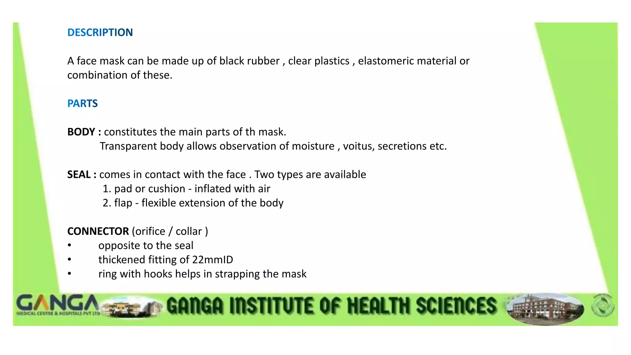
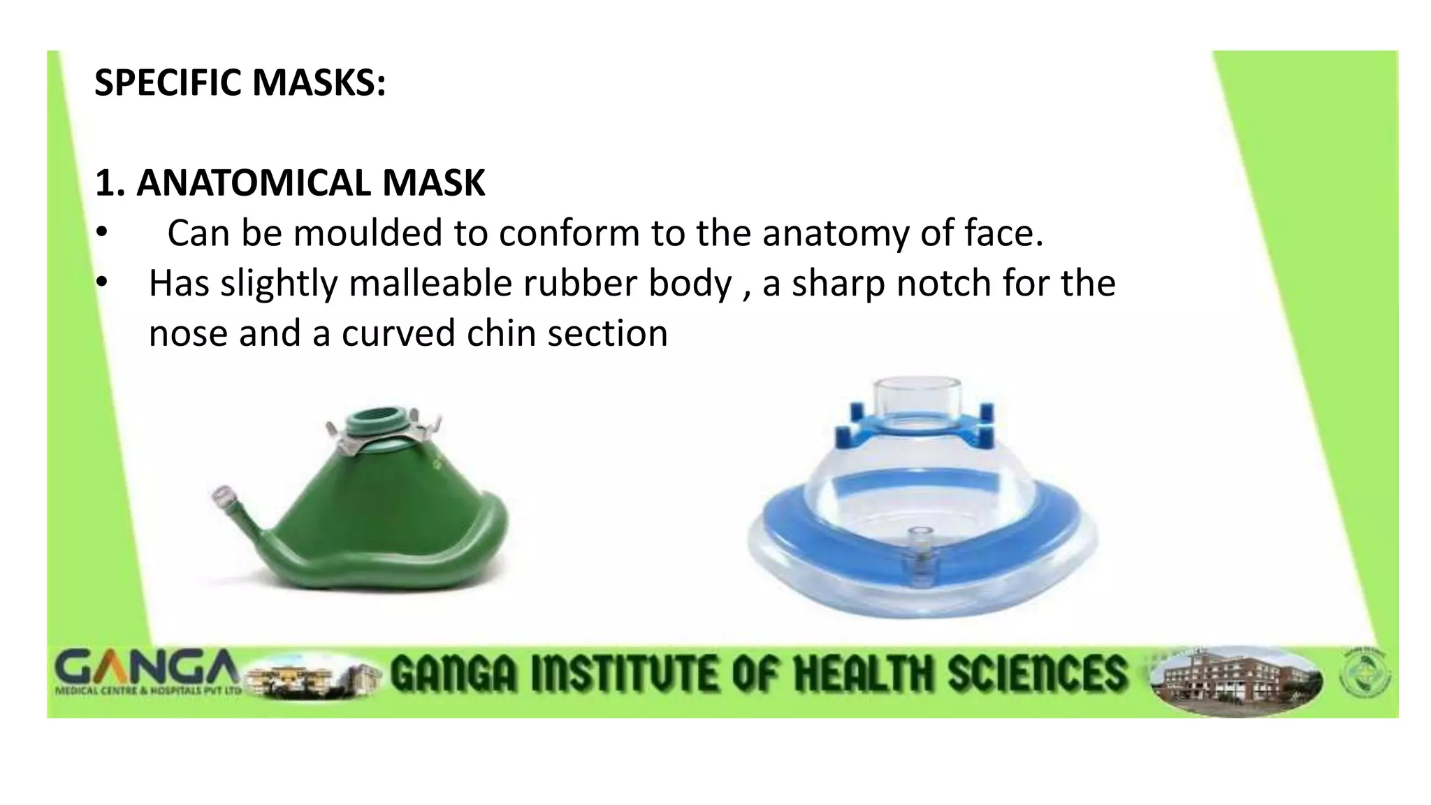
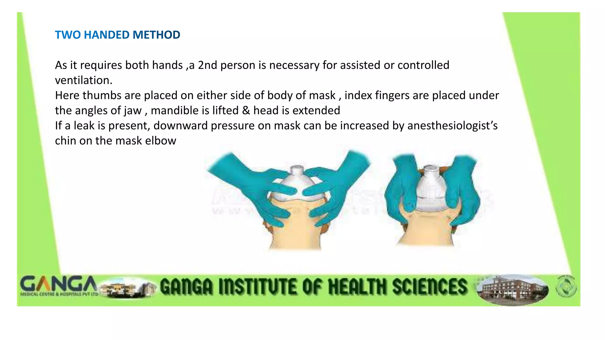
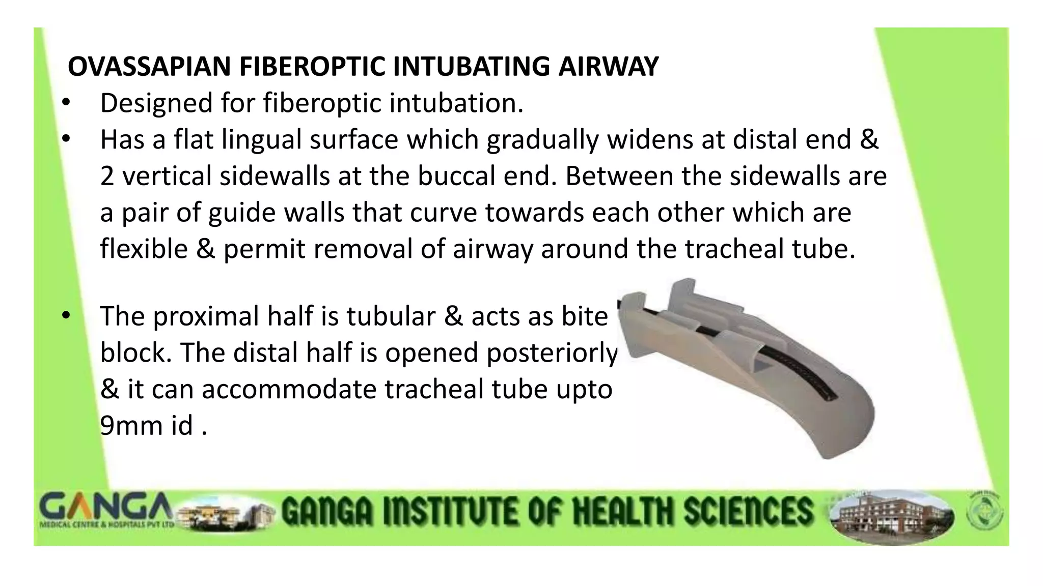
M.PRARTHANA
LECTURER
DEPARTMENT OF OPERATION THEATRE AND
ANESTHESIA TECHNOLOGY
It is the device which allowsadministration of
gases to the patient from breathing system
without intriducing any apparatus to the patients
mouth.
A face mask can be made up of black rubber , clear plastics , elastomeric material or
combination of these.
BODY : constitutes the main parts of th mask.
Transparent body allows observation of moisture , voitus, secretions etc.
SEAL : comes in contact with the face . Two types are available
1. pad or cushion - inflated with air
2. flap - flexible extension of the body
CONNECTOR (orifice / collar )
• opposite to the seal
• thickened fitting of 22mmID
• ring with hooks helps in strapping the mask
SPECIFIC MASKS:
1. ANATOMICAL MASK
• Can be moulded to conform to the anatomy of face.
• Has slightly malleable rubber body , a sharp notch for the
nose and a curved chin section
2. RENDELL-BAKER-SOUCEK(RBC) MASK
• Designed for paediatric patients below 10years .
• It has triangular body and low dead space.
• Used in tracheostomy and acromegaly patients
ENDOSCOPIC MASK
• Designed to allow mask ventilation during endoscopy
• It has port or diaphragm in the body to allow fiberscope
insertion.
SCENTED MASK
• Scent incorporated into the mask by manufacturer or
Anesthesia provider.
• Added for better acceptability and pleasant experience
during induction
• Disadvantage - ethyl alcohol in some flavour may affect
accuracy of gas monitors.
SIZE AGE GROUP DEAD SPACE
0 Preterm 3 cu.mm
1 infant 4 cu.mm
2 1 to 3 years 8 cu.mm
3 4 to 10 years 12 cu.mm
The face mask should form tight seal on the patients face while fitting comfortably in the
users hand .
The smallest mask is most desirable because it will cause least increase in dead space , easy
to hold & less likely to result in pressure on eyes.
• One hand method
• two hand method
• two hand jaw thrust
• claw hand technique
• The thumb & index finger of the left hand are place on mask body on oppposite sides
of connector push downward to prevent leak .
• the remaining 3 finger are placed on the mandible such the middle finger is applied to
the mentum, ring finger on body of mandible & little finger at angle of mandible to give
jaw thrust anteriorly
As it requires both hands ,a 2nd person is necessary for assisted or controlled
ventilation.
Here thumbs are placed on either side of body of mask , index fingers are placed under
the angles of jaw , mandible is lifted & head is extended
If a leak is present, downward pressure on mask can be increased by anesthesiologist’s
chin on the mask elbow
• 1 person stands at head end of patient & performs jaw thrust with his left hand at angle of
left mandible while his right hand compressesthe resevoir bag .
• The 2nd person stands at patient shoulder facing 1st person . this person right hand covers
the left hand of the 1st person and the left hand achieves right sided jaw thrust & mask
seal .
• It is useful for children undegoing short duration procedures.
• The anesthesiologist stands at the side of bed facing the child . the face mask is applied to
face by using the right hand with the palmar surface facing upward . the ring & middle
finger are placed under the angle of jaw & thumb encircle the body of mask.
Face mask and its adaptor increase dead space and it may contribute 30% or more of tidal volume
in neonates and infants .
It can be reduced by :
a) using smaller size mask .
b) increasing pressure on mask.
c) blowing a jet of fresh gas into the mask.
d) changing the volume of seal cushion.
• LOWER INCIDENCE OF THROAT
• REQUIRES LESS ANESTHETIC DEPTH THAN TRACHEAL TUBE OR SUPRAGLOTTIC DEVICE.
• NO NEED OF MUSCLE RELAXANTS
• THE FACE MASK MAY BE THE MOST COST EFFICIENT METHOD FOR SHORT CASES.
• ANESTHESILOGIST’S HANDS ARE TIEDUP
• HIGHER FRESH GAS FLOWS ARE OFTEN NEEDED
• DURING REMOTE ANESTHESIA , AIRWAY ACCESS IS DIFFICULT(CT & MRI)
• OFTEN MORE EPISODES OF OXYGEN DESATIRATION & REQUIRE MORE
INTRAOPERATIVE AIRWAY MANIPULATIONS.
• Skin problems - dermatitis, pressure necrosis.
• nerve injury
• eye injury - conjunctival chemosis , eyelid edema& corneal injuries .
• gastric inflation
• latex allergy
• lack of co-relation between arterial & end tidal CO2.
• environmental pollution with anesthetic gases
• jaw pain & user fatigue.
• A fundamental responsibility of anesthesia providers is to maintain a patients airway
• the airway passage has a rigid posterior wall and a collapsible anterior wall consisting of
the toungue and epiglottis.
• under anesthesia the toungue & epiglottis falls back into the posterior pharynx occluding
the airway .
• the purpose of airway is to lift the toungue & epiglottis away from the posterior
pharyngeal wall & maintain a patent airway.
TERMINOLOGY :
ARTIFICIAL AIRWAY - any device that aims to maintain oral or nasal air passages . It may be
SIMPLE SUPRAGLOTTIC DEVICE
such as oropharyngeal or nasopharyngeal airways. These may not be sufficient to maintain
the patency of airway on therir own & may require patient jaw to be supported as well
AUGMENTED SUPRAGLOTTIC DEVICE
such as LMA & airway management device
INFRAGOTTIC DEVICES
such as endotracheal tubes , tracheostomy, jet ventilation catheters.
OROPHARYNGEAL AIRWAY
It extends from lips to pharynx, fitting between tongue & posterior
pharyngeal wall. Made up of elastomeric or plastic material. Parts are
FLANGE - it is at buccal end to prevent it from moving deeper into mouth &
may also serve to fix airway in place
BITE PORTION - it is straight & fits between teeth & gums.
CURVED PORTION - extends backwards to correspond the shape of tongue &
palate.
The American National Standard specifies that the size of oral
airways be designated by a no. i.e the length in cms.
SIZE COLOUR LENGTH
000 Violet 3.5
00 Blue 4.5
0 Black 5.5
1 White 6.5
2 Green 7.5
3 Orange 8.5
4 Red 9.5
5 Yellow 10.5
SPECIFIC AIRWAYS
GUEDEL AIRWAY
Most frequently used Airway & has large flange & reinforced bite
portion with tubular channel for air exchange & suctioning.
CUFFED ORO-PHARYNGEAL AIRWAY
• It is Guedel's airway with an inflatable CUFFED designed to seal
the oropharynx. It has an integral bite block & a 15mm
connector for attachment of the breathing circuit.
• The cuff is inflated with air to displace the base of tongue &
form a low pressure seal with the pharynx & provide an open
airway.
PATIL- SYRACUSE ENDOSCOPIC AIRWAY
• It was designed to aid fiber - optic intubation. It is made of
aluminium.
• It has lateral channel & a central groove on the lingual surface to
allow passage of fiberscope.A slit in distal end allows fiberscope
to be manipulated in antero- posterior direction but limits lateral
movement.
BERMAN INTUBATING AIRWAY
It is tubular along its entire length. It is open on 1 side so that
it can be split & removed from around a tracheal tube.It can
be used as an oral airway or as an aid to fiberoptic or blind
oro-tracheal intubation.
WILLIAM AIRWAY INTUBATOR
• Designed for blind tracheal intubation & can also be used
for fiber optic intubation or as an oral airway.
• It is available in 2 size #9&#10 which will admit upto &
8.5mm tracheal tube respectively.
• The proximal half is cylindrical while distal half is open on
lingual surface.
OVASSAPIAN FIBEROPTIC INTUBATING AIRWAY
• Designed for fiberoptic intubation.
• Has a flat lingual surface which gradually widens at distal end &
2 vertical sidewalls at the buccal end. Between the sidewalls are
a pair of guide walls that curve towards each other which are
flexible & permit removal of airway around the tracheal tube.
• The proximal half is tubular & acts as bite
block. The distal half is opened posteriorly
& it can accommodate tracheal tube upto
9mm id .
METHOD OF INSERTION
• Pharyngeal & laryngeal reflexes should be depressed before an
airwy is inserted to avoid coughing & laryngospasm.
• Selecting the correct size is important. correct size is estimated
by holding the airway next to patient mouth & the tip should be
at the angle of mandible.
• The best criteria for proper size & position is unobstructed gas
exchange.
Oral airway may be inserted in 2ways
• The jaw opened with left hand.
• The airway is inserted with its concave side towards the upper lip &
when the junction of bite portion & curved section is near the
incisors, the airway is rotated 180° & slipped behind the tongue in
the final position.
• A tongue blade is used to push forward and depress the
tongue & the airway is inserted with concave side towards
the tongue & is advanced.
OTHER USES
• May be used to prevent patient from biting & occluding
ETT.
• protect the tongue from biting.
• To facilitate suctioning.
• To obtain better mask fit.
NASOPHARYNGEAL AIRWAY (nasal trumpet/nasal airway)
• It extends from nose to pharynx, the pharyngeal end should be
below base of tongue but above the epiglottis.
• A nasal airway is better tolerated than oral airway if patient has
intact airway reflexes
• DESCRIPTION
• It resembles a shortened tracheal tube with a flange at the
outer end to prevent it from completely passing into the nares.
It is made up of plastic or rubber & the size is designated by the
inner diameter in mm.
SPECIFIC TYPES
LINDER NASOPHARYNGEAL AIRWAY
• It is plastic with large flange & flat distal end and is supplied with
introducer which has balloon on its tip.
• Before insertion, the introducer is inserted into the airway until
the tip of balloon is just past the end
• Air is injected until the balloon tip is inflated to approx outer
diameter of tube.
• The complete assembly is lubricated & inserted through the
nostril.
• After it is in place, the balloon is deflated & introducer is
removed.
CUFFED NASOPHARYNGEAL AIRWAY
It is similar to a short, Cuffed tracheal tube. It is inserted through
the nose into the pharynx; Cuff inflated & then is pulled back until
resistance is felt.
BINASAL AIRWAY
It consists of 2 nasal airway joined together by an adaptor for
attachment to the breathing system. Can be used to administer
anesthesia.
INSERTION
• The diameter of nasal airway should be the same as
needed to insert the tracheal tube (0.5-1.0mm smaller than
oral tracheal tube).
• Before insertion it should be lubricated thoroughly along its
entire length. The airway is held with the bevel against
septum & gently advanced posteriorly while being rotated
back & forth.
COMPLICATIONS
• Airway obstruction
• Epistaxis
• Ulceration & necrosis
• Dental damage
• CNS trauma
• Laryngospasm & coughing.
• Aspiration or swallowing of part or all of the airway
• Latex allergy
• Gastric distension.
THANK YOU

FACE MASK AND AIRWAY.pptx

  • 1.
    M.PRARTHANA LECTURER DEPARTMENT OF OPERATIONTHEATRE AND ANESTHESIA TECHNOLOGY
  • 2.
    It is thedevice which allowsadministration of gases to the patient from breathing system without intriducing any apparatus to the patients mouth.
  • 3.
    A face maskcan be made up of black rubber , clear plastics , elastomeric material or combination of these. BODY : constitutes the main parts of th mask. Transparent body allows observation of moisture , voitus, secretions etc. SEAL : comes in contact with the face . Two types are available 1. pad or cushion - inflated with air 2. flap - flexible extension of the body CONNECTOR (orifice / collar ) • opposite to the seal • thickened fitting of 22mmID • ring with hooks helps in strapping the mask
  • 4.
    SPECIFIC MASKS: 1. ANATOMICALMASK • Can be moulded to conform to the anatomy of face. • Has slightly malleable rubber body , a sharp notch for the nose and a curved chin section
  • 5.
    2. RENDELL-BAKER-SOUCEK(RBC) MASK •Designed for paediatric patients below 10years . • It has triangular body and low dead space. • Used in tracheostomy and acromegaly patients
  • 6.
    ENDOSCOPIC MASK • Designedto allow mask ventilation during endoscopy • It has port or diaphragm in the body to allow fiberscope insertion.
  • 7.
    SCENTED MASK • Scentincorporated into the mask by manufacturer or Anesthesia provider. • Added for better acceptability and pleasant experience during induction • Disadvantage - ethyl alcohol in some flavour may affect accuracy of gas monitors.
  • 8.
    SIZE AGE GROUPDEAD SPACE 0 Preterm 3 cu.mm 1 infant 4 cu.mm 2 1 to 3 years 8 cu.mm 3 4 to 10 years 12 cu.mm
  • 9.
    The face maskshould form tight seal on the patients face while fitting comfortably in the users hand . The smallest mask is most desirable because it will cause least increase in dead space , easy to hold & less likely to result in pressure on eyes. • One hand method • two hand method • two hand jaw thrust • claw hand technique
  • 10.
    • The thumb& index finger of the left hand are place on mask body on oppposite sides of connector push downward to prevent leak . • the remaining 3 finger are placed on the mandible such the middle finger is applied to the mentum, ring finger on body of mandible & little finger at angle of mandible to give jaw thrust anteriorly
  • 11.
    As it requiresboth hands ,a 2nd person is necessary for assisted or controlled ventilation. Here thumbs are placed on either side of body of mask , index fingers are placed under the angles of jaw , mandible is lifted & head is extended If a leak is present, downward pressure on mask can be increased by anesthesiologist’s chin on the mask elbow
  • 12.
    • 1 personstands at head end of patient & performs jaw thrust with his left hand at angle of left mandible while his right hand compressesthe resevoir bag . • The 2nd person stands at patient shoulder facing 1st person . this person right hand covers the left hand of the 1st person and the left hand achieves right sided jaw thrust & mask seal . • It is useful for children undegoing short duration procedures. • The anesthesiologist stands at the side of bed facing the child . the face mask is applied to face by using the right hand with the palmar surface facing upward . the ring & middle finger are placed under the angle of jaw & thumb encircle the body of mask.
  • 13.
    Face mask andits adaptor increase dead space and it may contribute 30% or more of tidal volume in neonates and infants . It can be reduced by : a) using smaller size mask . b) increasing pressure on mask. c) blowing a jet of fresh gas into the mask. d) changing the volume of seal cushion.
  • 14.
    • LOWER INCIDENCEOF THROAT • REQUIRES LESS ANESTHETIC DEPTH THAN TRACHEAL TUBE OR SUPRAGLOTTIC DEVICE. • NO NEED OF MUSCLE RELAXANTS • THE FACE MASK MAY BE THE MOST COST EFFICIENT METHOD FOR SHORT CASES. • ANESTHESILOGIST’S HANDS ARE TIEDUP • HIGHER FRESH GAS FLOWS ARE OFTEN NEEDED • DURING REMOTE ANESTHESIA , AIRWAY ACCESS IS DIFFICULT(CT & MRI) • OFTEN MORE EPISODES OF OXYGEN DESATIRATION & REQUIRE MORE INTRAOPERATIVE AIRWAY MANIPULATIONS.
  • 15.
    • Skin problems- dermatitis, pressure necrosis. • nerve injury • eye injury - conjunctival chemosis , eyelid edema& corneal injuries . • gastric inflation • latex allergy • lack of co-relation between arterial & end tidal CO2. • environmental pollution with anesthetic gases • jaw pain & user fatigue.
  • 16.
    • A fundamentalresponsibility of anesthesia providers is to maintain a patients airway • the airway passage has a rigid posterior wall and a collapsible anterior wall consisting of the toungue and epiglottis. • under anesthesia the toungue & epiglottis falls back into the posterior pharynx occluding the airway . • the purpose of airway is to lift the toungue & epiglottis away from the posterior pharyngeal wall & maintain a patent airway.
  • 17.
    TERMINOLOGY : ARTIFICIAL AIRWAY- any device that aims to maintain oral or nasal air passages . It may be SIMPLE SUPRAGLOTTIC DEVICE such as oropharyngeal or nasopharyngeal airways. These may not be sufficient to maintain the patency of airway on therir own & may require patient jaw to be supported as well AUGMENTED SUPRAGLOTTIC DEVICE such as LMA & airway management device INFRAGOTTIC DEVICES such as endotracheal tubes , tracheostomy, jet ventilation catheters.
  • 18.
    OROPHARYNGEAL AIRWAY It extendsfrom lips to pharynx, fitting between tongue & posterior pharyngeal wall. Made up of elastomeric or plastic material. Parts are FLANGE - it is at buccal end to prevent it from moving deeper into mouth & may also serve to fix airway in place BITE PORTION - it is straight & fits between teeth & gums. CURVED PORTION - extends backwards to correspond the shape of tongue & palate.
  • 19.
    The American NationalStandard specifies that the size of oral airways be designated by a no. i.e the length in cms. SIZE COLOUR LENGTH 000 Violet 3.5 00 Blue 4.5 0 Black 5.5 1 White 6.5 2 Green 7.5 3 Orange 8.5 4 Red 9.5 5 Yellow 10.5
  • 20.
    SPECIFIC AIRWAYS GUEDEL AIRWAY Mostfrequently used Airway & has large flange & reinforced bite portion with tubular channel for air exchange & suctioning.
  • 21.
    CUFFED ORO-PHARYNGEAL AIRWAY •It is Guedel's airway with an inflatable CUFFED designed to seal the oropharynx. It has an integral bite block & a 15mm connector for attachment of the breathing circuit. • The cuff is inflated with air to displace the base of tongue & form a low pressure seal with the pharynx & provide an open airway.
  • 22.
    PATIL- SYRACUSE ENDOSCOPICAIRWAY • It was designed to aid fiber - optic intubation. It is made of aluminium. • It has lateral channel & a central groove on the lingual surface to allow passage of fiberscope.A slit in distal end allows fiberscope to be manipulated in antero- posterior direction but limits lateral movement.
  • 23.
    BERMAN INTUBATING AIRWAY Itis tubular along its entire length. It is open on 1 side so that it can be split & removed from around a tracheal tube.It can be used as an oral airway or as an aid to fiberoptic or blind oro-tracheal intubation.
  • 24.
    WILLIAM AIRWAY INTUBATOR •Designed for blind tracheal intubation & can also be used for fiber optic intubation or as an oral airway. • It is available in 2 size #9&#10 which will admit upto & 8.5mm tracheal tube respectively. • The proximal half is cylindrical while distal half is open on lingual surface.
  • 25.
    OVASSAPIAN FIBEROPTIC INTUBATINGAIRWAY • Designed for fiberoptic intubation. • Has a flat lingual surface which gradually widens at distal end & 2 vertical sidewalls at the buccal end. Between the sidewalls are a pair of guide walls that curve towards each other which are flexible & permit removal of airway around the tracheal tube. • The proximal half is tubular & acts as bite block. The distal half is opened posteriorly & it can accommodate tracheal tube upto 9mm id .
  • 26.
    METHOD OF INSERTION •Pharyngeal & laryngeal reflexes should be depressed before an airwy is inserted to avoid coughing & laryngospasm. • Selecting the correct size is important. correct size is estimated by holding the airway next to patient mouth & the tip should be at the angle of mandible. • The best criteria for proper size & position is unobstructed gas exchange.
  • 27.
    Oral airway maybe inserted in 2ways • The jaw opened with left hand. • The airway is inserted with its concave side towards the upper lip & when the junction of bite portion & curved section is near the incisors, the airway is rotated 180° & slipped behind the tongue in the final position.
  • 28.
    • A tongueblade is used to push forward and depress the tongue & the airway is inserted with concave side towards the tongue & is advanced.
  • 29.
    OTHER USES • Maybe used to prevent patient from biting & occluding ETT. • protect the tongue from biting. • To facilitate suctioning. • To obtain better mask fit.
  • 30.
    NASOPHARYNGEAL AIRWAY (nasaltrumpet/nasal airway) • It extends from nose to pharynx, the pharyngeal end should be below base of tongue but above the epiglottis. • A nasal airway is better tolerated than oral airway if patient has intact airway reflexes • DESCRIPTION • It resembles a shortened tracheal tube with a flange at the outer end to prevent it from completely passing into the nares. It is made up of plastic or rubber & the size is designated by the inner diameter in mm.
  • 32.
    SPECIFIC TYPES LINDER NASOPHARYNGEALAIRWAY • It is plastic with large flange & flat distal end and is supplied with introducer which has balloon on its tip. • Before insertion, the introducer is inserted into the airway until the tip of balloon is just past the end • Air is injected until the balloon tip is inflated to approx outer diameter of tube. • The complete assembly is lubricated & inserted through the nostril. • After it is in place, the balloon is deflated & introducer is removed.
  • 33.
    CUFFED NASOPHARYNGEAL AIRWAY Itis similar to a short, Cuffed tracheal tube. It is inserted through the nose into the pharynx; Cuff inflated & then is pulled back until resistance is felt. BINASAL AIRWAY It consists of 2 nasal airway joined together by an adaptor for attachment to the breathing system. Can be used to administer anesthesia.
  • 34.
    INSERTION • The diameterof nasal airway should be the same as needed to insert the tracheal tube (0.5-1.0mm smaller than oral tracheal tube). • Before insertion it should be lubricated thoroughly along its entire length. The airway is held with the bevel against septum & gently advanced posteriorly while being rotated back & forth.
  • 35.
    COMPLICATIONS • Airway obstruction •Epistaxis • Ulceration & necrosis • Dental damage • CNS trauma • Laryngospasm & coughing. • Aspiration or swallowing of part or all of the airway • Latex allergy • Gastric distension.
  • 37.