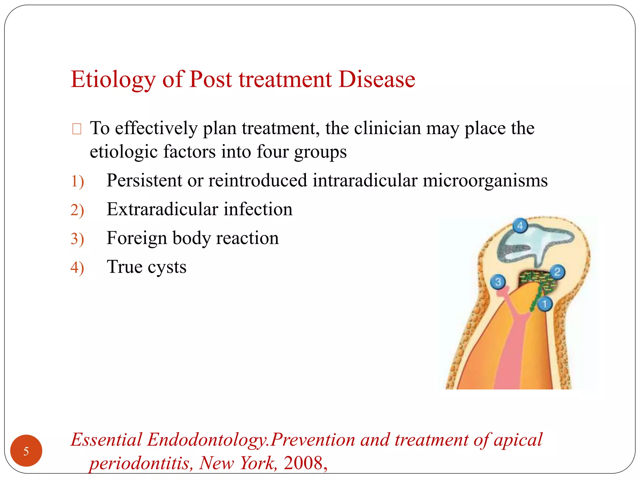
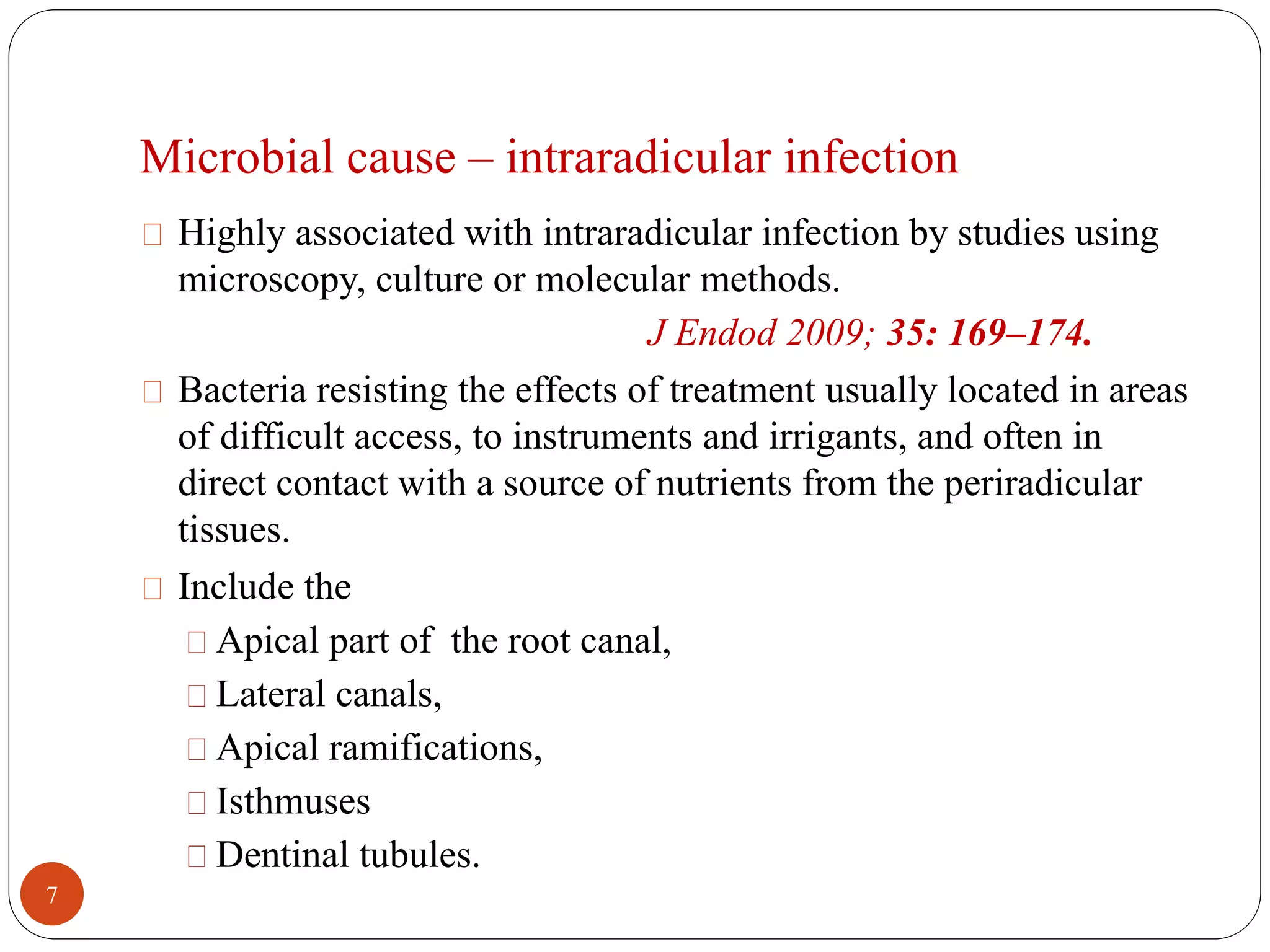
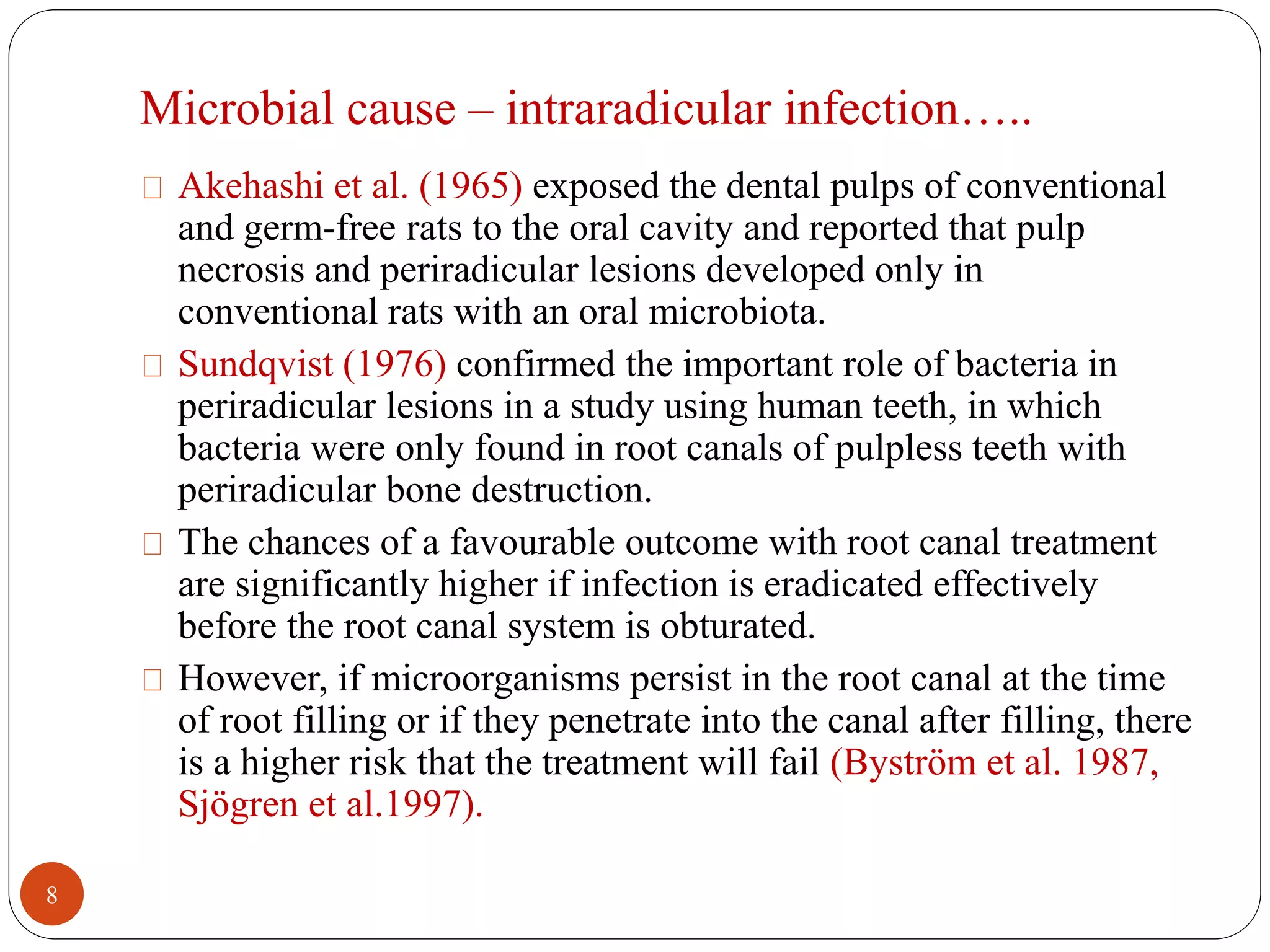
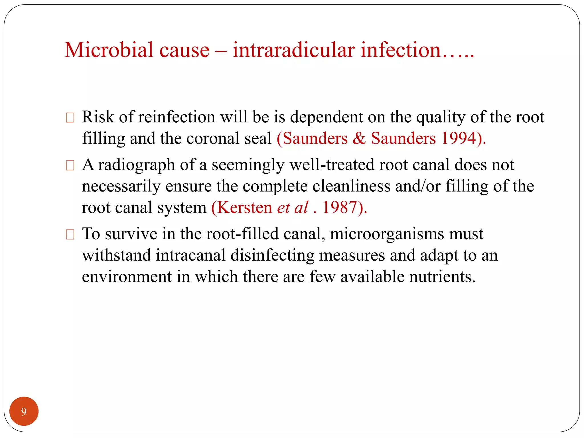
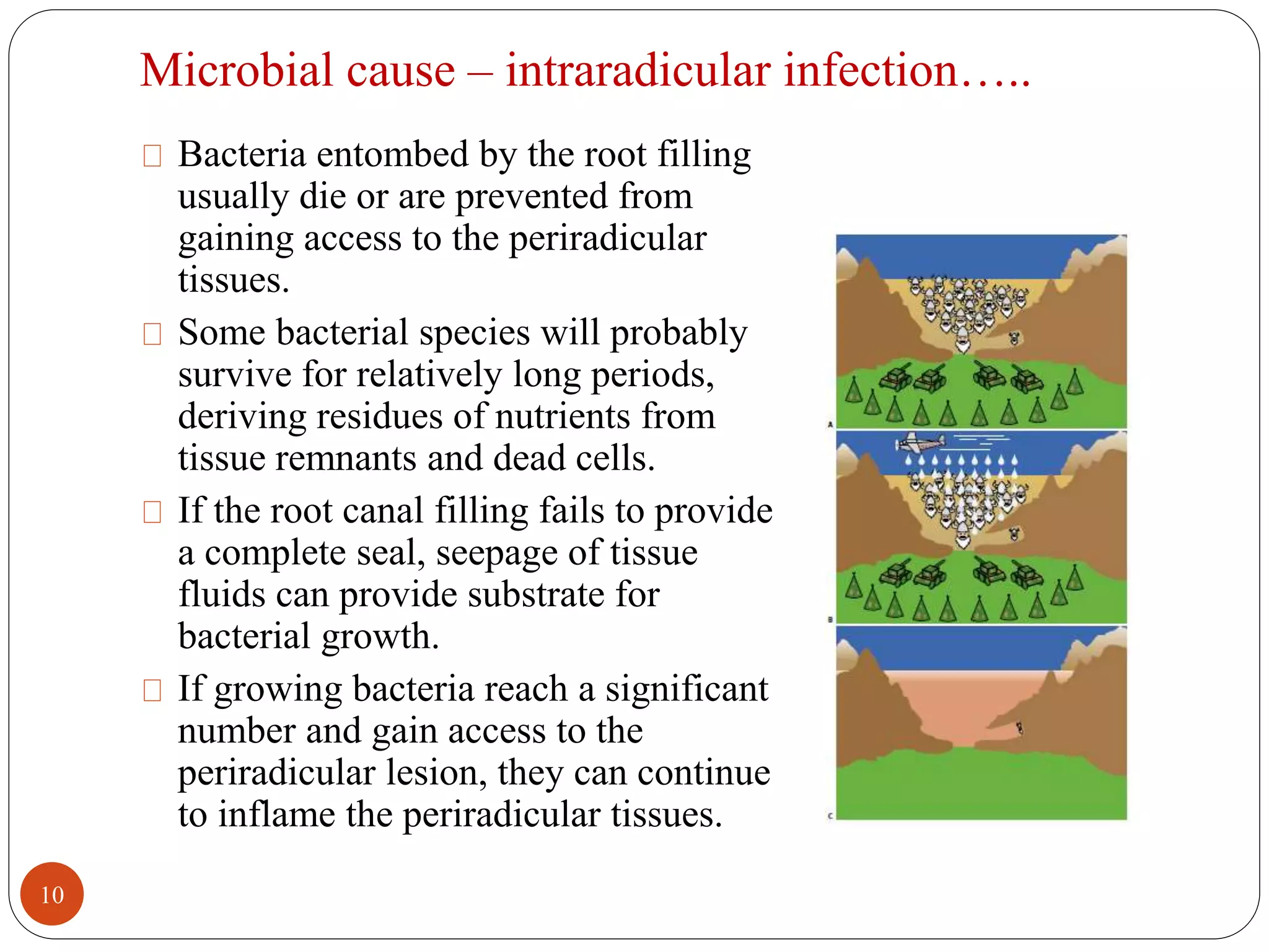
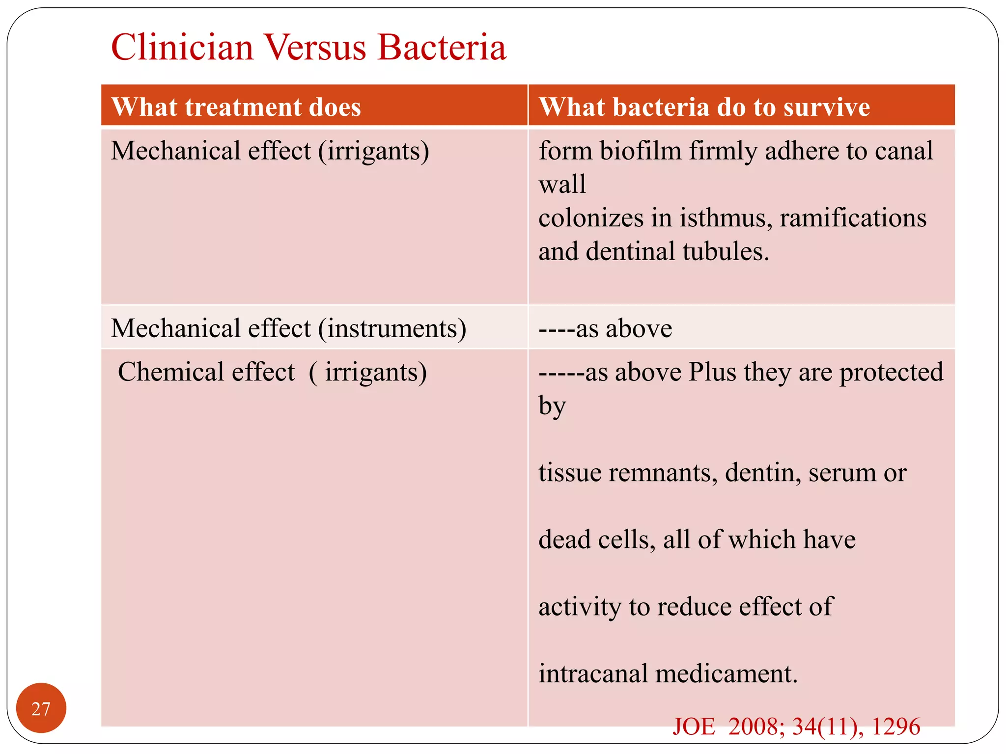
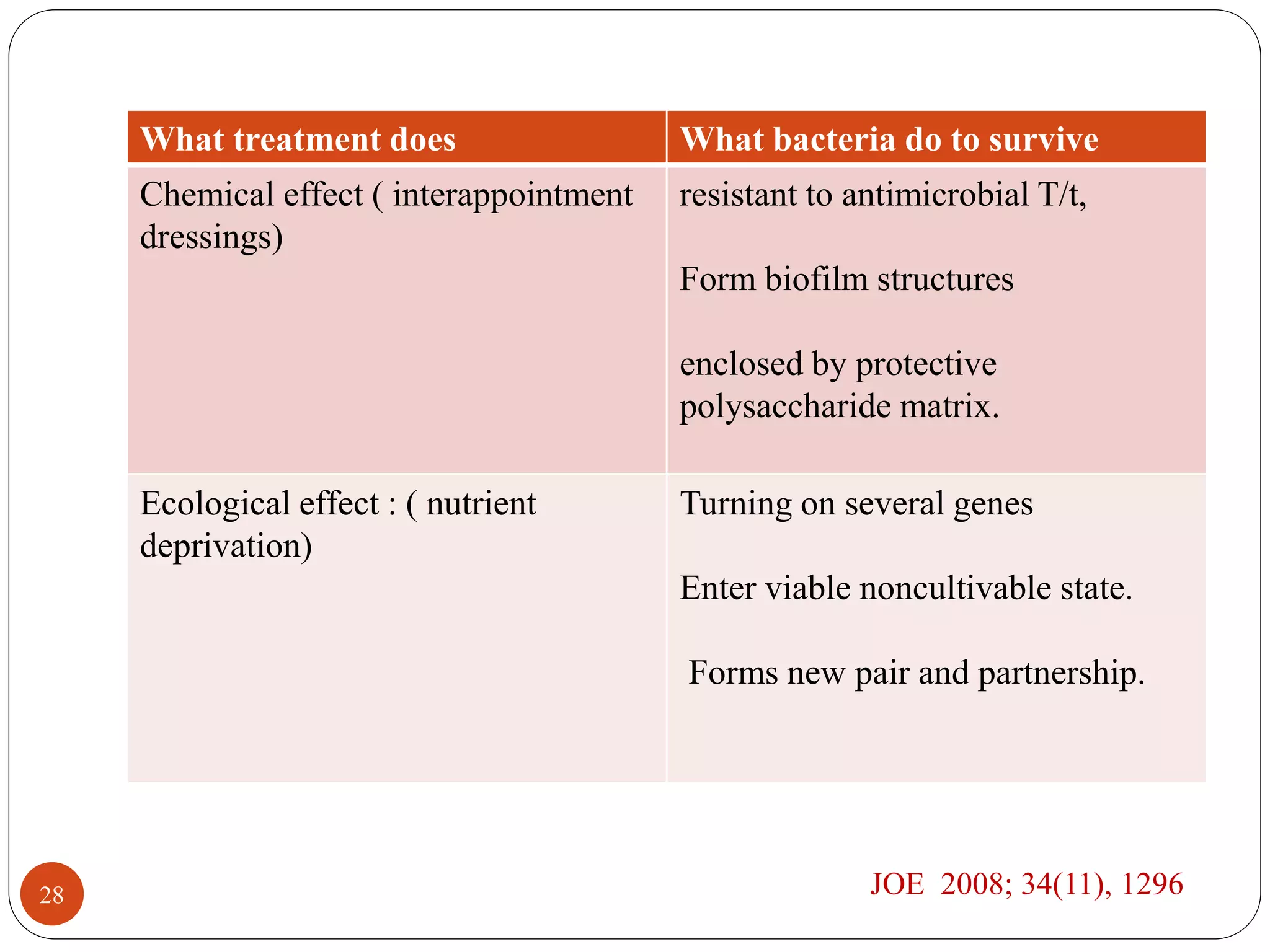
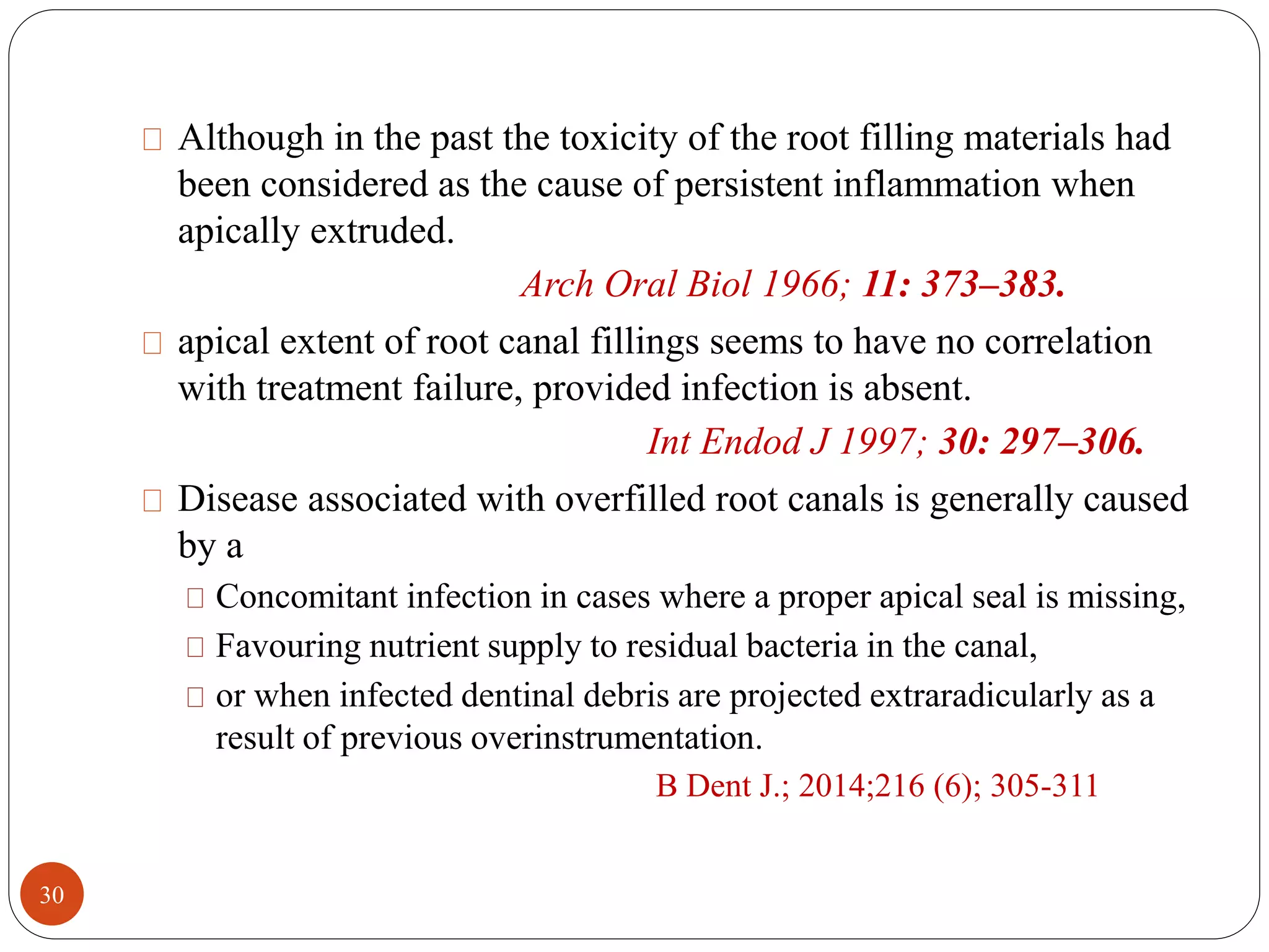
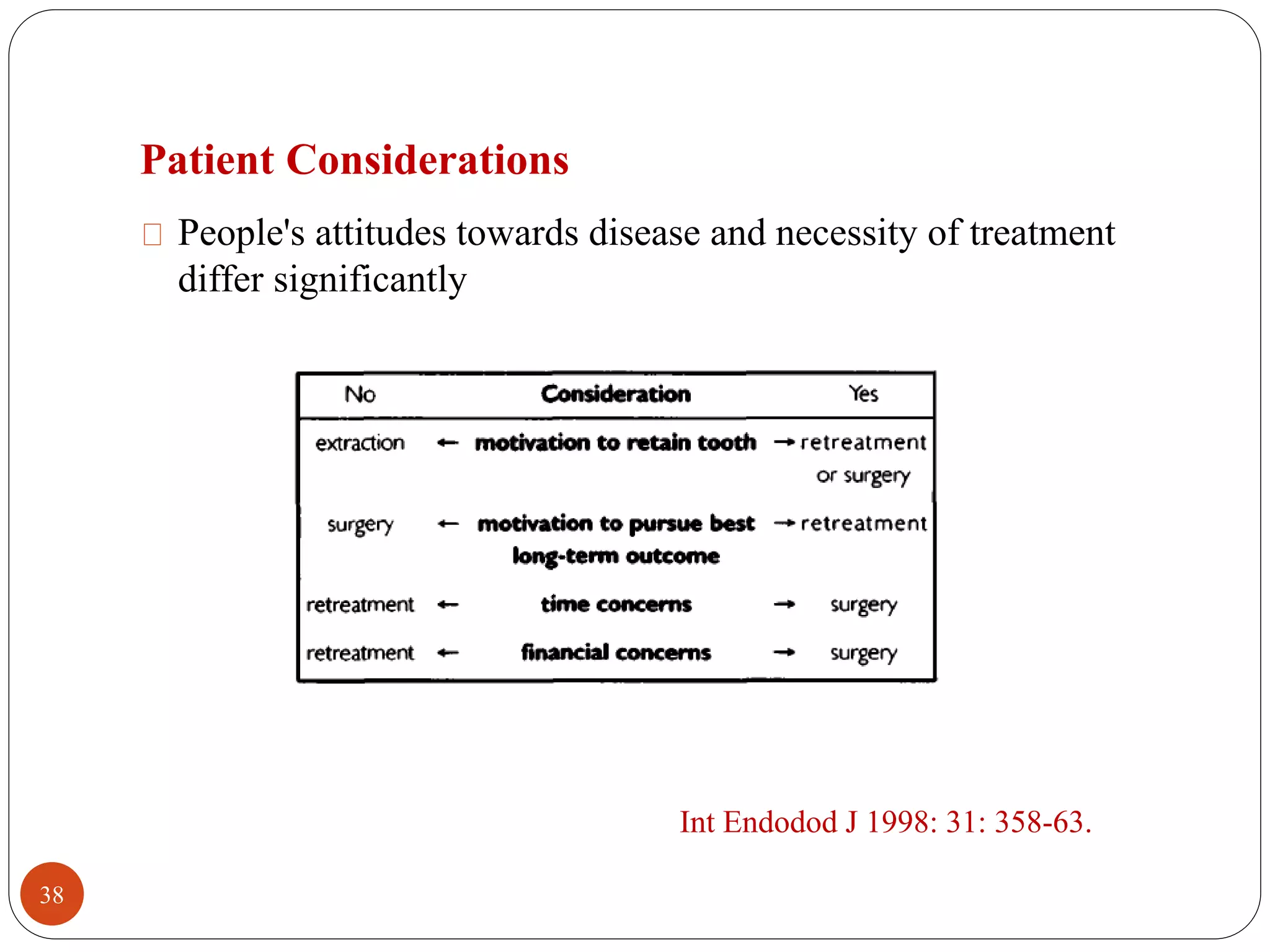
Causes and management of post treatment 
Praveen Jangid. 
endodontic disease 
1
Introduction 
In recent years the number of people seeking endodontic 
treatment has dramatically increased because of conservative 
tendency towards root canal treatment over tooth extraction 
(Ruddle 2002) 
The aim of root canal treatment is to clean and disinfect the root 
canal system in order to reduce the number of micro-organisms, 
remove necrotic tissue, and finally seal the system to prevent 
recontamination. 
Success rates up to 97% have been reported for endodontic 
initial treatment (Friedman 2004) 
2
Prevalence of apical periodontitis and other post-treatment 
periradicular diseases can exceed 30% of all root-filled teeth. 
(Boucher 2002; Eriksen 2002; Friedman 2002; Dugas 2003) 
In the past, undesirable outcomes of endodontic therapy were 
described as failures. 
Friedman states that “most patients can relate to the concept of 
disease-treatment-healing, whereas failure, apart from being a 
negative and relative term, does not imply the necessity to pursue 
treatment.” 
He has suggested using the term posttreatment disease to describe 
those cases that would previously have been referred to as treatment 
failures. 
Principles and practice of endodontics, ed 4, St. Louis, 2009. 
3
Post-treatment apical periodontitis, 
which can be categorised as- 
• If developed after 
treatment 
Emergent 
• If persisted despite 
treatment 
Persistent 
• If developed after 
having healed 
Recurrent 
B Dent J.; 2014;216 (6); 305-311 
4
Etiology of Post treatment Disease 
To effectively plan treatment, the clinician may place the 
etiologic factors into four groups 
1) Persistent or reintroduced intraradicular microorganisms 
2) Extraradicular infection 
3) Foreign body reaction 
4) True cysts 
Essential Endodontology.Prevention and treatment of apical 
periodontitis, New York, 2008, 
5
Causes of post-treatment apical periodontitis 
Microbial cause – intraradicular infection 
Microbial cause – extraradicular infection 
Non-microbial cause 
Procedural errors and 
6
Microbial cause – intraradicular infection 
Highly associated with intraradicular infection by studies using 
microscopy, culture or molecular methods. 
J Endod 2009; 35: 169–174. 
Bacteria resisting the effects of treatment usually located in areas 
of difficult access, to instruments and irrigants, and often in 
direct contact with a source of nutrients from the periradicular 
tissues. 
Include the 
Apical part of the root canal, 
Lateral canals, 
Apical ramifications, 
Isthmuses 
Dentinal tubules. 
7
Microbial cause – intraradicular infection….. 
Akehashi et al. (1965) exposed the dental pulps of conventional 
and germ-free rats to the oral cavity and reported that pulp 
necrosis and periradicular lesions developed only in 
conventional rats with an oral microbiota. 
Sundqvist (1976) confirmed the important role of bacteria in 
periradicular lesions in a study using human teeth, in which 
bacteria were only found in root canals of pulpless teeth with 
periradicular bone destruction. 
The chances of a favourable outcome with root canal treatment 
are significantly higher if infection is eradicated effectively 
before the root canal system is obturated. 
However, if microorganisms persist in the root canal at the time 
of root filling or if they penetrate into the canal after filling, there 
is a higher risk that the treatment will fail (Byström et al. 1987, 
Sjögren et al.1997). 
8
Microbial cause – intraradicular infection….. 
Risk of reinfection will be is dependent on the quality of the root 
filling and the coronal seal (Saunders & Saunders 1994). 
A radiograph of a seemingly well-treated root canal does not 
necessarily ensure the complete cleanliness and/or filling of the 
root canal system (Kersten et al . 1987). 
To survive in the root-filled canal, microorganisms must 
withstand intracanal disinfecting measures and adapt to an 
environment in which there are few available nutrients. 
9
Microbial cause – intraradicular infection….. 
Bacteria entombed by the root filling 
usually die or are prevented from 
gaining access to the periradicular 
tissues. 
Some bacterial species will probably 
survive for relatively long periods, 
deriving residues of nutrients from 
tissue remnants and dead cells. 
If the root canal filling fails to provide 
a complete seal, seepage of tissue 
fluids can provide substrate for 
bacterial growth. 
If growing bacteria reach a significant 
number and gain access to the 
periradicular lesion, they can continue 
to inflame the periradicular tissues. 
10
Microbial cause – intraradicular infection….. 
Several regulatory systems play essential roles in the ability of 
bacteria to withstand nutrient depletion. 
These systems are under the control of determined genes, whose 
transcription is activated under conditions of starvation. 
Under conditions of nitrogen starvation, the activation of the 
Ntr gene system enables bacteria that require ammonia as a 
nitrogen source to scavenge even small traces of ammonia. 
Under high concentration of ammonia, the Ntr gene system is 
uncoupled. 
Int Endod J 2001; 34, 1–10. 
11
Microbial cause – intraradicular infection….. 
Some facultative bacteria may activate the Arc system 
(aerobic respiration regulatory ), under conditions of low 
concentrations of molecular oxygen. 
So that a shift can occur from aerobic to anaerobic 
metabolism. 
Under low concentrations of glucose, some bacteria can 
activate the catabolite repressor system, under control of the 
genes Cya (adenylate cyclase) and Crp (catabolite repressor 
protein), which induce the synthesis of enzymes for the 
utilization of various other organic carbon sources. 
12
Microbial cause – intraradicular infection….. 
Under conditions of phosphate starvation triggered by low 
concentrations of inorganic phosphate, cells turn on genes for 
utilization of organic phosphate compounds and for the 
scavenging of trace amounts of inorganic phosphate. 
Int Endod J 2001; 34, 1–10. 
Heat-shock proteins are family of highly conserved proteins 
whose main role is to allow microorganism to survive under 
stressful conditions. 
13
Microbial cause – intraradicular infection….. 
The microbiota associated with failed cases differs markedly 
from that reported in untreated teeth (primary root canal 
infection). 
T.denticola, P.alactolyticus, T.forsythia, P.gingivalis, T. 
socranskii, P.endodontalis. 
Fungi are not found. 
The reduction of Gram-negative organisms following endodontic 
treatment and the subsequent proportional increase of Gram-positives 
facultatives. 
Streptococci ( Streptococcus mitis, Streptocoocus gordonii, 
streptococcus anginosus, Streptococcus sanguinis and 
streptococuus oralis), Parvimonas micra, Actinomyces species, 
Propionobacter species, Pseudoramibacter alactolyticus, 
lactobacilli, E faecalis. 
14 JOE 2008; 34(11), 1296
Microbial cause – intraradicular infection….. 
15
Microbial cause – intraradicular infection….. 
Möller (1966), reported a mean of 1.6 bacterial species per root 
canal. Anaerobic bacteria corresponded to 51% of the isolates. 
Enterococcus faecalis was found in 29% of the cases. 
E. faecalis strains have been demonstrated to be extremely 
resistant to several medicaments, including calcium hydroxide 
(Jett et al . 1994, Siqueira & Uzeda 1996, Siqueira & Lopes 1999). 
It has been shown that some bacteria, such as E. faecalis, can 
enter a viable but noncultivable state, which is a survival 
mechanism adopted by many bacteria when exposed to 
environmental stresses like low nutrients concentrations, high 
salinity, and extreme pH 
16
Microbial cause – intraradicular infection….. 
Sedgley et al showed that E. faecalis has the capacity to recover 
from prolonged starvation state in the root canal treated teeth; 
when inoculated into the canals, this bacterium maintain viability 
for 12 months without additional nutrients. 
Thus viable E. faecalis entombed at the time of root canal filling 
may provide a long term nidus for subsequent infection. 
Yeast-like microorganisms have also been found in root canals of 
obturated teeth in which treatment has failed (Nair et al. 1990a). 
In fact, it has been demonstrated that Candida spp. are resistant 
to some medicaments commonly used in endodontics (Waltimo 
et al . 1999). 
17
Microbial cause – intraradicular infection….. 
Persistent infections are the major cause of post-treatment apical 
periodontitis does not preclude secondary infections due to 
coronal leakage. 
Cross-sectional studies indicate that the best outcome is achieved 
in teeth with adequate root canal fillings associated with 
adequate coronal restorations. 
J Endod 2013; 39: 600–604. 
It is advisable to treat the tooth as a continuum, placing a well-adapted 
permanent coronal restoration as soon as possible after 
finishing root canal treatment. 
18
Microbial cause – extraradicular infection 
Extraradicular infection may be associated with chronic 
inflammation and lead to endodontic treatment failure. 
By defending themselves against the action of the complement 
system, avoiding destruction by phagocytes, causing 
immunosuppression, changing antigenic coats, and inducing 
proteolysis of antibody molecules (Siqueira 1997). 
Int Endod J, 2001; 34, 1–10, 
May be associated with – 
A biofilm formation on the external root surface 
Endod Dent Traumatol 1990; 6: 73–77. 
Sometimes showing calculus-like calcifications, 
Int Endod J 2005; 38: 262–271. 
or forming cohesive actinomycotic colonies within the body of the 
lesion. 
Endod Dent Traumatol 1986; 2: 205–209. 
19
Microbial cause – extraradicular infection….. 
The extraradicular infectious process can be dependent on or 
independent of the intraradicular infection. 
Dependent infection- maintained by constant proliferation and 
invasion of the periradicular tissues by bacteria present in the 
intraradicular infection. 
Cannot sustain itself without the intraradicular component. 
Independent infections - that are no longer fostered by an 
intraradicular infection and as such may not respond to adequate 
root canal treatment. 
Endodontic Topics 2003; 6: 78–95. 
20
Microbial cause – extraradicular infection….. 
One of the most significant mechanisms of evasion from 
the host defence system is the microbial arrangement in a 
biofilm. 
A biofilm can be defined as a microbial population attached to an 
organic or inorganic substrate, surrounded by microbial 
extracellular products, which form an intermicrobial matrix 
(Costerton et al ) 
Biofilm formation is a step-wise procedure ,its formation occurs 
in the presence of microorganisms, fluid and solid surface. 
21
Microbial cause – extraradicular infection….. 
Organized in biofilms, microorganisms show higher resistance to 
both antimicrobial agents and host defence mechanisms when 
compared with planktonic cells. 
(Costerton et al 1987, 1994, Gilbert et al. 1997). 
By examining teeth refractory to root canal treatment, Tronstad 
et al. (1990) reported the occurrence of bacterial biofilms 
adjacent to the apical foramen and bacterial colonies located 
inside periradicular granulomas. 
Int Endod J, 2001; 34, 1–10. 
22
Microbial cause – extraradicular infection….. 
The major consideration regarding treatment of periradicular 
biofilms is that the clinician cannot detect a biofilm in any 
particular clinical case. 
Theoretically, microbiological sample could inform the clinician 
if the root canal is bacteria free or if there are persistent 
intracanal microorganisms. 
Once root canal samples yield negative cultures, the canal is 
obturated. If subsequent healing does not occur, then one may 
suspect extraradicular infection. 
It is well known that intracanal disinfection procedures or 
systemically administrated antibiotics can not easily affect 
bacteria located outside the apical foramen. 
23
Microbial cause – extraradicular infection….. 
The placement of endodontic medicaments into the periradicular 
tissues in order to eliminate microorganisms and to decompose 
periradicular biofilms does not appear to be an adequate 
procedure. 
First, it is currently difficult or even impossible to clinically 
diagnose extraradicular infections. 
Secondly, most endodontic medicaments are cytotoxic and/or 
may have their antimicrobial effects neutralized after apical 
extrusion. 
The development of a nonsurgical strategy to combat biofilms 
appears questionable. 
Therefore, extraradicular infections, if present, must be treated 
by means of periradicular surgery. 
24
Microbial cause – extraradicular infection….. 
So far, there is no clear evidence that an extraradicular infection 
can exist as a self-sustained process independent of the 
intraradicular infection. 
J Endod 2008; 34: 1124–1129 
Ricucci et al. evaluated several treated teeth with post-treatment 
apical periodontitis and could not detect any case of independent 
extraradicular infection. 
J Endod 2009; 35: 493–502. 
25
Sjogren’s study on Success vs. Failures 
Sjögren and his associates - study of 356 endodontic patients, re-examined 
8 to 10 years later 
Reported a 96% success rate if the teeth had vital pulps prior to 
treatment. 
The success rate dropped to 86% if the pulps were necrotic with 
periradicular lesions. 
They dropped still lower to 62% if the teeth had been re-treated. 
They concluded by stating that “teeth with pulp necrosis and 
periradicular lesions and those with periradicular lesions 
undergoing re-treatment constitute major therapeutic problems… 
J Endod 1990;16:498–504 
26
Clinician Versus Bacteria 
What treatment does What bacteria do to survive 
Mechanical effect (irrigants) form biofilm firmly adhere to canal 
wall 
colonizes in isthmus, ramifications 
and dentinal tubules. 
Mechanical effect (instruments) ----as above 
Chemical effect ( irrigants) -----as above Plus they are protected 
by 
tissue remnants, dentin, serum or 
dead cells, all of which have 
activity to reduce effect of 
intracanal medicament. 
JOE 2008; 34(11), 1296 
27
What treatment does What bacteria do to survive 
Chemical effect ( interappointment 
resistant to antimicrobial T/t, 
dressings) 
Form biofilm structures 
enclosed by protective 
polysaccharide matrix. 
Ecological effect : ( nutrient 
deprivation) 
Turning on several genes 
Enter viable noncultivable state. 
Forms new pair and partnership. 
28 JOE 2008; 34(11), 1296
Non-microbial cause – fact or myth? 
There are few case reports that suggest that some lesions may not 
heal because of endogenous or exogenous non-microbial factors. 
Crit Rev Oral Biol Med 2004; 15: 348–381. 
Endogenous causes include cholesterol crystals and true cysts, 
Exogenous causes comprise foreign-body reactions to apically 
extruded filling materials, paper points or food. 
J Endod 2009; 35: 493–502. 
In most of these cases it is very difficult to rule out the 
concomitant presence of infection as the cause of disease. 
Therefore, the participation of non-microbial factors as the 
exclusive cause of treatment failure has still to be consistently 
proven. 
29
Although in the past the toxicity of the root filling materials had 
been considered as the cause of persistent inflammation when 
apically extruded. 
Arch Oral Biol 1966; 11: 373–383. 
apical extent of root canal fillings seems to have no correlation 
with treatment failure, provided infection is absent. 
Int Endod J 1997; 30: 297–306. 
Disease associated with overfilled root canals is generally caused 
by a 
Concomitant infection in cases where a proper apical seal is missing, 
Favouring nutrient supply to residual bacteria in the canal, 
or when infected dentinal debris are projected extraradicularly as a 
result of previous overinstrumentation. 
B Dent J.; 2014;216 (6); 305-311 
30
Procedural errors and post-treatment disease 
The major problem with a procedural accident arising during 
chemomechanical procedures is when it prevents or makes it 
difficult for the clinician to properly disinfect the apical part of 
the root canal. 
Such as- 
Missing canal, 
Fractured instrument, 
Ledge 
Perforation 
Overfilling 
31
Management of post-treatment apical 
periodontitis 
Teeth with post-treatment apical periodontitis can be managed 
by either nonsurgical endodontic retreatment or periradicular 
surgery, some teeth are extracted, but many remain untreated in 
spite of the evident pathosis. 
These two approaches differ significantly in rationale – 
retreatment is an attempt to eliminate root canal infection, 
whereas apical surgery is an attempt to enclose the infection within 
the canal. 
To foster confidence and appropriate management, definitive 
criteria are required to select cases for extraction, retreatment or 
apical surgery 
32
Considerations are unique to retreatment cases 
(i) An extensive restoration may have to be sacrificed and remade; 
(ii) Potential for future disease may have to be considered ; 
(iii) Morphologic alterations resulting from the previous treatment 
may present unusual technical and therapeutic challenges ; 
(iv)The healing rate is generally lower than after initial treatment. 
because of greater difficulty in eliminating the infection ; 
(v) Patients may be more apprehensive than with the "routine“ 
initial treatment. 
The above considerations complicate the subjective process of 
case selection 
J. O. E. 1986; 12: 28-33. 
33
Case Selection 
Diagnosis. 
Presence or absence of disease is determined according to 
clinical and radiographic findings. 
Assessment of outcome of root canal treatment 
Should be assessed at least after 1 year and subsequently as 
required. 
Favorable outcome: 
Absence of pain, swelling and other symptoms, no sinus tract, 
no loss of function. 
Radiological- normal periodontal ligament space around the 
root. 
Quality guidelines European Society of Endodontology 
34 IEJ,39, 921–930, 2006
Uncertain outcome: 
lesion has remained the same size or has only diminished in size. 
In this situation it is advised to assess the lesion further until it 
has resolved or for a minimum period of 4 years. 
If a lesion persists after4 years the root canal treatment is usually 
considered to be associated with post-treatment disease. 
Unfavorable outcome: 
1) The tooth is associated with signs and symptoms of Infection. 
2) A radiologically visible lesion has appeared subsequent to 
treatment or a pre-existing lesion has increased in size. 
3) A lesion has remained the same size or has only diminished in 
size during the 4-year assessment period. 
4) Signs of continuing root resorption are present. 
In these situations it is advised that the tooth requires further 
treatment. 
IEJ,39, 921–930, 2006 
35
Non-endodontic disease or a healing process should be carefully 
considered as a differential diagnosis . 
The case history is reviewed, 
Noting previous radiographs when available. 
Past symptoms. 
Time elapsed since previous treatment and 
Previous attempts at retreatment or apical surgery 
36
In the past, it was the clinician's responsibility to select and 
then provide the most appropriate treatment. 
Currently, it is the patient who selects the treatment. 
The clinician's responsibility is to communicate the 
information and thus facilitate the patient's decision making 
process, and to provide the selected treatment. 
37
Patient Considerations 
People's attitudes towards disease and necessity of treatment 
differ significantly 
Int Endodod J 1998: 31: 358-63. 
38
Tooth Considerations 
Site of infection- 
For root canal infection prognosis is best with retreatment. 
For periapical (extraradicular) infection independent of the root 
canal flora, prognosis is best with apical surgery. 
In contrast, when a vertical crack or fracture is present, prognosis is 
hopeless with both procedures . 
Differential diagnosis is required, therefore, to establish the likely 
site of infection. 
Typical manifestation of periapical actinomycosis - one or more 
sinus tracts, and that of vertical crack/ fracture - isolated, narrow 
defect along the root 
39
Root canal complexities 
The potential of retreatment to 
capacitate healing is actualised mainly 
when root canal patency can be regained 
throughout. 
Feasibility of overcoming these 
complexities must be assessed. 
40
Perforation 
Perforation of the pulp chamber or root comprises a pathway of 
infection and impairs the prognosis. 
Retreatment in conjunction with internal repair of the perforation 
is warranted to curtail the infection. 
when healing appears unlikely or does not occur, surgery may be 
required, including external repair of the perforation and 
possibly an attempt at guided tissue regeneration. 
Restorative, periodontal and aesthetic factors. 
Teeth considered to have hopeless restorative or periodontal 
prognosis should be extracted. 
With compromised periodontal support, surgery may result in an 
unfavourable crown-root ratio; therefore, retreatment is selected. 
41
Clinician considerations 
Clinicians vary with regards to- 
Capability- Procedure that can be performed best by the 
attending clinician is selected, 
Armamentarium- The instruments required to perform either 
procedure are available, that procedure is selected. 
Time availability- In specific circumstances (remote areas, 
community clinics) an excessive practice load prevents a 
clinician from undertaking a lengthy retreatment of one complex 
case, surgery is selected 
42
Previous Treatment Attempts 
If a previous retreatment or apical surgery procedure did not 
result in healing, the quality of the treatment provided should be 
accessed. 
If it is considered that the initial case selection was appropriate 
but the quality can be significantly improved, the same 
procedure is selected again. 
Otherwise, the alternative procedure is selected, considering that 
it may better address the site of the infection and capacitate 
healing 
43
Prevention of potential disease 
Endodontically treated teeth may appear to be free of any signs 
of disease and yet harbor microorganisms in the canal. 
The factors that may affect emergence of post-treatment 
endodontic disease are listed below: 
 The adequacy of the coronal seal; 
 The adequacy of the root filling 
 The need for a new restoration 
44 Endodontic Topics 2002, 1, 54–78
When both the root filling and the coronal seal are suspect, and a 
new restoration is needed. 
In these cases retreatment is indicated, as it offers the benefit of 
preventing post treatment disease. 
When a new restoration is not needed and only the root filling is 
suspect, emergence of post-treatment disease is less likely, and 
retreatment offers a lesser or no benefit. 
In these cases only follow-up is indicated; retreatment, and 
associated possible complications, can be avoided. 
45 Endodontic Topics 2002, 1, 54–78
Endodontically treated tooth 
Evaluation of treatment results 
Established failure Potential failure 
Feasibility of coronal access 
unfeasible 
Surgery 
Feasible 
Evaluation of obturation quality 
Needed Not needed 
Retreatment new restoration 
Satisfactory 
Follow up 
Unsatisfactory 
Needs new restoration 
Follow up 
46
Outcome of endodontic retreatment 
success rate of retreatment as revealed by well-controlled studies 
ranges from 62% to 84%. 
J Endod 2004;30: 745–50 
The approximately 10-20% lower success rate to be related to 
the following: 
Inability of completely removing the previous obturation or 
Correcting previous errors, which may limit access to residual 
bacteria; 
Difficulties to reach persistent bacteria located in areas distant 
from the main root canal; and 
Resistance of persistent bacteria to the antimicrobials used. 
47
Outcome of periradicular surgery 
Teeth that had previous root canal obturation categorised as 
inadequate, presents a significantly better outcome than well-treated 
teeth. 
J Endod 2004; 30: 627–633. 
Inadequately treated teeth may not respond so well to 
retreatment, periradicular surgery may then arise as a good 
therapeutic alternative. 
Studies have reported a high success rate for surgery (87%-92%) 
when performed using magnification, ultrasonic root-end 
preparation, and root-end fillings with materials such as mineral 
trioxide aggregate (MTA), intermediate restorative material 
(IRM) or Super ethoxy benzoic acid (SuperEBA). 
48 Int Endod J 2003; 36: 520–526.
Rubinstein and Kim reported a 91.5% success rate over 5–7 
years. These percentages are significantly better than those 
quoted in the earlier studies of endodontic periapical surgery 
Int Endod J 2003; 36: 193–8 
In summary, the best success rate can be achieved if orthograde 
retreatment is done first followed by periapical surgery, if 
indicated. 
49
Nonsurgical Vs surgical retreatment 
At the 1-year follow up the success rate for surgical treatment was 
slightly better than non-surgical. 
When the follow up was extended to 4 years the outcome for the two 
procedures became similar. 
Cochrane 50 Database Syst Rev. 2007 Jul 18;(3):CD005511
Interestingly, the percentage of teeth still in ‘function’ ranged 
from 78% to 97%. This is a similar term to ‘implant survival’ 
which many implant studies have used as a measure of implant 
treatment outcomes. 
The survival rate of dental implants has been reported as ranging 
from 76% to 94% ‘Survival’ or ‘functional’, however, do not 
necessarily equate to biological success. 
Clin Oral Implants Res 2004; 15: 8–17 
51
Conclusion 
The main aim of root canal treatment is to clean and disinfect the root 
canal system, thereby reducing the bacterial load, removing necrotic 
tissue and creating an environment in which periapical healing can 
occur. 
Identify the possible causes of post-treatment apical periodontitis and 
approach these cases accordingly. 
Nonsurgical retreatment is considered to provide a better opportunity to 
disinfect the root canal system than a surgical approach, as it is 
generally not possible to clean the coronal part of the root canal system 
during endodontic surgery. 
The best success rate can be achieved if orthograde retreatment is done 
first followed by periapical surgery, if indicated. 
52
Thank you 
53

causes and mangment of post endodontic disease

  • 1.
    Causes and managementof post treatment Praveen Jangid. endodontic disease 1
  • 2.
    Introduction In recentyears the number of people seeking endodontic treatment has dramatically increased because of conservative tendency towards root canal treatment over tooth extraction (Ruddle 2002) The aim of root canal treatment is to clean and disinfect the root canal system in order to reduce the number of micro-organisms, remove necrotic tissue, and finally seal the system to prevent recontamination. Success rates up to 97% have been reported for endodontic initial treatment (Friedman 2004) 2
  • 3.
    Prevalence of apicalperiodontitis and other post-treatment periradicular diseases can exceed 30% of all root-filled teeth. (Boucher 2002; Eriksen 2002; Friedman 2002; Dugas 2003) In the past, undesirable outcomes of endodontic therapy were described as failures. Friedman states that “most patients can relate to the concept of disease-treatment-healing, whereas failure, apart from being a negative and relative term, does not imply the necessity to pursue treatment.” He has suggested using the term posttreatment disease to describe those cases that would previously have been referred to as treatment failures. Principles and practice of endodontics, ed 4, St. Louis, 2009. 3
  • 4.
    Post-treatment apical periodontitis, which can be categorised as- • If developed after treatment Emergent • If persisted despite treatment Persistent • If developed after having healed Recurrent B Dent J.; 2014;216 (6); 305-311 4
  • 5.
    Etiology of Posttreatment Disease To effectively plan treatment, the clinician may place the etiologic factors into four groups 1) Persistent or reintroduced intraradicular microorganisms 2) Extraradicular infection 3) Foreign body reaction 4) True cysts Essential Endodontology.Prevention and treatment of apical periodontitis, New York, 2008, 5
  • 6.
    Causes of post-treatmentapical periodontitis Microbial cause – intraradicular infection Microbial cause – extraradicular infection Non-microbial cause Procedural errors and 6
  • 7.
    Microbial cause –intraradicular infection Highly associated with intraradicular infection by studies using microscopy, culture or molecular methods. J Endod 2009; 35: 169–174. Bacteria resisting the effects of treatment usually located in areas of difficult access, to instruments and irrigants, and often in direct contact with a source of nutrients from the periradicular tissues. Include the Apical part of the root canal, Lateral canals, Apical ramifications, Isthmuses Dentinal tubules. 7
  • 8.
    Microbial cause –intraradicular infection….. Akehashi et al. (1965) exposed the dental pulps of conventional and germ-free rats to the oral cavity and reported that pulp necrosis and periradicular lesions developed only in conventional rats with an oral microbiota. Sundqvist (1976) confirmed the important role of bacteria in periradicular lesions in a study using human teeth, in which bacteria were only found in root canals of pulpless teeth with periradicular bone destruction. The chances of a favourable outcome with root canal treatment are significantly higher if infection is eradicated effectively before the root canal system is obturated. However, if microorganisms persist in the root canal at the time of root filling or if they penetrate into the canal after filling, there is a higher risk that the treatment will fail (Byström et al. 1987, Sjögren et al.1997). 8
  • 9.
    Microbial cause –intraradicular infection….. Risk of reinfection will be is dependent on the quality of the root filling and the coronal seal (Saunders & Saunders 1994). A radiograph of a seemingly well-treated root canal does not necessarily ensure the complete cleanliness and/or filling of the root canal system (Kersten et al . 1987). To survive in the root-filled canal, microorganisms must withstand intracanal disinfecting measures and adapt to an environment in which there are few available nutrients. 9
  • 10.
    Microbial cause –intraradicular infection….. Bacteria entombed by the root filling usually die or are prevented from gaining access to the periradicular tissues. Some bacterial species will probably survive for relatively long periods, deriving residues of nutrients from tissue remnants and dead cells. If the root canal filling fails to provide a complete seal, seepage of tissue fluids can provide substrate for bacterial growth. If growing bacteria reach a significant number and gain access to the periradicular lesion, they can continue to inflame the periradicular tissues. 10
  • 11.
    Microbial cause –intraradicular infection….. Several regulatory systems play essential roles in the ability of bacteria to withstand nutrient depletion. These systems are under the control of determined genes, whose transcription is activated under conditions of starvation. Under conditions of nitrogen starvation, the activation of the Ntr gene system enables bacteria that require ammonia as a nitrogen source to scavenge even small traces of ammonia. Under high concentration of ammonia, the Ntr gene system is uncoupled. Int Endod J 2001; 34, 1–10. 11
  • 12.
    Microbial cause –intraradicular infection….. Some facultative bacteria may activate the Arc system (aerobic respiration regulatory ), under conditions of low concentrations of molecular oxygen. So that a shift can occur from aerobic to anaerobic metabolism. Under low concentrations of glucose, some bacteria can activate the catabolite repressor system, under control of the genes Cya (adenylate cyclase) and Crp (catabolite repressor protein), which induce the synthesis of enzymes for the utilization of various other organic carbon sources. 12
  • 13.
    Microbial cause –intraradicular infection….. Under conditions of phosphate starvation triggered by low concentrations of inorganic phosphate, cells turn on genes for utilization of organic phosphate compounds and for the scavenging of trace amounts of inorganic phosphate. Int Endod J 2001; 34, 1–10. Heat-shock proteins are family of highly conserved proteins whose main role is to allow microorganism to survive under stressful conditions. 13
  • 14.
    Microbial cause –intraradicular infection….. The microbiota associated with failed cases differs markedly from that reported in untreated teeth (primary root canal infection). T.denticola, P.alactolyticus, T.forsythia, P.gingivalis, T. socranskii, P.endodontalis. Fungi are not found. The reduction of Gram-negative organisms following endodontic treatment and the subsequent proportional increase of Gram-positives facultatives. Streptococci ( Streptococcus mitis, Streptocoocus gordonii, streptococcus anginosus, Streptococcus sanguinis and streptococuus oralis), Parvimonas micra, Actinomyces species, Propionobacter species, Pseudoramibacter alactolyticus, lactobacilli, E faecalis. 14 JOE 2008; 34(11), 1296
  • 15.
    Microbial cause –intraradicular infection….. 15
  • 16.
    Microbial cause –intraradicular infection….. Möller (1966), reported a mean of 1.6 bacterial species per root canal. Anaerobic bacteria corresponded to 51% of the isolates. Enterococcus faecalis was found in 29% of the cases. E. faecalis strains have been demonstrated to be extremely resistant to several medicaments, including calcium hydroxide (Jett et al . 1994, Siqueira & Uzeda 1996, Siqueira & Lopes 1999). It has been shown that some bacteria, such as E. faecalis, can enter a viable but noncultivable state, which is a survival mechanism adopted by many bacteria when exposed to environmental stresses like low nutrients concentrations, high salinity, and extreme pH 16
  • 17.
    Microbial cause –intraradicular infection….. Sedgley et al showed that E. faecalis has the capacity to recover from prolonged starvation state in the root canal treated teeth; when inoculated into the canals, this bacterium maintain viability for 12 months without additional nutrients. Thus viable E. faecalis entombed at the time of root canal filling may provide a long term nidus for subsequent infection. Yeast-like microorganisms have also been found in root canals of obturated teeth in which treatment has failed (Nair et al. 1990a). In fact, it has been demonstrated that Candida spp. are resistant to some medicaments commonly used in endodontics (Waltimo et al . 1999). 17
  • 18.
    Microbial cause –intraradicular infection….. Persistent infections are the major cause of post-treatment apical periodontitis does not preclude secondary infections due to coronal leakage. Cross-sectional studies indicate that the best outcome is achieved in teeth with adequate root canal fillings associated with adequate coronal restorations. J Endod 2013; 39: 600–604. It is advisable to treat the tooth as a continuum, placing a well-adapted permanent coronal restoration as soon as possible after finishing root canal treatment. 18
  • 19.
    Microbial cause –extraradicular infection Extraradicular infection may be associated with chronic inflammation and lead to endodontic treatment failure. By defending themselves against the action of the complement system, avoiding destruction by phagocytes, causing immunosuppression, changing antigenic coats, and inducing proteolysis of antibody molecules (Siqueira 1997). Int Endod J, 2001; 34, 1–10, May be associated with – A biofilm formation on the external root surface Endod Dent Traumatol 1990; 6: 73–77. Sometimes showing calculus-like calcifications, Int Endod J 2005; 38: 262–271. or forming cohesive actinomycotic colonies within the body of the lesion. Endod Dent Traumatol 1986; 2: 205–209. 19
  • 20.
    Microbial cause –extraradicular infection….. The extraradicular infectious process can be dependent on or independent of the intraradicular infection. Dependent infection- maintained by constant proliferation and invasion of the periradicular tissues by bacteria present in the intraradicular infection. Cannot sustain itself without the intraradicular component. Independent infections - that are no longer fostered by an intraradicular infection and as such may not respond to adequate root canal treatment. Endodontic Topics 2003; 6: 78–95. 20
  • 21.
    Microbial cause –extraradicular infection….. One of the most significant mechanisms of evasion from the host defence system is the microbial arrangement in a biofilm. A biofilm can be defined as a microbial population attached to an organic or inorganic substrate, surrounded by microbial extracellular products, which form an intermicrobial matrix (Costerton et al ) Biofilm formation is a step-wise procedure ,its formation occurs in the presence of microorganisms, fluid and solid surface. 21
  • 22.
    Microbial cause –extraradicular infection….. Organized in biofilms, microorganisms show higher resistance to both antimicrobial agents and host defence mechanisms when compared with planktonic cells. (Costerton et al 1987, 1994, Gilbert et al. 1997). By examining teeth refractory to root canal treatment, Tronstad et al. (1990) reported the occurrence of bacterial biofilms adjacent to the apical foramen and bacterial colonies located inside periradicular granulomas. Int Endod J, 2001; 34, 1–10. 22
  • 23.
    Microbial cause –extraradicular infection….. The major consideration regarding treatment of periradicular biofilms is that the clinician cannot detect a biofilm in any particular clinical case. Theoretically, microbiological sample could inform the clinician if the root canal is bacteria free or if there are persistent intracanal microorganisms. Once root canal samples yield negative cultures, the canal is obturated. If subsequent healing does not occur, then one may suspect extraradicular infection. It is well known that intracanal disinfection procedures or systemically administrated antibiotics can not easily affect bacteria located outside the apical foramen. 23
  • 24.
    Microbial cause –extraradicular infection….. The placement of endodontic medicaments into the periradicular tissues in order to eliminate microorganisms and to decompose periradicular biofilms does not appear to be an adequate procedure. First, it is currently difficult or even impossible to clinically diagnose extraradicular infections. Secondly, most endodontic medicaments are cytotoxic and/or may have their antimicrobial effects neutralized after apical extrusion. The development of a nonsurgical strategy to combat biofilms appears questionable. Therefore, extraradicular infections, if present, must be treated by means of periradicular surgery. 24
  • 25.
    Microbial cause –extraradicular infection….. So far, there is no clear evidence that an extraradicular infection can exist as a self-sustained process independent of the intraradicular infection. J Endod 2008; 34: 1124–1129 Ricucci et al. evaluated several treated teeth with post-treatment apical periodontitis and could not detect any case of independent extraradicular infection. J Endod 2009; 35: 493–502. 25
  • 26.
    Sjogren’s study onSuccess vs. Failures Sjögren and his associates - study of 356 endodontic patients, re-examined 8 to 10 years later Reported a 96% success rate if the teeth had vital pulps prior to treatment. The success rate dropped to 86% if the pulps were necrotic with periradicular lesions. They dropped still lower to 62% if the teeth had been re-treated. They concluded by stating that “teeth with pulp necrosis and periradicular lesions and those with periradicular lesions undergoing re-treatment constitute major therapeutic problems… J Endod 1990;16:498–504 26
  • 27.
    Clinician Versus Bacteria What treatment does What bacteria do to survive Mechanical effect (irrigants) form biofilm firmly adhere to canal wall colonizes in isthmus, ramifications and dentinal tubules. Mechanical effect (instruments) ----as above Chemical effect ( irrigants) -----as above Plus they are protected by tissue remnants, dentin, serum or dead cells, all of which have activity to reduce effect of intracanal medicament. JOE 2008; 34(11), 1296 27
  • 28.
    What treatment doesWhat bacteria do to survive Chemical effect ( interappointment resistant to antimicrobial T/t, dressings) Form biofilm structures enclosed by protective polysaccharide matrix. Ecological effect : ( nutrient deprivation) Turning on several genes Enter viable noncultivable state. Forms new pair and partnership. 28 JOE 2008; 34(11), 1296
  • 29.
    Non-microbial cause –fact or myth? There are few case reports that suggest that some lesions may not heal because of endogenous or exogenous non-microbial factors. Crit Rev Oral Biol Med 2004; 15: 348–381. Endogenous causes include cholesterol crystals and true cysts, Exogenous causes comprise foreign-body reactions to apically extruded filling materials, paper points or food. J Endod 2009; 35: 493–502. In most of these cases it is very difficult to rule out the concomitant presence of infection as the cause of disease. Therefore, the participation of non-microbial factors as the exclusive cause of treatment failure has still to be consistently proven. 29
  • 30.
    Although in thepast the toxicity of the root filling materials had been considered as the cause of persistent inflammation when apically extruded. Arch Oral Biol 1966; 11: 373–383. apical extent of root canal fillings seems to have no correlation with treatment failure, provided infection is absent. Int Endod J 1997; 30: 297–306. Disease associated with overfilled root canals is generally caused by a Concomitant infection in cases where a proper apical seal is missing, Favouring nutrient supply to residual bacteria in the canal, or when infected dentinal debris are projected extraradicularly as a result of previous overinstrumentation. B Dent J.; 2014;216 (6); 305-311 30
  • 31.
    Procedural errors andpost-treatment disease The major problem with a procedural accident arising during chemomechanical procedures is when it prevents or makes it difficult for the clinician to properly disinfect the apical part of the root canal. Such as- Missing canal, Fractured instrument, Ledge Perforation Overfilling 31
  • 32.
    Management of post-treatmentapical periodontitis Teeth with post-treatment apical periodontitis can be managed by either nonsurgical endodontic retreatment or periradicular surgery, some teeth are extracted, but many remain untreated in spite of the evident pathosis. These two approaches differ significantly in rationale – retreatment is an attempt to eliminate root canal infection, whereas apical surgery is an attempt to enclose the infection within the canal. To foster confidence and appropriate management, definitive criteria are required to select cases for extraction, retreatment or apical surgery 32
  • 33.
    Considerations are uniqueto retreatment cases (i) An extensive restoration may have to be sacrificed and remade; (ii) Potential for future disease may have to be considered ; (iii) Morphologic alterations resulting from the previous treatment may present unusual technical and therapeutic challenges ; (iv)The healing rate is generally lower than after initial treatment. because of greater difficulty in eliminating the infection ; (v) Patients may be more apprehensive than with the "routine“ initial treatment. The above considerations complicate the subjective process of case selection J. O. E. 1986; 12: 28-33. 33
  • 34.
    Case Selection Diagnosis. Presence or absence of disease is determined according to clinical and radiographic findings. Assessment of outcome of root canal treatment Should be assessed at least after 1 year and subsequently as required. Favorable outcome: Absence of pain, swelling and other symptoms, no sinus tract, no loss of function. Radiological- normal periodontal ligament space around the root. Quality guidelines European Society of Endodontology 34 IEJ,39, 921–930, 2006
  • 35.
    Uncertain outcome: lesionhas remained the same size or has only diminished in size. In this situation it is advised to assess the lesion further until it has resolved or for a minimum period of 4 years. If a lesion persists after4 years the root canal treatment is usually considered to be associated with post-treatment disease. Unfavorable outcome: 1) The tooth is associated with signs and symptoms of Infection. 2) A radiologically visible lesion has appeared subsequent to treatment or a pre-existing lesion has increased in size. 3) A lesion has remained the same size or has only diminished in size during the 4-year assessment period. 4) Signs of continuing root resorption are present. In these situations it is advised that the tooth requires further treatment. IEJ,39, 921–930, 2006 35
  • 36.
    Non-endodontic disease ora healing process should be carefully considered as a differential diagnosis . The case history is reviewed, Noting previous radiographs when available. Past symptoms. Time elapsed since previous treatment and Previous attempts at retreatment or apical surgery 36
  • 37.
    In the past,it was the clinician's responsibility to select and then provide the most appropriate treatment. Currently, it is the patient who selects the treatment. The clinician's responsibility is to communicate the information and thus facilitate the patient's decision making process, and to provide the selected treatment. 37
  • 38.
    Patient Considerations People'sattitudes towards disease and necessity of treatment differ significantly Int Endodod J 1998: 31: 358-63. 38
  • 39.
    Tooth Considerations Siteof infection- For root canal infection prognosis is best with retreatment. For periapical (extraradicular) infection independent of the root canal flora, prognosis is best with apical surgery. In contrast, when a vertical crack or fracture is present, prognosis is hopeless with both procedures . Differential diagnosis is required, therefore, to establish the likely site of infection. Typical manifestation of periapical actinomycosis - one or more sinus tracts, and that of vertical crack/ fracture - isolated, narrow defect along the root 39
  • 40.
    Root canal complexities The potential of retreatment to capacitate healing is actualised mainly when root canal patency can be regained throughout. Feasibility of overcoming these complexities must be assessed. 40
  • 41.
    Perforation Perforation ofthe pulp chamber or root comprises a pathway of infection and impairs the prognosis. Retreatment in conjunction with internal repair of the perforation is warranted to curtail the infection. when healing appears unlikely or does not occur, surgery may be required, including external repair of the perforation and possibly an attempt at guided tissue regeneration. Restorative, periodontal and aesthetic factors. Teeth considered to have hopeless restorative or periodontal prognosis should be extracted. With compromised periodontal support, surgery may result in an unfavourable crown-root ratio; therefore, retreatment is selected. 41
  • 42.
    Clinician considerations Cliniciansvary with regards to- Capability- Procedure that can be performed best by the attending clinician is selected, Armamentarium- The instruments required to perform either procedure are available, that procedure is selected. Time availability- In specific circumstances (remote areas, community clinics) an excessive practice load prevents a clinician from undertaking a lengthy retreatment of one complex case, surgery is selected 42
  • 43.
    Previous Treatment Attempts If a previous retreatment or apical surgery procedure did not result in healing, the quality of the treatment provided should be accessed. If it is considered that the initial case selection was appropriate but the quality can be significantly improved, the same procedure is selected again. Otherwise, the alternative procedure is selected, considering that it may better address the site of the infection and capacitate healing 43
  • 44.
    Prevention of potentialdisease Endodontically treated teeth may appear to be free of any signs of disease and yet harbor microorganisms in the canal. The factors that may affect emergence of post-treatment endodontic disease are listed below:  The adequacy of the coronal seal;  The adequacy of the root filling  The need for a new restoration 44 Endodontic Topics 2002, 1, 54–78
  • 45.
    When both theroot filling and the coronal seal are suspect, and a new restoration is needed. In these cases retreatment is indicated, as it offers the benefit of preventing post treatment disease. When a new restoration is not needed and only the root filling is suspect, emergence of post-treatment disease is less likely, and retreatment offers a lesser or no benefit. In these cases only follow-up is indicated; retreatment, and associated possible complications, can be avoided. 45 Endodontic Topics 2002, 1, 54–78
  • 46.
    Endodontically treated tooth Evaluation of treatment results Established failure Potential failure Feasibility of coronal access unfeasible Surgery Feasible Evaluation of obturation quality Needed Not needed Retreatment new restoration Satisfactory Follow up Unsatisfactory Needs new restoration Follow up 46
  • 47.
    Outcome of endodonticretreatment success rate of retreatment as revealed by well-controlled studies ranges from 62% to 84%. J Endod 2004;30: 745–50 The approximately 10-20% lower success rate to be related to the following: Inability of completely removing the previous obturation or Correcting previous errors, which may limit access to residual bacteria; Difficulties to reach persistent bacteria located in areas distant from the main root canal; and Resistance of persistent bacteria to the antimicrobials used. 47
  • 48.
    Outcome of periradicularsurgery Teeth that had previous root canal obturation categorised as inadequate, presents a significantly better outcome than well-treated teeth. J Endod 2004; 30: 627–633. Inadequately treated teeth may not respond so well to retreatment, periradicular surgery may then arise as a good therapeutic alternative. Studies have reported a high success rate for surgery (87%-92%) when performed using magnification, ultrasonic root-end preparation, and root-end fillings with materials such as mineral trioxide aggregate (MTA), intermediate restorative material (IRM) or Super ethoxy benzoic acid (SuperEBA). 48 Int Endod J 2003; 36: 520–526.
  • 49.
    Rubinstein and Kimreported a 91.5% success rate over 5–7 years. These percentages are significantly better than those quoted in the earlier studies of endodontic periapical surgery Int Endod J 2003; 36: 193–8 In summary, the best success rate can be achieved if orthograde retreatment is done first followed by periapical surgery, if indicated. 49
  • 50.
    Nonsurgical Vs surgicalretreatment At the 1-year follow up the success rate for surgical treatment was slightly better than non-surgical. When the follow up was extended to 4 years the outcome for the two procedures became similar. Cochrane 50 Database Syst Rev. 2007 Jul 18;(3):CD005511
  • 51.
    Interestingly, the percentageof teeth still in ‘function’ ranged from 78% to 97%. This is a similar term to ‘implant survival’ which many implant studies have used as a measure of implant treatment outcomes. The survival rate of dental implants has been reported as ranging from 76% to 94% ‘Survival’ or ‘functional’, however, do not necessarily equate to biological success. Clin Oral Implants Res 2004; 15: 8–17 51
  • 52.
    Conclusion The mainaim of root canal treatment is to clean and disinfect the root canal system, thereby reducing the bacterial load, removing necrotic tissue and creating an environment in which periapical healing can occur. Identify the possible causes of post-treatment apical periodontitis and approach these cases accordingly. Nonsurgical retreatment is considered to provide a better opportunity to disinfect the root canal system than a surgical approach, as it is generally not possible to clean the coronal part of the root canal system during endodontic surgery. The best success rate can be achieved if orthograde retreatment is done first followed by periapical surgery, if indicated. 52
  • 53.