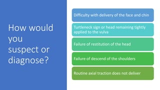
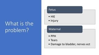
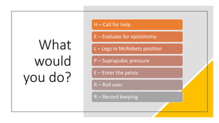
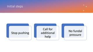
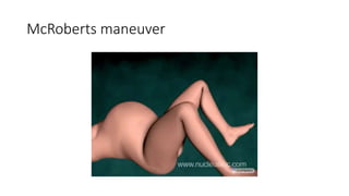
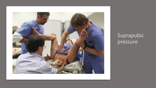
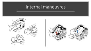
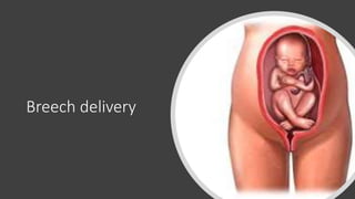
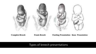
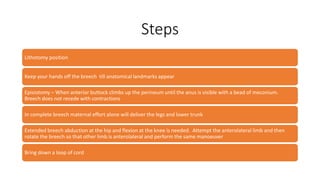
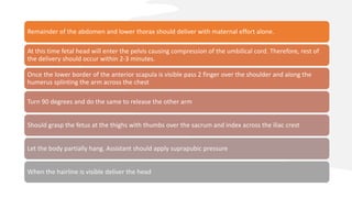
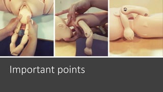
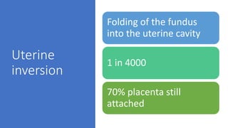
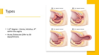
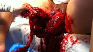
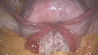
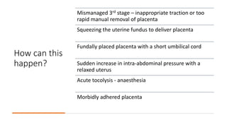
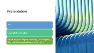
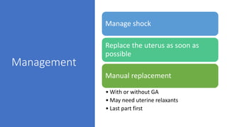
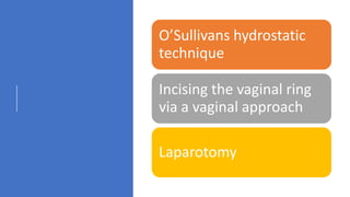
Emergencies in
Obstetrics
Dr. Indunil Piyadigama
Consultant Obstetrician and Gynaecologist
Pre-intern Progam
Ministry of Health, Sri Lanka
4th October 2021
• Your 1st week at the obstetric unit. You are oncall at the
labour ward. 1725 the labour ward nursing sister calls you
• 34 year old diabetic mother. Induction of labour at 39
weeks of getation. She was induced with prostaglandin
yesterday. ARM done at 0517. Was on syntocinon for
augmentation due to slow progress.
• Babies head was delivered. But the body has not followed.
Shoulder
dystocia
How would
you
suspect or
diagnose?
Difficulty with delivery of the face and chin
Turtleneck sign or head remaining tightly
applied to the vulva
Failure of restitution of the head
Failure of descend of the shoulders
Routine axial traction does not deliver
What is the
problem?
• HIE
• Injury
Fetus
• PPH
• Tears
• Damage to bladder, nerves ect
Maternal
What
would
you do?
H – Call for help
E – Evaluate for episiotomy
L – Legs in McRobets position
P – Suprapubic pressure
E – Enter the pelvis
R – Roll over
R – Record keeping
Initial steps
Stop pushing
Call for
additional
help
No fundal
pressure
McRoberts maneuver
Suprapubic
pressure
Internal maneuvres
Records
• Medico legal issues
• Contemporanous documentation
• Times and the intervensions
• The anterior and posterior shoulders should be documented
Post procedure
PPH
Neonatal
resuscitation
Breech delivery
Types of breech presentations
Steps
Lithotomy position
Keep your hands off the breech till anatomical landmarks appear
Episiotomy – When anterior buttock climbs up the perineum until the anus is visible with a bead of meconium.
Breech does not recede with contractions
In complete breech maternal effort alone will deliver the legs and lower trunk
Extended breech abduction at the hip and flexion at the knee is needed. Attempt the anterolateral limb and then
rotate the breech so that other limb is anterolateral and perform the same manoeuver
Bring down a loop of cord
Remainder of the abdomen and lower thorax should deliver with maternal effort alone.
At this time fetal head will enter the pelvis causing compression of the umbilical cord. Therefore, rest of
the delivery should occur within 2-3 minutes.
Once the lower border of the anterior scapula is visible pass 2 finger over the shoulder and along the
humerus splinting the arm across the chest
Turn 90 degrees and do the same to release the other arm
Should grasp the fetus at the thighs with thumbs over the sacrum and index across the iliac crest
Let the body partially hang. Assistant should apply suprapubic pressure
When the hairline is visible deliver the head
Important points
Uterine
inversion
Folding of the fundus
into the uterine cavity
1 in 4000
70% placenta still
attached
Types
• 1-4th degree – Cervix, introitus, 4th
within the vagina
• Acute /Subacute (24hr to 30
days)/Chronic
How can this
happen?
Mismanaged 3rd stage – inappropriate traction or too
rapid manual removal of placenta
Squeezing the uterine fundus to deliver placenta
Fundally placed placenta with a short umbilical cord
Sudden increase in intra-abdominal pressure with a
relaxed uterus
Acute tocolysis - anaesthesia
Morbidly adhered placenta
Presentation
PPH
Mass at the introitus
Shock without vaginal bleeding - Neurogenic
due to traction on adjacent structures
Management
Manage shock
Replace the uterus as soon as
possible
Manual replacement
• With or without GA
• May need uterine relaxants
• Last part first
O’Sullivans hydrostatic
technique
Incising the vaginal ring
via a vaginal approach
Laparotomy
Thank you

Emergencies in obstetrics

  • 1.
    Emergencies in Obstetrics Dr. IndunilPiyadigama Consultant Obstetrician and Gynaecologist Pre-intern Progam Ministry of Health, Sri Lanka 4th October 2021
  • 2.
    • Your 1stweek at the obstetric unit. You are oncall at the labour ward. 1725 the labour ward nursing sister calls you • 34 year old diabetic mother. Induction of labour at 39 weeks of getation. She was induced with prostaglandin yesterday. ARM done at 0517. Was on syntocinon for augmentation due to slow progress. • Babies head was delivered. But the body has not followed.
  • 3.
  • 4.
    How would you suspect or diagnose? Difficultywith delivery of the face and chin Turtleneck sign or head remaining tightly applied to the vulva Failure of restitution of the head Failure of descend of the shoulders Routine axial traction does not deliver
  • 5.
    What is the problem? •HIE • Injury Fetus • PPH • Tears • Damage to bladder, nerves ect Maternal
  • 6.
    What would you do? H –Call for help E – Evaluate for episiotomy L – Legs in McRobets position P – Suprapubic pressure E – Enter the pelvis R – Roll over R – Record keeping
  • 7.
    Initial steps Stop pushing Callfor additional help No fundal pressure
  • 8.
  • 9.
  • 10.
  • 11.
    Records • Medico legalissues • Contemporanous documentation • Times and the intervensions • The anterior and posterior shoulders should be documented
  • 12.
  • 13.
  • 14.
    Types of breechpresentations
  • 16.
    Steps Lithotomy position Keep yourhands off the breech till anatomical landmarks appear Episiotomy – When anterior buttock climbs up the perineum until the anus is visible with a bead of meconium. Breech does not recede with contractions In complete breech maternal effort alone will deliver the legs and lower trunk Extended breech abduction at the hip and flexion at the knee is needed. Attempt the anterolateral limb and then rotate the breech so that other limb is anterolateral and perform the same manoeuver Bring down a loop of cord
  • 17.
    Remainder of theabdomen and lower thorax should deliver with maternal effort alone. At this time fetal head will enter the pelvis causing compression of the umbilical cord. Therefore, rest of the delivery should occur within 2-3 minutes. Once the lower border of the anterior scapula is visible pass 2 finger over the shoulder and along the humerus splinting the arm across the chest Turn 90 degrees and do the same to release the other arm Should grasp the fetus at the thighs with thumbs over the sacrum and index across the iliac crest Let the body partially hang. Assistant should apply suprapubic pressure When the hairline is visible deliver the head
  • 18.
  • 19.
    Uterine inversion Folding of thefundus into the uterine cavity 1 in 4000 70% placenta still attached
  • 20.
    Types • 1-4th degree– Cervix, introitus, 4th within the vagina • Acute /Subacute (24hr to 30 days)/Chronic
  • 23.
    How can this happen? Mismanaged3rd stage – inappropriate traction or too rapid manual removal of placenta Squeezing the uterine fundus to deliver placenta Fundally placed placenta with a short umbilical cord Sudden increase in intra-abdominal pressure with a relaxed uterus Acute tocolysis - anaesthesia Morbidly adhered placenta
  • 24.
    Presentation PPH Mass at theintroitus Shock without vaginal bleeding - Neurogenic due to traction on adjacent structures
  • 25.
    Management Manage shock Replace theuterus as soon as possible Manual replacement • With or without GA • May need uterine relaxants • Last part first
  • 26.
    O’Sullivans hydrostatic technique Incising thevaginal ring via a vaginal approach Laparotomy
  • 27.