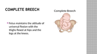
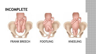
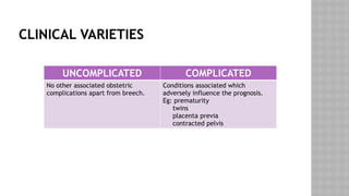
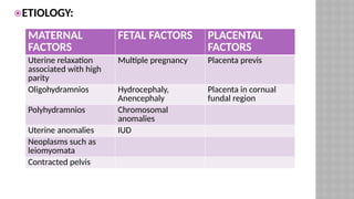
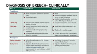
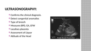
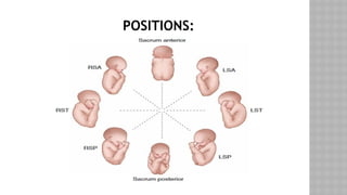
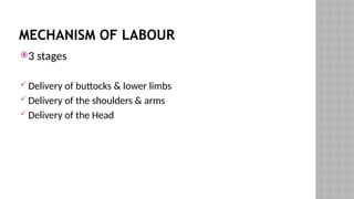
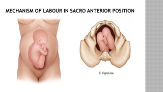
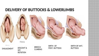
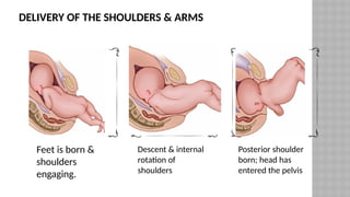
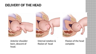
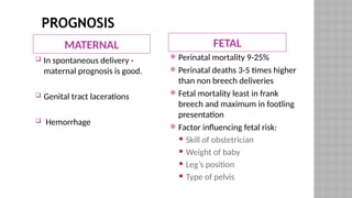
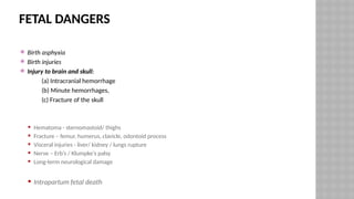
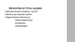
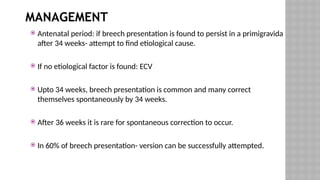
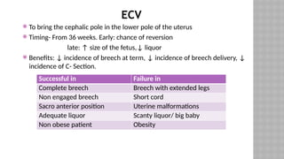
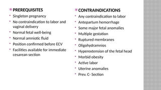
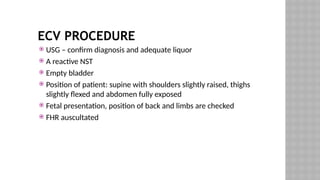
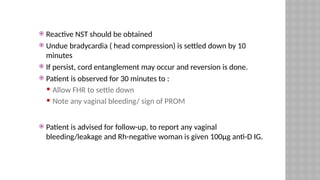
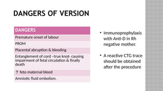
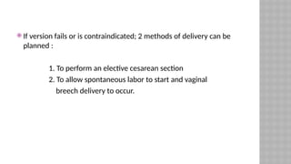
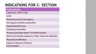
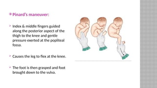
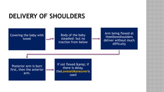
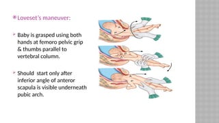
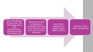
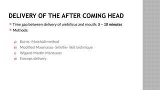
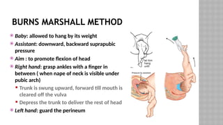
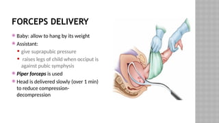
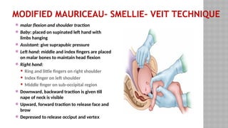
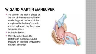
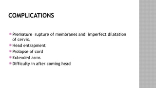
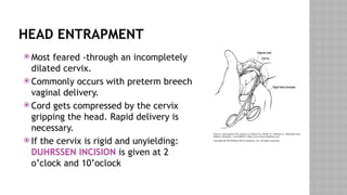
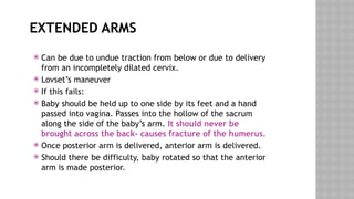
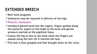
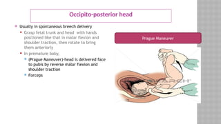
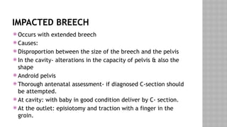
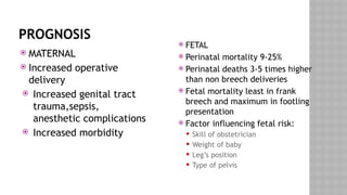
BREECH
BREECH PRESENTATION
 Defined as longitudinal lie with variation in polarity.
 Denominator – Sacrum
 Engaging diameter – Bitrochanteric diameter
 Frequency: depends on period of gestation
at 32 weeks: 16%
at term : 3-5%
COMPLETE BREECH
 Fetus maintains the attitude of
universal flexion with the
thighs flexed at hips and the
legs at the knees.
INCOMPLETE
FRANK BREECH FOOTLING KNEELING
CLINICAL VARIETIES
UNCOMPLICATED COMPLICATED
No other associated obstetric
complications apart from breech.
Conditions associated which
adversely influence the prognosis.
Eg: prematurity
twins
placenta previa
contracted pelvis
ETIOLOGY:
MATERNAL
FACTORS
FETAL FACTORS PLACENTAL
FACTORS
Uterine relaxation
associated with high
parity
Multiple pregnancy Placenta previs
Oligohydramnios Hydrocephaly,
Anencephaly
Placenta in cornual
fundal region
Polyhydramnios Chromosomal
anomalies
Uterine anomalies IUD
Neoplasms such as
leiomyomata
Contracted pelvis
DIAGNOSIS OF BREECH- CLINICALLY
ULTRASONOGRAPHY:
 Confirms the clinical diagnosis.
 Detect congenital anomalies
 Type of breech
 Measures BPD, GA, EFW
 Localises placenta
 Assessment of Liquor
 Attitude of the Head
POSITIONS:
MECHANISM OF LABOUR
3 stages
 Delivery of buttocks & lower limbs
 Delivery of the shoulders & arms
 Delivery of the Head
MECHANISM OF LABOUR IN SACRO ANTERIOR POSITION
DELIVERY OF BUTTOCKS & LOWERLIMBS
ENGAGEMENT DESCENT &
INT.
ROTATION
BREECH
CLIMBING
BIRTH OF
POST. BUTTOCK
BIRTH OF ANT.
BUTTOCK
DELIVERY OF THE SHOULDERS & ARMS
Feet is born &
shoulders
engaging.
Descent & internal
rotation of
shoulders
Posterior shoulder
born; head has
entered the pelvis
DELIVERY OF THE HEAD
Anterior shoulder
born, descent of
head.
Internal rotation &
flexion of head
Flexion of the head
complete
PROGNOSIS
MATERNAL FETAL
 In spontaneous delivery -
maternal prognosis is good.
 Genital tract lacerations
 Hemorrhage
 Perinatal mortality 9-25%
 Perinatal deaths 3-5 times higher
than non breech deliveries
 Fetal mortality least in frank
breech and maximum in footling
presentation
 Factor influencing fetal risk:
ï‚Ą Skill of obstetrician
ï‚Ą Weight of baby
ï‚Ą Leg’s position
ï‚Ą Type of pelvis
FETAL DANGERS
 Birth asphyxia
 Birth injuries
 Injury to brain and skull:
(a) Intracranial hemorrhage
(b) Minute hemorrhages,
(c) Fracture of the skull
ï‚Ą Hematoma - sternomastoid/ thighs
ï‚Ą Fracture – femur, humerus, clavicle, odontoid process
ï‚Ą Visceral injuries - liver/ kidney / lungs rupture
ï‚Ą Nerve – Erb’s / Klumpke’s palsy
ï‚Ą Long-term neurological damage
ï‚Ą Intrapartum fetal death
PREVENTION OF FETAL HAZARDS
 Minimize breech incidence by ECV
 Delivery by cesarean section
 Vaginal breech delivery by
skilled obstetrician,
anesthesist,
neonatologist
MANAGEMENT
 Antenatal period: if breech presentation is found to persist in a primigravida
after 34 weeks- attempt to find etiological cause.
 If no etiological factor is found: ECV
 Upto 34 weeks, breech presentation is common and many correct
themselves spontaneously by 34 weeks.
 After 36 weeks it is rare for spontaneous correction to occur.
 In 60% of breech presentation- version can be successfully attempted.
ECV
 To bring the cephalic pole in the lower pole of the uterus
 Timing- From 36 weeks. Early: chance of reversion
late: ↑ size of the fetus,↓ liquor
 Benefits: ↓ incidence of breech at term, ↓ incidence of breech delivery, ↓
incidence of C- Section.
Successful in Failure in
Complete breech Breech with extended legs
Non engaged breech Short cord
Sacro anterior position Uterine malformations
Adequate liquor Scanty liquor/ big baby
Non obese patient Obesity
 PREREQUISITES
 Singleton pregnancy
 No contraindication to labor and
vaginal delivery
 Normal fetal well-being
 Normal amniotic fluid
 Position confirmed before ECV
 Facilities available for immediate
cesarean section
 CONTRAINDICATIONS
 Any contraindication to labor
 Antepartum hemorrhage
 Some major fetal anomalies
 Multiple gestation
 Ruptured membranes
 Oligohydramnios
 Hyperextension of the fetal head
 Morbid obesity
 Active labor
 Uterine anomalies
 Prev. C- Section
ECV PROCEDURE
 USG – confirm diagnosis and adequate liquor
 A reactive NST
 Empty bladder
 Position of patient: supine with shoulders slightly raised, thighs
slightly flexed and abdomen fully exposed
 Fetal presentation, position of back and limbs are checked
 FHR auscultated
‘FORWARD ROLL’ MOVEMENT
STEP 1
 Mobilize breech using both hands towards which back of fetus lie
 Right hand : grasp podalic pole
 Left hand: grasp head
 STEP-2
 L:Pressure exerted to head to push breech
 R:Pressure in opposite direction to guide vertex
 Intermittent pressure given till lie become transverse
 FHR is checked
 Changing of hands
 Intermittent pressure exerted till head is brought to lower pole of uterus
 STEP-3 :Changing of hands
 Intermittent pressure exerted till head is at lower pole of uterus.
 Reactive NST should be obtained
 Undue bradycardia ( head compression) is settled down by 10
minutes
 If persist, cord entanglement may occur and reversion is done.
 Patient is observed for 30 minutes to :
ï‚Ą Allow FHR to settle down
ï‚Ą Note any vaginal bleeding/ sign of PROM
 Patient is advised for follow-up, to report any vaginal
bleeding/leakage and Rh-negative woman is given 100”g anti-D IG.
DANGERS OF VERSION
‱ Immunoprophylaxis
with Anti-D in Rh
negative mother.
‱ A reactive CTG trace
should be obtained
after the procedure
DANGERS
Premature onset of labour
PROM
Placental abruption & bleeding
Entanglement of cord –true knot- causing
impairment of fetal circulation & finally
death
↑ feto maternal bleed
Amniotic fluid embolism.
 If version fails or is contraindicated; 2 methods of delivery can be
planned :
1. To perform an elective cesarean section
2. To allow spontaneous labor to start and vaginal
breech delivery to occur.
INDICATIONS FOR C- SECTION
Indications
Large fetus, EFW>3.5kg
IUGR
Placenta previa of any degree
Any degree of pelvic contraction
Hyperextended head
Footling presentation
Previous perinatal death/ H/O birth trauma
Need to terminate pregnancy- fetal/ maternal indication
Placental insufficiency
Failure of descent of breech
Cord prolapse
ZATUCHNI-ANDROS BREECH SCORING
 If the score is 0-4, cesarean delivery is recommended
Add 0 Points Add 1 Point Add 2 Points
Parity 0 1 2
Gestational age
(wk)
39+ 38 <37
EFW (lb) 8 7-8 <7
Previous breech 0 1 2
Dilatation 2 3 4
Station -3 -2 -1
VAGINAL DELIVERY
 Spontaneous breech delivery
 Assisted breech delivery
Total breech extraction
MANAGEMENT OF THE FIRST STAGE
 Rest in bed- due to fear of cord prolapse in the event of rupture
of membranes
 FHR should be observed atleast every 15 minutes. Especially
after ROM.
 With increasing uterine contraction, breech begins to descend.
 An IV line is started with RL
 When membranes rupture advisable to conduct a vaginal
examination.
MANAGEMENT OF SECOND STAGE
Depending on the level of assistance required
 Spontaneous (10%):
ï‚Ą not preferred
ï‚Ą Usually with very LBW & dead babies
 Assisted breech : The fetus is delivered spontaneously as far as the umbilicus, but
the remainder of the body is extracted or delivered with operator traction and
assisted maneuvers, with or without maternal expulsive efforts.
 Breech extraction :
ï‚Ą entire body of fetus is extracted by obstetrician
ï‚Ą Indications:
ï‚ą delivery of 2nd
twin after IPV
ï‚ą Cord prolapse
ï‚ą Extended legs
ASSISTED
BREECH
DELIVERY
NEED FOR PLANNING
 Before starting to assist the breech delivery, all arrangements must be
ready to treat neonatal asphyxia.
 No necessity to hurry and complete the delivery.
 episiotomy is given when between contractions the baby’s anus is visible
at the vulval outlet- CLIMBING ON THE PERINEUM
 Golden rule in assisted breech delivery: Keep the hands off the breech and
allow the breech to deliver spontaneously till the umbilicus.
 Delivery of breech
 The patient is encouraged to bear down with the contractions but must
rest between them
 As long as there is no fetal or maternal distress, spontaneous delivery to
the umbilicus is awaited.
 The legs usually deliver spontaneously; if not they are easily extracted. Do
not extract the legs until the popliteal fossae are visible (Pinard’s
maneuver)
 The baby is covered with a warm towel, and the body is supported
 A loop of umbilical cord is pulled aside.
 Pinard’s maneuver:
 Index & middle fingers guided
along the posterior aspect of the
thigh to the knee and gentle
pressure exerted at the popliteal
fossa.
 Causes the leg to flex at the knee.
 The foot is then grasped and foot
brought down to the vulva.
DELIVERY OF SHOULDERS
Covering the baby with
towel
Body of the baby
steadied- but no
traction from below
Arm being flexed at
theelbowshoulders
deliver without much
difficulty
Posterior arm is born
first, then the anterior
arm.
If not flexed &amp; if
there is delay,
theLovesetManeuveris
used
 Loveset’s maneuver:
 Baby is grasped using both
hands at femoro pelvic grip
& thumbs parallel to
vertebral column.
 Should start only after
inferior angle of anteror
scapula is visible underneath
pubic arch.
After the delivery
of both arms, the
child hangs down
from
thevulvaloutlet,
with back facing
the obstetrician
Allowing the child
to hang down-
facilitates the entry
of the head into the
pelvis by the action
of gravity
Any further
assistance is
needed- supra
pubic pressure
Delivery of the
after coming head
DELIVERY OF THE AFTER COMING HEAD
 Time gap between delivery of umbilicus and mouth: 5 – 10 minutes
 Methods:
a) Burns- Marshall method
b) Modified Mauriceau- Smellie- Veit technique
c) Wigand Martin Maneuver
d) Forceps delivery
BURNS MARSHALL METHOD
 Baby: allowed to hang by its weight
 Assistant: downward, backward suprapubic
pressure
 Aim : to promote flexion of head
 Right hand: grasp ankles with a finger in
between ( when nape of neck is visible under
pubic arch)
ï‚Ą Trunk is swung upward, forward till mouth is
cleared off the vulva
ï‚Ą Depress the trunk to deliver the rest of head
 Left hand: guard the perineum
FORCEPS DELIVERY
 Baby: allow to hang by its weight
 Assistant:
ï‚Ą give suprapubic pressure
ï‚Ą raises legs of child when occiput is
against pubic symphysis
 Piper forceps is used
 Head is delivered slowly (over 1 min)
to reduce compression-
decompression
MODIFIED MAURICEAU- SMELLIE- VEIT TECHNIQUE
 malar flexion and shoulder traction
 Baby: placed on supinated left hand with
limbs hanging
 Assistant: give suprapubic pressure
 Left hand: middle and index fingers are placed
on malar bones to maintain head flexion
 Right hand:
ï‚Ą Ring and little fingers on right shoulder
ï‚Ą Index finger on left shoulder
ï‚Ą Middle finger on sub-occipital region
 Downward, backward traction is given till
nape of neck is visible
 Upward, forward traction to release face and
brow
 Depressed to release occiput and vertex
WIGAND MARTIN MANEUVER
 The body of the baby is placed on
the arm of the operator with the
middle finger of the hand of that
arm placed in the baby’s mouth
and the index and ring fingers on
the malar bones
 Maintain flexion.
 With the other hand, the
obstetrician exerts suprapubic
pressure on the head through the
mother’s abdomen
COMPLICATIONS
 Premature rupture of membranes and imperfect dilatation
of cervix.
 Head entrapment
 Prolapse of cord
 Extended arms
 Difficulty in after coming head
PREMATURE RUPTURE OF MEMBRANES AND
IMPERFECT DILATATION OF THE CERVIX
 VERY COMMON
 More common in footling & complete breech
 Foot slips out in a fully flexed breech presentation.
 No attempt should be made to extract the breech, when the
cervix is not fully dilated- tears of the cervix, difficulties in
delivering the arm and head- stillbirth. Better deliver by C-
section.
HEAD ENTRAPMENT
 Most feared -through an incompletely
dilated cervix.
 Commonly occurs with preterm breech
vaginal delivery.
 Cord gets compressed by the cervix
gripping the head. Rapid delivery is
necessary.
 If the cervix is rigid and unyielding:
DUHRSSEN INCISION is given at 2
o’clock and 10’oclock
PROLAPSE OF THE CORD
 Common in complete breech, more common in footling
presentation.
 Immediate C- section
 If completely dilated cervix- BREECH EXTRACTION
EXTENDED ARMS
 Can be due to undue traction from below or due to delivery
from an incompletely dilated cervix.
 Lovset’s maneuver
 If this fails:
 Baby should be held up to one side by its feet and a hand
passed into vagina. Passes into the hollow of the sacrum
along the side of the baby’s arm. It should never be
brought across the back- causes fracture of the humerus.
 Once posterior arm is delivered, anterior arm is delivered.
 Should there be difficulty, baby rotated so that the anterior
arm is made posterior.
DIFFICULTY IN AFTERCOMING HEAD
 Deflexion of the head
 Undiagnosed disproportion between the head and the pelvic
brim
 Delivery through an imperfectly dilated and retracted cervix.
 Narrow subpubic arch.
EXTENDED BREECH
 Best fetal prognosis
 Assistance may be required in delivery of the legs.
 Pinard’s maneuver:
 Introduce gloved hand into the vagina, fingers guided along
the posterior aspect of the thigh to the knee and gentle
pressure exerted at the popliteal fossa.
 Causes the leg to flex at the knee when the fingers are
passed along the shin till it reaches the foot.
 The foot is then grasped and foot brought down to the vulva.
Occipito-posterior head
 Usually in spontaneous breech delivery
ï‚Ą Grasp fetal trunk and head with hands
positioned like that in malar flexion and
shoulder traction, then rotate to bring
them anteriorly
ï‚Ą In premature baby,
ï‚ą (Prague Maneuver)-head is delivered face
to pubis by reverse malar flexion and
shoulder traction
ï‚ą Forceps
Prague Maneuver
IMPACTED BREECH
 Occurs with extended breech
 Causes:
 Disproportion between the size of the breech and the pelvis
 In the cavity- alterations in the capacity of pelvis & also the
shape
 Android pelvis
 Thorough antenatal assessment- if diagnosed C-section should
be attempted.
 At cavity: with baby in good condition deliver by C- section.
 At the outlet: episiotomy and traction with a finger in the
groin.
PROGNOSIS
 MATERNAL
 Increased operative
delivery
 Increased genital tract
trauma,sepsis,
anesthetic complications
 Increased morbidity
 FETAL
 Perinatal mortality 9-25%
 Perinatal deaths 3-5 times higher
than non breech deliveries
 Fetal mortality least in frank
breech and maximum in footling
presentation
 Factor influencing fetal risk:
ï‚Ą Skill of obstetrician
ï‚Ą Weight of baby
ï‚Ą Leg’s position
ï‚Ą Type of pelvis
THANK YOU

Breech presentation obstetrics .pptx

  • 1.
  • 2.
    BREECH PRESENTATION  Definedas longitudinal lie with variation in polarity.  Denominator – Sacrum  Engaging diameter – Bitrochanteric diameter  Frequency: depends on period of gestation at 32 weeks: 16% at term : 3-5%
  • 3.
    COMPLETE BREECH  Fetusmaintains the attitude of universal flexion with the thighs flexed at hips and the legs at the knees.
  • 4.
  • 5.
    CLINICAL VARIETIES UNCOMPLICATED COMPLICATED Noother associated obstetric complications apart from breech. Conditions associated which adversely influence the prognosis. Eg: prematurity twins placenta previa contracted pelvis
  • 6.
    ETIOLOGY: MATERNAL FACTORS FETAL FACTORS PLACENTAL FACTORS Uterinerelaxation associated with high parity Multiple pregnancy Placenta previs Oligohydramnios Hydrocephaly, Anencephaly Placenta in cornual fundal region Polyhydramnios Chromosomal anomalies Uterine anomalies IUD Neoplasms such as leiomyomata Contracted pelvis
  • 7.
  • 8.
    ULTRASONOGRAPHY:  Confirms theclinical diagnosis.  Detect congenital anomalies  Type of breech  Measures BPD, GA, EFW  Localises placenta  Assessment of Liquor  Attitude of the Head
  • 9.
  • 10.
    MECHANISM OF LABOUR 3stages  Delivery of buttocks & lower limbs  Delivery of the shoulders & arms  Delivery of the Head
  • 11.
    MECHANISM OF LABOURIN SACRO ANTERIOR POSITION
  • 12.
    DELIVERY OF BUTTOCKS& LOWERLIMBS ENGAGEMENT DESCENT & INT. ROTATION BREECH CLIMBING BIRTH OF POST. BUTTOCK BIRTH OF ANT. BUTTOCK
  • 13.
    DELIVERY OF THESHOULDERS & ARMS Feet is born & shoulders engaging. Descent & internal rotation of shoulders Posterior shoulder born; head has entered the pelvis
  • 14.
    DELIVERY OF THEHEAD Anterior shoulder born, descent of head. Internal rotation & flexion of head Flexion of the head complete
  • 15.
    PROGNOSIS MATERNAL FETAL  Inspontaneous delivery - maternal prognosis is good.  Genital tract lacerations  Hemorrhage  Perinatal mortality 9-25%  Perinatal deaths 3-5 times higher than non breech deliveries  Fetal mortality least in frank breech and maximum in footling presentation  Factor influencing fetal risk: ï‚Ą Skill of obstetrician ï‚Ą Weight of baby ï‚Ą Leg’s position ï‚Ą Type of pelvis
  • 16.
    FETAL DANGERS  Birthasphyxia  Birth injuries  Injury to brain and skull: (a) Intracranial hemorrhage (b) Minute hemorrhages, (c) Fracture of the skull ï‚Ą Hematoma - sternomastoid/ thighs ï‚Ą Fracture – femur, humerus, clavicle, odontoid process ï‚Ą Visceral injuries - liver/ kidney / lungs rupture ï‚Ą Nerve – Erb’s / Klumpke’s palsy ï‚Ą Long-term neurological damage ï‚Ą Intrapartum fetal death
  • 17.
    PREVENTION OF FETALHAZARDS  Minimize breech incidence by ECV  Delivery by cesarean section  Vaginal breech delivery by skilled obstetrician, anesthesist, neonatologist
  • 18.
    MANAGEMENT  Antenatal period:if breech presentation is found to persist in a primigravida after 34 weeks- attempt to find etiological cause.  If no etiological factor is found: ECV  Upto 34 weeks, breech presentation is common and many correct themselves spontaneously by 34 weeks.  After 36 weeks it is rare for spontaneous correction to occur.  In 60% of breech presentation- version can be successfully attempted.
  • 19.
    ECV  To bringthe cephalic pole in the lower pole of the uterus  Timing- From 36 weeks. Early: chance of reversion late: ↑ size of the fetus,↓ liquor  Benefits: ↓ incidence of breech at term, ↓ incidence of breech delivery, ↓ incidence of C- Section. Successful in Failure in Complete breech Breech with extended legs Non engaged breech Short cord Sacro anterior position Uterine malformations Adequate liquor Scanty liquor/ big baby Non obese patient Obesity
  • 20.
     PREREQUISITES  Singletonpregnancy  No contraindication to labor and vaginal delivery  Normal fetal well-being  Normal amniotic fluid  Position confirmed before ECV  Facilities available for immediate cesarean section  CONTRAINDICATIONS  Any contraindication to labor  Antepartum hemorrhage  Some major fetal anomalies  Multiple gestation  Ruptured membranes  Oligohydramnios  Hyperextension of the fetal head  Morbid obesity  Active labor  Uterine anomalies  Prev. C- Section
  • 21.
    ECV PROCEDURE  USG– confirm diagnosis and adequate liquor  A reactive NST  Empty bladder  Position of patient: supine with shoulders slightly raised, thighs slightly flexed and abdomen fully exposed  Fetal presentation, position of back and limbs are checked  FHR auscultated
  • 22.
    ‘FORWARD ROLL’ MOVEMENT STEP1  Mobilize breech using both hands towards which back of fetus lie  Right hand : grasp podalic pole  Left hand: grasp head  STEP-2  L:Pressure exerted to head to push breech  R:Pressure in opposite direction to guide vertex  Intermittent pressure given till lie become transverse  FHR is checked  Changing of hands  Intermittent pressure exerted till head is brought to lower pole of uterus  STEP-3 :Changing of hands  Intermittent pressure exerted till head is at lower pole of uterus.
  • 23.
     Reactive NSTshould be obtained  Undue bradycardia ( head compression) is settled down by 10 minutes  If persist, cord entanglement may occur and reversion is done.  Patient is observed for 30 minutes to : ï‚Ą Allow FHR to settle down ï‚Ą Note any vaginal bleeding/ sign of PROM  Patient is advised for follow-up, to report any vaginal bleeding/leakage and Rh-negative woman is given 100”g anti-D IG.
  • 24.
    DANGERS OF VERSION ‱Immunoprophylaxis with Anti-D in Rh negative mother. ‱ A reactive CTG trace should be obtained after the procedure DANGERS Premature onset of labour PROM Placental abruption & bleeding Entanglement of cord –true knot- causing impairment of fetal circulation & finally death ↑ feto maternal bleed Amniotic fluid embolism.
  • 25.
     If versionfails or is contraindicated; 2 methods of delivery can be planned : 1. To perform an elective cesarean section 2. To allow spontaneous labor to start and vaginal breech delivery to occur.
  • 26.
    INDICATIONS FOR C-SECTION Indications Large fetus, EFW>3.5kg IUGR Placenta previa of any degree Any degree of pelvic contraction Hyperextended head Footling presentation Previous perinatal death/ H/O birth trauma Need to terminate pregnancy- fetal/ maternal indication Placental insufficiency Failure of descent of breech Cord prolapse
  • 27.
    ZATUCHNI-ANDROS BREECH SCORING If the score is 0-4, cesarean delivery is recommended Add 0 Points Add 1 Point Add 2 Points Parity 0 1 2 Gestational age (wk) 39+ 38 <37 EFW (lb) 8 7-8 <7 Previous breech 0 1 2 Dilatation 2 3 4 Station -3 -2 -1
  • 28.
    VAGINAL DELIVERY  Spontaneousbreech delivery  Assisted breech delivery Total breech extraction
  • 29.
    MANAGEMENT OF THEFIRST STAGE  Rest in bed- due to fear of cord prolapse in the event of rupture of membranes  FHR should be observed atleast every 15 minutes. Especially after ROM.  With increasing uterine contraction, breech begins to descend.  An IV line is started with RL  When membranes rupture advisable to conduct a vaginal examination.
  • 30.
    MANAGEMENT OF SECONDSTAGE Depending on the level of assistance required  Spontaneous (10%): ï‚Ą not preferred ï‚Ą Usually with very LBW & dead babies  Assisted breech : The fetus is delivered spontaneously as far as the umbilicus, but the remainder of the body is extracted or delivered with operator traction and assisted maneuvers, with or without maternal expulsive efforts.  Breech extraction : ï‚Ą entire body of fetus is extracted by obstetrician ï‚Ą Indications: ï‚ą delivery of 2nd twin after IPV ï‚ą Cord prolapse ï‚ą Extended legs
  • 31.
  • 32.
    NEED FOR PLANNING Before starting to assist the breech delivery, all arrangements must be ready to treat neonatal asphyxia.  No necessity to hurry and complete the delivery.  episiotomy is given when between contractions the baby’s anus is visible at the vulval outlet- CLIMBING ON THE PERINEUM  Golden rule in assisted breech delivery: Keep the hands off the breech and allow the breech to deliver spontaneously till the umbilicus.
  • 33.
     Delivery ofbreech  The patient is encouraged to bear down with the contractions but must rest between them  As long as there is no fetal or maternal distress, spontaneous delivery to the umbilicus is awaited.  The legs usually deliver spontaneously; if not they are easily extracted. Do not extract the legs until the popliteal fossae are visible (Pinard’s maneuver)  The baby is covered with a warm towel, and the body is supported  A loop of umbilical cord is pulled aside.
  • 34.
     Pinard’s maneuver: Index & middle fingers guided along the posterior aspect of the thigh to the knee and gentle pressure exerted at the popliteal fossa.  Causes the leg to flex at the knee.  The foot is then grasped and foot brought down to the vulva.
  • 35.
    DELIVERY OF SHOULDERS Coveringthe baby with towel Body of the baby steadied- but no traction from below Arm being flexed at theelbowshoulders deliver without much difficulty Posterior arm is born first, then the anterior arm. If not flexed &amp; if there is delay, theLovesetManeuveris used
  • 36.
     Loveset’s maneuver: Baby is grasped using both hands at femoro pelvic grip & thumbs parallel to vertebral column.  Should start only after inferior angle of anteror scapula is visible underneath pubic arch.
  • 37.
    After the delivery ofboth arms, the child hangs down from thevulvaloutlet, with back facing the obstetrician Allowing the child to hang down- facilitates the entry of the head into the pelvis by the action of gravity Any further assistance is needed- supra pubic pressure Delivery of the after coming head
  • 38.
    DELIVERY OF THEAFTER COMING HEAD  Time gap between delivery of umbilicus and mouth: 5 – 10 minutes  Methods: a) Burns- Marshall method b) Modified Mauriceau- Smellie- Veit technique c) Wigand Martin Maneuver d) Forceps delivery
  • 39.
    BURNS MARSHALL METHOD Baby: allowed to hang by its weight  Assistant: downward, backward suprapubic pressure  Aim : to promote flexion of head  Right hand: grasp ankles with a finger in between ( when nape of neck is visible under pubic arch) ï‚Ą Trunk is swung upward, forward till mouth is cleared off the vulva ï‚Ą Depress the trunk to deliver the rest of head  Left hand: guard the perineum
  • 40.
    FORCEPS DELIVERY  Baby:allow to hang by its weight  Assistant: ï‚Ą give suprapubic pressure ï‚Ą raises legs of child when occiput is against pubic symphysis  Piper forceps is used  Head is delivered slowly (over 1 min) to reduce compression- decompression
  • 41.
    MODIFIED MAURICEAU- SMELLIE-VEIT TECHNIQUE  malar flexion and shoulder traction  Baby: placed on supinated left hand with limbs hanging  Assistant: give suprapubic pressure  Left hand: middle and index fingers are placed on malar bones to maintain head flexion  Right hand: ï‚Ą Ring and little fingers on right shoulder ï‚Ą Index finger on left shoulder ï‚Ą Middle finger on sub-occipital region  Downward, backward traction is given till nape of neck is visible  Upward, forward traction to release face and brow  Depressed to release occiput and vertex
  • 42.
    WIGAND MARTIN MANEUVER The body of the baby is placed on the arm of the operator with the middle finger of the hand of that arm placed in the baby’s mouth and the index and ring fingers on the malar bones  Maintain flexion.  With the other hand, the obstetrician exerts suprapubic pressure on the head through the mother’s abdomen
  • 43.
    COMPLICATIONS  Premature ruptureof membranes and imperfect dilatation of cervix.  Head entrapment  Prolapse of cord  Extended arms  Difficulty in after coming head
  • 44.
    PREMATURE RUPTURE OFMEMBRANES AND IMPERFECT DILATATION OF THE CERVIX  VERY COMMON  More common in footling & complete breech  Foot slips out in a fully flexed breech presentation.  No attempt should be made to extract the breech, when the cervix is not fully dilated- tears of the cervix, difficulties in delivering the arm and head- stillbirth. Better deliver by C- section.
  • 45.
    HEAD ENTRAPMENT  Mostfeared -through an incompletely dilated cervix.  Commonly occurs with preterm breech vaginal delivery.  Cord gets compressed by the cervix gripping the head. Rapid delivery is necessary.  If the cervix is rigid and unyielding: DUHRSSEN INCISION is given at 2 o’clock and 10’oclock
  • 46.
    PROLAPSE OF THECORD  Common in complete breech, more common in footling presentation.  Immediate C- section  If completely dilated cervix- BREECH EXTRACTION
  • 47.
    EXTENDED ARMS  Canbe due to undue traction from below or due to delivery from an incompletely dilated cervix.  Lovset’s maneuver  If this fails:  Baby should be held up to one side by its feet and a hand passed into vagina. Passes into the hollow of the sacrum along the side of the baby’s arm. It should never be brought across the back- causes fracture of the humerus.  Once posterior arm is delivered, anterior arm is delivered.  Should there be difficulty, baby rotated so that the anterior arm is made posterior.
  • 48.
    DIFFICULTY IN AFTERCOMINGHEAD  Deflexion of the head  Undiagnosed disproportion between the head and the pelvic brim  Delivery through an imperfectly dilated and retracted cervix.  Narrow subpubic arch.
  • 49.
    EXTENDED BREECH  Bestfetal prognosis  Assistance may be required in delivery of the legs.  Pinard’s maneuver:  Introduce gloved hand into the vagina, fingers guided along the posterior aspect of the thigh to the knee and gentle pressure exerted at the popliteal fossa.  Causes the leg to flex at the knee when the fingers are passed along the shin till it reaches the foot.  The foot is then grasped and foot brought down to the vulva.
  • 50.
    Occipito-posterior head  Usuallyin spontaneous breech delivery ï‚Ą Grasp fetal trunk and head with hands positioned like that in malar flexion and shoulder traction, then rotate to bring them anteriorly ï‚Ą In premature baby, ï‚ą (Prague Maneuver)-head is delivered face to pubis by reverse malar flexion and shoulder traction ï‚ą Forceps Prague Maneuver
  • 51.
    IMPACTED BREECH  Occurswith extended breech  Causes:  Disproportion between the size of the breech and the pelvis  In the cavity- alterations in the capacity of pelvis & also the shape  Android pelvis  Thorough antenatal assessment- if diagnosed C-section should be attempted.  At cavity: with baby in good condition deliver by C- section.  At the outlet: episiotomy and traction with a finger in the groin.
  • 52.
    PROGNOSIS  MATERNAL  Increasedoperative delivery  Increased genital tract trauma,sepsis, anesthetic complications  Increased morbidity  FETAL  Perinatal mortality 9-25%  Perinatal deaths 3-5 times higher than non breech deliveries  Fetal mortality least in frank breech and maximum in footling presentation  Factor influencing fetal risk: ï‚Ą Skill of obstetrician ï‚Ą Weight of baby ï‚Ą Leg’s position ï‚Ą Type of pelvis
  • 53.