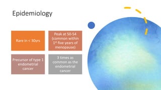
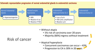
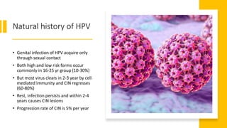
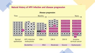
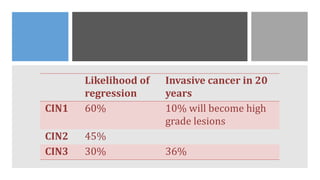
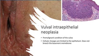
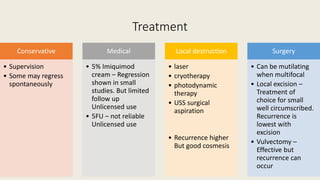
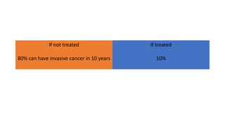
Pre-malignant
Gynaecological
Conditions
Dr. Indunil Piyadigama
Endometrial
hyperplasia
• Irregular proliferation of the
endometrial glands with an
increased gland to stromal ratio
(more than 1:1)
• Lesions that mimic EH
• Disordered proliferation
• Secretory endometrium
• Benign polyps
Epidemiology
Rare in < 30yrs
Peak at 50-54
(common within
1st five years of
menopause)
Precursor of type 1
endometrial
cancer
3 times as
common as the
endometrial
cancer
Risk factors
Increased endogenous oestrogen
• Anovulation
• Obesity
• Diabetes
• PCOS
• Granulosa cell tumours
Exogenous oestrogen
• HRT - Cyclical use – Endometrial hyperplasia in 0.7%
• Tamoxifen
• Risk of hyperplasia 2-3 times higher than general population.
Currently restricted for 5 years use only
• Pretreatment screening, prophylactic hysterectomy or
endometrial sampling at a low threshold are suggested
interventions.
• Aromatase inhibitors – No increase in risk of cancer
Immunosuppression
Infection
Current classification -
WHO 2014
Hyperplasia
without
atypia
Atypical
hyperplasia
Risk of cancer
• Without atypia
• 5% risk of carcinoma over 20 years
• Majority (80%) regress without treatment
• Atypical hyperplasia
• Concurrent carcinomas can occur – 43%
• Progression to CA is 30% in 20 years
Clinical presentation
• AUB – 15% are diagnosed endometrial hyperplasia
• Intermenstrual bleeding
• Post menopausal bleeding
• Small number can be asymptomatic
Diagnosis
TVS
• In premenopausal is restricted to finding structural causes
• But for PCOS with absent withdrawal bleeding or AUB ET is
recommended
• <7mm unlikely to have endometrial hyperplasia
Pipelle biopsy
Despite negative biopsy 2% of women still can have endometrial
hyperplasia
Sensitivity
Premenopausal 80% Postmenopausal 75%
D&C
• Inpatient
• Up to 10% of endometrial pathologies missed
Hysteroscopy and biopsy
• Indications
• When EB fails
• Persistent AUB
• If intrauterine structural anomalies or
polyps suspected – Focal endometrial
pathology is seen in about 10% of
population
• Better at detecting rather than
excluding endometrial pathology
• 98% sensitive for hyperplasia
Treatment - Hyperplasia without atypia
Risk factor modification
• Weight
• Exogenous hormones
Progesterone
Indications
•Fail to regress following observation
•Symptomatic women
Type
•Continuous oral progesterone – MPA 10-20mg/day, NET 10-15mg/day
•LNG- IUS
Action
•Antimitotic on endometrial cells
•Modulating growth stimulatory signals of E2
•Reduces oestrogen secretion by HPO axis
•Reduces receptors
•Increases metabolism of E2
Higher disease regression rate (95%) compared to observation
alone (80%)
6 months use better than 3 months
Follow up
• EB with a minimum of 6 months treatment - 2 biopsies negative
shows cure and discharge. But on an off biopsies for 2 years
• High risk women (BMI>35 on oral progestogens) needs 6 monthly
assessment for 2 years and annual screening there after for follow up
• If bleeding needs further evaluation
Hysterectomy + BSO
• Indications
• Progression to atypical hyperplasia
• No regression following 12 months of treatment
• Recurrence
• Symptomatic
• If does not wish to have surveillance or treatment
• Premenopausal individualize oophorectomy, but B/L salpingectomy.
Oestrogen replacement therapy should be considered.
• Subtotal avoided
Endometrial ablation
• Not recommended
• Complete or persistent endometrial destruction cannot be ensured
and adhesion formation may preclude further histological surveillance
Atypical hyperplasia
• Total hysterectomy
Women who
have fertility
wishes
• MDT
• Counsel about the risk of underlying
malignancy
• Coexisting cancers
• Ovarian cancer – 4%
• Higher than stage1 ECa – 2%
• Metastatic disease – 0.5%
• Prior need
• Hysteroscopy
• MRI/CT
• Ca 125
LNG IUS or Oral progesterone
Follow up
• with 3 monthly EB
• Once 2 consecutive samples negative – Every
6monthly to 1year biopsies till hysterectomy
25% of live pregnancy rate
Results are better if
• Disease optimally suppressed
• Assisted reproduction techniques
Cervical
cancer
• Preventable
• Curable
• Long natural history
• Most common female malignancy in developing
countries
• 7th most common cancer in women worldwide
• Good screening had led to a dramatic reduction
in cervical cancer – 30% decrease from 1993-
2003 in UK
Cervical pre-invasive
disease
Natural history of HPV
• Genital infection of HPV acquire only
through sexual contact
• Both high and low risk forms occur
commonly in 16-25 yr group (10-30%)
• But most virus clears in 2-3 year by cell
mediated immunity and CIN regresses
(60-80%)
• Rest, infection persists and within 2-4
years causes CIN lesions
• Progression rate of CIN is 5% per year
Likelihood of
regression
Invasive cancer in 20
years
CIN1 60% 10% will become high
grade lesions
CIN2 45%
CIN3 30% 36%
• Virus enters the epithelium through a breach in skin integrity caused
by microtrauma
• It remain in the cytoplasm as episomes and can be cleared
• But sometimes enter the nucleus and this is called integration
• The cell no longer undergo apoptosis after 40-60 cell cycles and
instead become immortal. E6 and E7 oncoproteins which binds to p53
and RB respectively are responsible for this
• Prevalence of HPV in cervical cancers - 99.7%
• HPV infection is necessary
• But persistence is important
• 70% of cancers have High risk 16 and 18
• Above 30 yrs HPV incidence decline to about 5% but the regression
rate is much lower
• In a woman over 30 years with high risk HPV positivity the risk of
developing CIN 3 is 116 times compared to a control
• The whole process of early lesion to invasive cancer may take 15-20
years
Risk factors
• early onset of sexual activity
• multiple sexual partners (of self or of the partner)
• low socioeconomic status
• Tobacco smoking (2-fold)
• oral contraceptives (2.5-fold)
• other sexually transmitted infections like herpes simplex virus,
Chlamydia and bacterial vaginosis might play a role in the
progression of HPV infection to dyskaryosis
• immuno-compromise, including human immunodeficiency virus (HIV)
(5-fold)
Screening
• Cytology based
• HPV based
• Histology to confirm –
colposcopy based
Routine
screening
Negative
Routine
recall
Inadequate
Rpt in 3/12
Borderline/
low grade
HPV
Negative Intermediate
Initial
borderline
Rpt Cytology
in 6/12
Initial LSIL
Colposcopy
Positive
Colposcopy
High grade
Colposcopy
Colposcopy
< CIN 1
Routine recall
CIN 1
Untreated
12/12
cytology
follow up
CIN 1/2/3
Treatment
Test of cure in
6/12
cGIN
• Associated particularly with HPV 18
• Cytology is unsatisfactory and no colposcopic features
• Diagnosis is by chance during treatment of squamous pre-invasive disease
• 90% of the lesions occur 1cm from SCJ. But can occur anywhere in the
endocervical canal
• Can have skip lesions higher up in the endocervical canal. Therefore even
after treatment 15% have recurrence.
• This is associated with 50% of CIN and 40% of adenocarcinoma
• Therefore cone biopsy is needed to exclude occult invasive
adenocarcinoma. Endometrial biopsy to rule out abnormality higher up
Management
• Colposcopy and biopsy – Because higher percentage has concomitant CIN
• Punch biopsy is inadequate since disease often occurs at the crypts of the
glands
• Therefore for diagnosis and treatment conization is used
• Endometrial sampling
• Local excision – excision margins should be clear
• Fertility sparing surgery is a possibility provided that the margins are clear
• These people are at higher risk of recurrence
• 15% when margins free
• 50% when margins involved
• Hysterectomy
Vulval intraepithelial
neoplasia
• Premalignant condition of the vulva
• Cellular changes are limited to the epithelium. Does not
breech the basement memebrane
Unifocal
• Postmenopausal
• Non HPV
• Differentiated histology
Multifocal
• Premenopausal
• HPV related
• Interlabial grooves, posterior
forchette, perineum
Clinical features
Pruritus
Perineal pain
Dysuria
50% asymptomatic
Mass, ulcer
Altered appearance
Most multifocal, non hairy part
raised/verrucous white, red, brown,
pink, gray, macular lesion
Investigations
Multiple biopsies needed - histology shows
loss of organisation of squamous epithelium
with a variable degree of cytological atypia
which is graded as undifferentiated or
differentiated and by depth
Colposcopy to exclude CIN and VIN
If perianal lesions anoscopy
Treatment
Conservative
• Supervision
• Some may regress
spontaneously
Medical
• 5% Imiquimod
cream – Regression
shown in small
studies. But limited
follow up
Unlicensed use
• 5FU – not reliable
Unlicensed use
Local destruction
• laser
• cryotherapy
• photodynamic
therapy
• USS surgical
aspiration
• Recurrence higher
But good cosmesis
Surgery
• Can be mutilating
when multifocal
• Local excision –
Treatment of
choice for small
well circumscribed.
Recurrence is
lowest with
excision
• Vulvectomy –
Effective but
recurrence can
occur
If not treated
80% can have invasive cancer in 10 years
If treated
10%
• Extramammary paget’s is a rare form of VIN. It is associated with
cancer of the apocrine gland
• Follow up - Specially for VIN 3
Thank you

Premalignant Gynaecological Conditions

  • 1.
  • 2.
    Endometrial hyperplasia • Irregular proliferationof the endometrial glands with an increased gland to stromal ratio (more than 1:1) • Lesions that mimic EH • Disordered proliferation • Secretory endometrium • Benign polyps
  • 3.
    Epidemiology Rare in <30yrs Peak at 50-54 (common within 1st five years of menopause) Precursor of type 1 endometrial cancer 3 times as common as the endometrial cancer
  • 4.
    Risk factors Increased endogenousoestrogen • Anovulation • Obesity • Diabetes • PCOS • Granulosa cell tumours Exogenous oestrogen • HRT - Cyclical use – Endometrial hyperplasia in 0.7% • Tamoxifen • Risk of hyperplasia 2-3 times higher than general population. Currently restricted for 5 years use only • Pretreatment screening, prophylactic hysterectomy or endometrial sampling at a low threshold are suggested interventions. • Aromatase inhibitors – No increase in risk of cancer Immunosuppression Infection
  • 5.
    Current classification - WHO2014 Hyperplasia without atypia Atypical hyperplasia
  • 7.
    Risk of cancer •Without atypia • 5% risk of carcinoma over 20 years • Majority (80%) regress without treatment • Atypical hyperplasia • Concurrent carcinomas can occur – 43% • Progression to CA is 30% in 20 years
  • 8.
    Clinical presentation • AUB– 15% are diagnosed endometrial hyperplasia • Intermenstrual bleeding • Post menopausal bleeding • Small number can be asymptomatic
  • 9.
    Diagnosis TVS • In premenopausalis restricted to finding structural causes • But for PCOS with absent withdrawal bleeding or AUB ET is recommended • <7mm unlikely to have endometrial hyperplasia
  • 10.
    Pipelle biopsy Despite negativebiopsy 2% of women still can have endometrial hyperplasia Sensitivity Premenopausal 80% Postmenopausal 75%
  • 11.
    D&C • Inpatient • Upto 10% of endometrial pathologies missed
  • 12.
    Hysteroscopy and biopsy •Indications • When EB fails • Persistent AUB • If intrauterine structural anomalies or polyps suspected – Focal endometrial pathology is seen in about 10% of population • Better at detecting rather than excluding endometrial pathology • 98% sensitive for hyperplasia
  • 13.
    Treatment - Hyperplasiawithout atypia Risk factor modification • Weight • Exogenous hormones
  • 14.
    Progesterone Indications •Fail to regressfollowing observation •Symptomatic women Type •Continuous oral progesterone – MPA 10-20mg/day, NET 10-15mg/day •LNG- IUS Action •Antimitotic on endometrial cells •Modulating growth stimulatory signals of E2 •Reduces oestrogen secretion by HPO axis •Reduces receptors •Increases metabolism of E2 Higher disease regression rate (95%) compared to observation alone (80%) 6 months use better than 3 months
  • 15.
    Follow up • EBwith a minimum of 6 months treatment - 2 biopsies negative shows cure and discharge. But on an off biopsies for 2 years • High risk women (BMI>35 on oral progestogens) needs 6 monthly assessment for 2 years and annual screening there after for follow up • If bleeding needs further evaluation
  • 16.
    Hysterectomy + BSO •Indications • Progression to atypical hyperplasia • No regression following 12 months of treatment • Recurrence • Symptomatic • If does not wish to have surveillance or treatment • Premenopausal individualize oophorectomy, but B/L salpingectomy. Oestrogen replacement therapy should be considered. • Subtotal avoided
  • 17.
    Endometrial ablation • Notrecommended • Complete or persistent endometrial destruction cannot be ensured and adhesion formation may preclude further histological surveillance
  • 18.
  • 19.
    Women who have fertility wishes •MDT • Counsel about the risk of underlying malignancy • Coexisting cancers • Ovarian cancer – 4% • Higher than stage1 ECa – 2% • Metastatic disease – 0.5% • Prior need • Hysteroscopy • MRI/CT • Ca 125
  • 20.
    LNG IUS orOral progesterone Follow up • with 3 monthly EB • Once 2 consecutive samples negative – Every 6monthly to 1year biopsies till hysterectomy 25% of live pregnancy rate Results are better if • Disease optimally suppressed • Assisted reproduction techniques
  • 21.
    Cervical cancer • Preventable • Curable •Long natural history • Most common female malignancy in developing countries • 7th most common cancer in women worldwide • Good screening had led to a dramatic reduction in cervical cancer – 30% decrease from 1993- 2003 in UK
  • 22.
  • 23.
    Natural history ofHPV • Genital infection of HPV acquire only through sexual contact • Both high and low risk forms occur commonly in 16-25 yr group (10-30%) • But most virus clears in 2-3 year by cell mediated immunity and CIN regresses (60-80%) • Rest, infection persists and within 2-4 years causes CIN lesions • Progression rate of CIN is 5% per year
  • 25.
    Likelihood of regression Invasive cancerin 20 years CIN1 60% 10% will become high grade lesions CIN2 45% CIN3 30% 36%
  • 26.
    • Virus entersthe epithelium through a breach in skin integrity caused by microtrauma • It remain in the cytoplasm as episomes and can be cleared • But sometimes enter the nucleus and this is called integration • The cell no longer undergo apoptosis after 40-60 cell cycles and instead become immortal. E6 and E7 oncoproteins which binds to p53 and RB respectively are responsible for this
  • 27.
    • Prevalence ofHPV in cervical cancers - 99.7% • HPV infection is necessary • But persistence is important • 70% of cancers have High risk 16 and 18 • Above 30 yrs HPV incidence decline to about 5% but the regression rate is much lower • In a woman over 30 years with high risk HPV positivity the risk of developing CIN 3 is 116 times compared to a control • The whole process of early lesion to invasive cancer may take 15-20 years
  • 28.
    Risk factors • earlyonset of sexual activity • multiple sexual partners (of self or of the partner) • low socioeconomic status • Tobacco smoking (2-fold) • oral contraceptives (2.5-fold) • other sexually transmitted infections like herpes simplex virus, Chlamydia and bacterial vaginosis might play a role in the progression of HPV infection to dyskaryosis • immuno-compromise, including human immunodeficiency virus (HIV) (5-fold)
  • 29.
    Screening • Cytology based •HPV based • Histology to confirm – colposcopy based
  • 30.
    Routine screening Negative Routine recall Inadequate Rpt in 3/12 Borderline/ lowgrade HPV Negative Intermediate Initial borderline Rpt Cytology in 6/12 Initial LSIL Colposcopy Positive Colposcopy High grade Colposcopy
  • 31.
    Colposcopy < CIN 1 Routinerecall CIN 1 Untreated 12/12 cytology follow up CIN 1/2/3 Treatment Test of cure in 6/12
  • 33.
    cGIN • Associated particularlywith HPV 18 • Cytology is unsatisfactory and no colposcopic features • Diagnosis is by chance during treatment of squamous pre-invasive disease • 90% of the lesions occur 1cm from SCJ. But can occur anywhere in the endocervical canal • Can have skip lesions higher up in the endocervical canal. Therefore even after treatment 15% have recurrence. • This is associated with 50% of CIN and 40% of adenocarcinoma • Therefore cone biopsy is needed to exclude occult invasive adenocarcinoma. Endometrial biopsy to rule out abnormality higher up
  • 34.
    Management • Colposcopy andbiopsy – Because higher percentage has concomitant CIN • Punch biopsy is inadequate since disease often occurs at the crypts of the glands • Therefore for diagnosis and treatment conization is used • Endometrial sampling • Local excision – excision margins should be clear • Fertility sparing surgery is a possibility provided that the margins are clear • These people are at higher risk of recurrence • 15% when margins free • 50% when margins involved • Hysterectomy
  • 35.
    Vulval intraepithelial neoplasia • Premalignantcondition of the vulva • Cellular changes are limited to the epithelium. Does not breech the basement memebrane
  • 36.
    Unifocal • Postmenopausal • NonHPV • Differentiated histology Multifocal • Premenopausal • HPV related • Interlabial grooves, posterior forchette, perineum
  • 37.
    Clinical features Pruritus Perineal pain Dysuria 50%asymptomatic Mass, ulcer Altered appearance Most multifocal, non hairy part raised/verrucous white, red, brown, pink, gray, macular lesion
  • 38.
    Investigations Multiple biopsies needed- histology shows loss of organisation of squamous epithelium with a variable degree of cytological atypia which is graded as undifferentiated or differentiated and by depth Colposcopy to exclude CIN and VIN If perianal lesions anoscopy
  • 39.
    Treatment Conservative • Supervision • Somemay regress spontaneously Medical • 5% Imiquimod cream – Regression shown in small studies. But limited follow up Unlicensed use • 5FU – not reliable Unlicensed use Local destruction • laser • cryotherapy • photodynamic therapy • USS surgical aspiration • Recurrence higher But good cosmesis Surgery • Can be mutilating when multifocal • Local excision – Treatment of choice for small well circumscribed. Recurrence is lowest with excision • Vulvectomy – Effective but recurrence can occur
  • 40.
    If not treated 80%can have invasive cancer in 10 years If treated 10%
  • 41.
    • Extramammary paget’sis a rare form of VIN. It is associated with cancer of the apocrine gland • Follow up - Specially for VIN 3
  • 42.