Embolisum
•Download as PPTX, PDF•
0 likes•74 views
the topic is about embolism, there is a video included as well, I hope ppt is useful for some one atleast
Report
Share
Report
Share

Recommended
Embolism
"Embolism"
this presentation is about embolism where some information about embolism is included. Hope this will help you in your work.
1. edema; hemodynamic disorders
The document discusses hemodynamic disorders including edema, hyperemia, hemorrhage, hemostasis, thrombosis, embolism, and infarction. Edema is defined as increased fluid in the interstitial tissue spaces and can be caused by increased hydrostatic pressure, decreased oncotic pressure, sodium retention, or inflammation. Treatment for edema focuses on reducing sodium intake, using diuretics to increase sodium excretion, and aldosterone antagonists.
Embolism
This document defines and describes different types of embolism. The most common type is thromboembolism, which occurs when a thrombus or part of a thrombus breaks off and is carried by the bloodstream. Pulmonary thromboembolism is a significant type that occurs when thrombi travel to the lungs and obstruct the pulmonary arteries. Other types include fat, air, gas and paradoxical embolisms. Air embolism can be venous, entering systemic veins during surgery or trauma, or arterial, entering the lungs during procedures like angiography. Decompression sickness is a form of gas embolism that affects divers or those exposed to changes in atmospheric pressure.
Shock
This document defines and classifies shock. Shock is characterized by inadequate perfusion of cells and tissues due to reduced circulating blood volume and blood pressure. Shock is classified as hypovolemic (caused by blood or plasma loss), cardiogenic (caused by heart disease), septic (caused by infection), or other types like traumatic or neurogenic shock. The pathophysiology of each type is described. Hypovolemic shock results from blood or fluid loss, cardiogenic shock from reduced cardiac output, and septic shock from activation of the immune system and inflammatory response to infection. Shock progresses through compensated, decompensated, and irreversible stages as the body attempts but fails to maintain adequate perfusion of vital
Embolism
This document defines and describes different types of embolism. It states that an embolism is a detached solid, liquid, or gas mass carried by the bloodstream to a distant site. It then lists and provides details on various types of embolisms including venous (pulmonary), arterial, paradoxical, fat, amniotic fluid, air, and septic embolisms. For pulmonary embolisms, it notes they most commonly arise from deep vein thromboses in the legs and can cause further recurrent embolic episodes. It also provides information on symptoms and causes of fat, air, and amniotic fluid embolisms.
Embolism
Embolism is the obstruction of blood vessels by foreign material carried by the bloodstream called an embolus. The most common type is thromboembolism caused by blood clots. Embolisms can be classified by the material causing the obstruction (solid, liquid, gas), whether infected or not, and the source of the embolus (cardiac, arterial, venous, lymphatic). Pulmonary embolism is a serious and potentially fatal condition where blood clots block arteries in the lungs. Risk factors include immobilization, and clots most often originate from veins in the legs. Consequences can include sudden death, lung infarction, hemorrhage and chronic lung disease.
7. infarction; hemodynamic disorders
This document discusses infarction, defined as ischemic necrosis of tissue due to occlusion of arterial or venous circulation. It notes that infarction is a common cause of death in the US, usually caused by arterial occlusion from thromboembolism. There are two morphological types of infarction - red (hemorrhagic) and white (anemic). Red infarctions occur in loose tissues or previously congested organs due to venous occlusion or dual blood supply. White infarctions occur in solid organs due to arterial occlusion and lack of collateral circulation. The final outcome of infarction is coagulative necrosis of the tissue.
2. hyperemia and congestion; hemodynamic disorders
This document discusses hemodynamic disorders including edema, hyperemia, congestion, hemorrhage, hemostasis, thrombosis, embolism, infarction, and shock. It defines key terms and compares hyperemia and congestion. Hyperemia is an active process involving arterial dilation that causes redness in tissue, while congestion is a passive process resulting from impaired venous outflow that causes blue-red discoloration. Congestion can lead to tissue hypoxia, degeneration, scarring, and fibrosis in organs like the lung and liver if not resolved.
Odema
This document provides an overview of edema, including its definition, types, pathogenesis, and examples of important types of edema. Edema is defined as the abnormal accumulation of free fluid in interstitial tissue spaces or body cavities. It can be localized to an organ or limb, or generalized throughout the body. The pathogenesis of edema involves mechanisms that interfere with fluid balance, plasma oncotic pressure, capillary hydrostatic pressure, lymphatic flow, and sodium and water retention. Examples of important types of edema discussed include renal edema, cardiac edema, pulmonary edema, cerebral edema, hepatic edema, nutritional edema, and myxoedema.
Thrombosis and Embolism
The document discusses various types of embolism and thrombosis. It describes the Virschow triad of factors that can lead to thrombosis - endothelial injury, changes in blood flow, and hypercoagulability. It then examines different causes and outcomes of thrombosis and embolism in various parts of the body, such as pulmonary embolism from deep vein thrombosis, systemic embolism from cardiac sources, and amniotic fluid embolism during childbirth.
Embolism
1. Embolism occurs when a detached mass such as a blood clot, air bubble, or fat globule travels through the blood vessels and blocks circulation.
2. There are several types of embolism including pulmonary (lung), systemic, fat, marrow, air, and amniotic embolism.
3. Pulmonary embolism is a blockage in the pulmonary artery often caused by deep vein thrombosis, and it can lead to conditions like saddle embolism, paradoxical embolism, or cor pulmonale if left untreated.
The cv system
The document discusses the cardiovascular system and high cholesterol. It describes the main parts of the cardiovascular system including capillaries, veins, the heart, and blood vessels. It then discusses several conditions related to high cholesterol, including coronary heart disease, arteriosclerosis, and atherosclerosis. Diagnostic procedures for detecting high cholesterol like serum lipoprotein levels are mentioned. The document concludes by outlining some surgical procedures used to treat conditions caused by high cholesterol, such as endarterectomy and intracoronary artery stents, as well as medications and lifestyle changes that can help treat and prevent coronary artery disease.
Embolism
An embolism is the obstruction of blood vessels by a mass or clot that has detached from its origin and traveled through the bloodstream. Embolisms can be classified based on the material causing the obstruction and the source and direction of blood flow. Common types of embolisms include pulmonary, fat, air, amniotic fluid, and tumor embolisms. Pulmonary embolisms originate in the lower leg veins and are caused by stasis, hypercoagulability, or a saddle embolism. Fat embolisms result from trauma or medical conditions and obstruct arterioles and capillaries. Decompression sickness is a form of gas embolism that affects scuba divers or caisson workers
Infarction
1) Infarction refers to the death of cells in a tissue due to an inadequate blood supply.
2) There are two main types of infarction - red and white - which are distinguished by their color and the type of tissue affected.
3) The development and severity of an infarction depends on factors like the anatomy of a tissue's blood supply, how quickly the blood flow is blocked, and the tissue's vulnerability to low oxygen levels.
Thrombosis
a haemodynamic disorder which has serious consequences. useful for pathology students and health-concerned people.
6 hemodynamic disorders
1. Hemodynamic disorders involve changes in intravascular volume, pressure, or protein content that affect fluid movement across vessel walls and can cause edema, hyperemia, congestion, hemorrhage, thrombosis, embolism, infarction, or shock.
2. Edema is increased fluid in tissues, caused by increased hydrostatic pressure, reduced plasma proteins, lymphatic obstruction, sodium retention, or inflammation.
3. Thrombosis is inappropriate blood clot formation from endothelial injury, blood stasis, or hypercoagulability per Virchow's triad, and thrombi can embolize or organize.
4. Embolism occurs when a detached mass is
EMBOLISM -1
Embolism occurs when a solid, liquid, or gaseous mass travels through the bloodstream and lodges in a blood vessel distant from the site of origin. Embolisms are classified based on direction of travel and composition. Pulmonary embolisms involve the lungs while systemic embolisms affect other organs. Common causes of embolism include blood clots, fat droplets, air bubbles, and infectious materials. Symptoms vary depending on the size and location of the embolism but may include dyspnea, chest pain, and coughing. Diagnosis involves blood tests, imaging, and scans. Consequences depend on factors like vessel size and collateral blood flow.
15 hemodynamic disorders
This document discusses various hemodynamic disorders including edema, hyperemia, congestion, hemorrhage, hemostasis, thrombosis, embolism, infarction, and shock. Edema is an excess of fluid in the interstitial spaces that can occur in response to injury or inflammation. Treatment focuses on addressing the underlying cause. Hyperemia is increased blood flow to an area, congestion is impaired outflow, and hemorrhage is blood escaping from vessels. Hemostasis is the body's natural response to stop bleeding through coagulation. Thrombosis and embolism involve blood clots forming and detaching within vessels. Infarction results from blocked blood flow causing tissue death. Shock is a
6. embolism; hemodynamic disordrs
This document discusses various hemodynamic disorders including edema, hyperemia, congestion, hemorrhage, hemostasis, thrombosis, embolism, infarction, and shock. Embolism can involve solid materials like thrombi, liquids like amniotic fluid, or gases like air. Embolisms are classified as systemic arterial, pulmonary, or venous and can cause vascular obstruction and infarction. Infarction results from ischemia and leads to coagulative necrosis, appearing as either red or white lesions. Septic shock is a type of shock caused by gram-positive bacteria through endotoxins like LPS, and can involve multiple organ failure.
Hemodynamic disorders
This document summarizes key concepts related to hemodynamic disorders, thrombosis, and shock. It discusses edema, including the mechanisms and clinical significance of edema. It also covers hyperemia and congestion, hemorrhage, and thrombosis. For edema, it describes how fluid moves between vascular and interstitial spaces and the causes of increased interstitial fluid. It discusses the pathologic features and clinical significance of pulmonary, subcutaneous, and brain edema. For thrombosis, hemorrhage, hyperemia and congestion, it outlines the mechanisms, morphological changes, and clinical implications.
64965 hemodyn[1] edema
The document discusses various types of hemodynamic disorders and edema. It describes how edema can be caused by increased hydrostatic pressure, decreased plasma oncotic pressure, inflammation, and lymphatic obstruction. The most common cause of generalized edema is congestive heart failure, which increases hydrostatic pressure through elevated central venous pressure and decreases renal perfusion. Other causes mentioned include nephrotic syndrome, cirrhosis, and protein malnutrition. The key organs involved in fluid homeostasis are the heart, liver, and kidneys.
Atherosclerosis
This document discusses atherosclerosis, a disease where plaque builds up inside arteries. It defines lipoproteins, which transport fats in the blood, and classifies different types based on density. The document then explains that atherosclerosis is caused by macrophages and fat accumulating in artery walls as plaques. These plaques can rupture, causing blood clots that block blood flow and lead to heart attacks or strokes. The progression of atherosclerosis involves endothelial dysfunction allowing LDL cholesterol to enter the artery wall and oxidative damage, followed by inflammation and plaque formation that narrows arteries over time.
Hemodynamic disorders
This document summarizes various hemodynamic disorders including edema, hyperemia, congestion, hemorrhage, and shock. It defines edema as increased fluid in tissues and discusses common sites of edema such as subcutaneous, pulmonary, and brain edema. It also defines hyperemia as a local increase in blood volume, congestion as passive hyperemia, and discusses common sites of congestion such as pulmonary and liver congestion. Additionally, it discusses hemorrhage as blood vessel rupture and various types including petechiae, purpura, and bruises. Finally, it defines shock as systemic hypoperfusion and discusses the different types and stages of shock.
Thrombosis, embolism and infarction
Thrombosis is the formation of a blood clot within a blood vessel or cavity of the heart. Virchow identified three main factors that contribute to thrombosis: endothelial injury, changes in blood flow, and hypercoagulability. Thrombi can propagate or embolize, becoming lodged in another vessel and resulting in infarction of downstream tissue. Infarctions appear pale/white in solid organs and red/hemorrhagic in lungs/other tissues. Over time, infarcted tissue progresses from coagulative necrosis to phagocytosis and scar formation.
Hemodynamic disorders - Edema, Hyperemia, Hemorrahge by DR. ROOPAM JAIN
Hemodynamic disorders can cause edema, hyperemia, and hemorrhage. Edema is the accumulation of fluid in tissues or body cavities due to increased hydrostatic pressure or decreased colloid osmotic pressure. Hyperemia is increased blood volume in tissues due to arteriolar dilation, while congestion is passive increased blood volume due to reduced outflow. Hemorrhage can occur from defects in platelets, coagulation factors, or vessel walls, manifesting as petechiae, purpura, ecchymoses, or hematomas. The document discusses the etiology, morphology, and clinical features of these hemodynamic disorders.
3. hemorrhage; hemodynamic disorders
This document discusses hemodynamic disorders including edema, hyperemia, congestion, hemorrhage, hemostasis, thrombosis, embolism, and infarction. It focuses on hemorrhage, defining it as blood escaping from vessels due to rupture. Common causes are listed as trauma, atherosclerosis, inflammation, and neoplasm. Specific types of hemorrhages like petechiae, purpura, and ecchymoses are described based on their size and location. Clinical features and significance are discussed depending on the rapidity and site of bleeding.
Hemodynamic Disorders, Thromboembolic Disease & Shock-HYPERCOAGULABLE STATES ...
Hypercoagulable states, also known as thrombophilia, are conditions that increase the risk of developing venous thrombosis. There are hereditary and acquired causes of thrombophilia. Microscopically, red thrombi found in veins contain more red blood cells, platelets, and leukocytes trapped in a fibrin meshwork, resembling blood clots. Over time, a thrombus may resolve through fibrinolysis, organize by developing connective tissue, propagate by continued deposition, or embolize to cause blockages elsewhere.
4. hemodyn disorders,thrombosis, shock
This document summarizes various hemodynamic disorders including edema, hyperemia, congestion, hemorrhage, hemostasis, thrombosis, embolism, and infarction. It discusses the key mechanisms and pathophysiology of each condition in 1-3 sentences. For example, it notes that edema can result from increased hydrostatic pressure, reduced oncotic pressure, lymphatic obstruction, or sodium/water retention. Congestion is defined as either an acute or chronic passive process in the lungs, liver or brain. Thrombosis formation is influenced by endothelial injury, abnormal blood flow, and hypercoagulability as per Virchow's triad.
5 embolism
An embolus is a solid, liquid, or gaseous mass that breaks off and travels through the bloodstream, lodging in and blocking smaller blood vessels. Pulmonary embolisms originate from deep leg vein thrombi in 95% of cases and can cause infarction or blockage of lung tissue. Systemic embolisms originate from heart mural thrombi in 80% of cases and commonly impact the brain or lower extremities. Fat embolisms occur after bone fractures and burns, causing pulmonary insufficiency, neurological issues, and thrombocytopenia. Air embolisms enter the circulation through chest or obstetric injuries and can block major blood vessels. Amniotic fluid embolisms are a rare
Infarction Path-201.pptx
1. Infarction occurs when there is a decrease in blood supply to an organ or tissue, causing localized ischemic necrosis. This can be caused by thrombi, emboli, vasospasm, expansion of atheroma, extrinsic vessel compression, vessel twisting, or traumatic vessel rupture.
2. There are three main types of infarction: red (hemorrhagic), white (anemic), and septic. Factors like vulnerability to hypoxia, oxygen content of blood, and blood supply nature influence whether infarction occurs.
3. Examples of infarctions include myocardial, pulmonary, and cerebral. Myocardial infarction due to coronary artery blockage can be fatal, while pulmonary infarction is usually
More Related Content
What's hot
Odema
This document provides an overview of edema, including its definition, types, pathogenesis, and examples of important types of edema. Edema is defined as the abnormal accumulation of free fluid in interstitial tissue spaces or body cavities. It can be localized to an organ or limb, or generalized throughout the body. The pathogenesis of edema involves mechanisms that interfere with fluid balance, plasma oncotic pressure, capillary hydrostatic pressure, lymphatic flow, and sodium and water retention. Examples of important types of edema discussed include renal edema, cardiac edema, pulmonary edema, cerebral edema, hepatic edema, nutritional edema, and myxoedema.
Thrombosis and Embolism
The document discusses various types of embolism and thrombosis. It describes the Virschow triad of factors that can lead to thrombosis - endothelial injury, changes in blood flow, and hypercoagulability. It then examines different causes and outcomes of thrombosis and embolism in various parts of the body, such as pulmonary embolism from deep vein thrombosis, systemic embolism from cardiac sources, and amniotic fluid embolism during childbirth.
Embolism
1. Embolism occurs when a detached mass such as a blood clot, air bubble, or fat globule travels through the blood vessels and blocks circulation.
2. There are several types of embolism including pulmonary (lung), systemic, fat, marrow, air, and amniotic embolism.
3. Pulmonary embolism is a blockage in the pulmonary artery often caused by deep vein thrombosis, and it can lead to conditions like saddle embolism, paradoxical embolism, or cor pulmonale if left untreated.
The cv system
The document discusses the cardiovascular system and high cholesterol. It describes the main parts of the cardiovascular system including capillaries, veins, the heart, and blood vessels. It then discusses several conditions related to high cholesterol, including coronary heart disease, arteriosclerosis, and atherosclerosis. Diagnostic procedures for detecting high cholesterol like serum lipoprotein levels are mentioned. The document concludes by outlining some surgical procedures used to treat conditions caused by high cholesterol, such as endarterectomy and intracoronary artery stents, as well as medications and lifestyle changes that can help treat and prevent coronary artery disease.
Embolism
An embolism is the obstruction of blood vessels by a mass or clot that has detached from its origin and traveled through the bloodstream. Embolisms can be classified based on the material causing the obstruction and the source and direction of blood flow. Common types of embolisms include pulmonary, fat, air, amniotic fluid, and tumor embolisms. Pulmonary embolisms originate in the lower leg veins and are caused by stasis, hypercoagulability, or a saddle embolism. Fat embolisms result from trauma or medical conditions and obstruct arterioles and capillaries. Decompression sickness is a form of gas embolism that affects scuba divers or caisson workers
Infarction
1) Infarction refers to the death of cells in a tissue due to an inadequate blood supply.
2) There are two main types of infarction - red and white - which are distinguished by their color and the type of tissue affected.
3) The development and severity of an infarction depends on factors like the anatomy of a tissue's blood supply, how quickly the blood flow is blocked, and the tissue's vulnerability to low oxygen levels.
Thrombosis
a haemodynamic disorder which has serious consequences. useful for pathology students and health-concerned people.
6 hemodynamic disorders
1. Hemodynamic disorders involve changes in intravascular volume, pressure, or protein content that affect fluid movement across vessel walls and can cause edema, hyperemia, congestion, hemorrhage, thrombosis, embolism, infarction, or shock.
2. Edema is increased fluid in tissues, caused by increased hydrostatic pressure, reduced plasma proteins, lymphatic obstruction, sodium retention, or inflammation.
3. Thrombosis is inappropriate blood clot formation from endothelial injury, blood stasis, or hypercoagulability per Virchow's triad, and thrombi can embolize or organize.
4. Embolism occurs when a detached mass is
EMBOLISM -1
Embolism occurs when a solid, liquid, or gaseous mass travels through the bloodstream and lodges in a blood vessel distant from the site of origin. Embolisms are classified based on direction of travel and composition. Pulmonary embolisms involve the lungs while systemic embolisms affect other organs. Common causes of embolism include blood clots, fat droplets, air bubbles, and infectious materials. Symptoms vary depending on the size and location of the embolism but may include dyspnea, chest pain, and coughing. Diagnosis involves blood tests, imaging, and scans. Consequences depend on factors like vessel size and collateral blood flow.
15 hemodynamic disorders
This document discusses various hemodynamic disorders including edema, hyperemia, congestion, hemorrhage, hemostasis, thrombosis, embolism, infarction, and shock. Edema is an excess of fluid in the interstitial spaces that can occur in response to injury or inflammation. Treatment focuses on addressing the underlying cause. Hyperemia is increased blood flow to an area, congestion is impaired outflow, and hemorrhage is blood escaping from vessels. Hemostasis is the body's natural response to stop bleeding through coagulation. Thrombosis and embolism involve blood clots forming and detaching within vessels. Infarction results from blocked blood flow causing tissue death. Shock is a
6. embolism; hemodynamic disordrs
This document discusses various hemodynamic disorders including edema, hyperemia, congestion, hemorrhage, hemostasis, thrombosis, embolism, infarction, and shock. Embolism can involve solid materials like thrombi, liquids like amniotic fluid, or gases like air. Embolisms are classified as systemic arterial, pulmonary, or venous and can cause vascular obstruction and infarction. Infarction results from ischemia and leads to coagulative necrosis, appearing as either red or white lesions. Septic shock is a type of shock caused by gram-positive bacteria through endotoxins like LPS, and can involve multiple organ failure.
Hemodynamic disorders
This document summarizes key concepts related to hemodynamic disorders, thrombosis, and shock. It discusses edema, including the mechanisms and clinical significance of edema. It also covers hyperemia and congestion, hemorrhage, and thrombosis. For edema, it describes how fluid moves between vascular and interstitial spaces and the causes of increased interstitial fluid. It discusses the pathologic features and clinical significance of pulmonary, subcutaneous, and brain edema. For thrombosis, hemorrhage, hyperemia and congestion, it outlines the mechanisms, morphological changes, and clinical implications.
64965 hemodyn[1] edema
The document discusses various types of hemodynamic disorders and edema. It describes how edema can be caused by increased hydrostatic pressure, decreased plasma oncotic pressure, inflammation, and lymphatic obstruction. The most common cause of generalized edema is congestive heart failure, which increases hydrostatic pressure through elevated central venous pressure and decreases renal perfusion. Other causes mentioned include nephrotic syndrome, cirrhosis, and protein malnutrition. The key organs involved in fluid homeostasis are the heart, liver, and kidneys.
Atherosclerosis
This document discusses atherosclerosis, a disease where plaque builds up inside arteries. It defines lipoproteins, which transport fats in the blood, and classifies different types based on density. The document then explains that atherosclerosis is caused by macrophages and fat accumulating in artery walls as plaques. These plaques can rupture, causing blood clots that block blood flow and lead to heart attacks or strokes. The progression of atherosclerosis involves endothelial dysfunction allowing LDL cholesterol to enter the artery wall and oxidative damage, followed by inflammation and plaque formation that narrows arteries over time.
Hemodynamic disorders
This document summarizes various hemodynamic disorders including edema, hyperemia, congestion, hemorrhage, and shock. It defines edema as increased fluid in tissues and discusses common sites of edema such as subcutaneous, pulmonary, and brain edema. It also defines hyperemia as a local increase in blood volume, congestion as passive hyperemia, and discusses common sites of congestion such as pulmonary and liver congestion. Additionally, it discusses hemorrhage as blood vessel rupture and various types including petechiae, purpura, and bruises. Finally, it defines shock as systemic hypoperfusion and discusses the different types and stages of shock.
Thrombosis, embolism and infarction
Thrombosis is the formation of a blood clot within a blood vessel or cavity of the heart. Virchow identified three main factors that contribute to thrombosis: endothelial injury, changes in blood flow, and hypercoagulability. Thrombi can propagate or embolize, becoming lodged in another vessel and resulting in infarction of downstream tissue. Infarctions appear pale/white in solid organs and red/hemorrhagic in lungs/other tissues. Over time, infarcted tissue progresses from coagulative necrosis to phagocytosis and scar formation.
Hemodynamic disorders - Edema, Hyperemia, Hemorrahge by DR. ROOPAM JAIN
Hemodynamic disorders can cause edema, hyperemia, and hemorrhage. Edema is the accumulation of fluid in tissues or body cavities due to increased hydrostatic pressure or decreased colloid osmotic pressure. Hyperemia is increased blood volume in tissues due to arteriolar dilation, while congestion is passive increased blood volume due to reduced outflow. Hemorrhage can occur from defects in platelets, coagulation factors, or vessel walls, manifesting as petechiae, purpura, ecchymoses, or hematomas. The document discusses the etiology, morphology, and clinical features of these hemodynamic disorders.
3. hemorrhage; hemodynamic disorders
This document discusses hemodynamic disorders including edema, hyperemia, congestion, hemorrhage, hemostasis, thrombosis, embolism, and infarction. It focuses on hemorrhage, defining it as blood escaping from vessels due to rupture. Common causes are listed as trauma, atherosclerosis, inflammation, and neoplasm. Specific types of hemorrhages like petechiae, purpura, and ecchymoses are described based on their size and location. Clinical features and significance are discussed depending on the rapidity and site of bleeding.
Hemodynamic Disorders, Thromboembolic Disease & Shock-HYPERCOAGULABLE STATES ...
Hypercoagulable states, also known as thrombophilia, are conditions that increase the risk of developing venous thrombosis. There are hereditary and acquired causes of thrombophilia. Microscopically, red thrombi found in veins contain more red blood cells, platelets, and leukocytes trapped in a fibrin meshwork, resembling blood clots. Over time, a thrombus may resolve through fibrinolysis, organize by developing connective tissue, propagate by continued deposition, or embolize to cause blockages elsewhere.
4. hemodyn disorders,thrombosis, shock
This document summarizes various hemodynamic disorders including edema, hyperemia, congestion, hemorrhage, hemostasis, thrombosis, embolism, and infarction. It discusses the key mechanisms and pathophysiology of each condition in 1-3 sentences. For example, it notes that edema can result from increased hydrostatic pressure, reduced oncotic pressure, lymphatic obstruction, or sodium/water retention. Congestion is defined as either an acute or chronic passive process in the lungs, liver or brain. Thrombosis formation is influenced by endothelial injury, abnormal blood flow, and hypercoagulability as per Virchow's triad.
What's hot (20)
Hemodynamic disorders - Edema, Hyperemia, Hemorrahge by DR. ROOPAM JAIN
Hemodynamic disorders - Edema, Hyperemia, Hemorrahge by DR. ROOPAM JAIN
Hemodynamic Disorders, Thromboembolic Disease & Shock-HYPERCOAGULABLE STATES ...
Hemodynamic Disorders, Thromboembolic Disease & Shock-HYPERCOAGULABLE STATES ...
Similar to Embolisum
5 embolism
An embolus is a solid, liquid, or gaseous mass that breaks off and travels through the bloodstream, lodging in and blocking smaller blood vessels. Pulmonary embolisms originate from deep leg vein thrombi in 95% of cases and can cause infarction or blockage of lung tissue. Systemic embolisms originate from heart mural thrombi in 80% of cases and commonly impact the brain or lower extremities. Fat embolisms occur after bone fractures and burns, causing pulmonary insufficiency, neurological issues, and thrombocytopenia. Air embolisms enter the circulation through chest or obstetric injuries and can block major blood vessels. Amniotic fluid embolisms are a rare
Infarction Path-201.pptx
1. Infarction occurs when there is a decrease in blood supply to an organ or tissue, causing localized ischemic necrosis. This can be caused by thrombi, emboli, vasospasm, expansion of atheroma, extrinsic vessel compression, vessel twisting, or traumatic vessel rupture.
2. There are three main types of infarction: red (hemorrhagic), white (anemic), and septic. Factors like vulnerability to hypoxia, oxygen content of blood, and blood supply nature influence whether infarction occurs.
3. Examples of infarctions include myocardial, pulmonary, and cerebral. Myocardial infarction due to coronary artery blockage can be fatal, while pulmonary infarction is usually
INFARCTION
This document discusses infarction, which is localized ischemic necrosis of tissue due to decreased blood supply. Infarction can be caused by thrombi, emboli, vasospasm, expansion of atheroma, extrinsic compression of vessels, vessel twisting, or traumatic vessel rupture. There are three main types of infarction: red (hemorrhagic), white (anemic), and septic. Factors that influence infarction development include vulnerability to hypoxia, blood oxygen content, rate of occlusion, and blood supply. Myocardial, pulmonary, and cerebral infarctions are provided as examples and their characteristics and outcomes described.
Circulitary disturbance
Edema can be caused by increased hydrostatic pressure, increased vascular permeability, decreased colloid osmotic pressure, decreased protein synthesis or increased protein loss, or lymphatic obstruction. The major mechanisms are increased hydrostatic pressure, as seen in congestive heart failure, or increased vascular permeability during inflammation. Edema fluid is usually a protein-poor transudate when caused by hydrostatic or oncotic pressure changes, but is a protein-rich exudate with inflammatory causes due to higher vascular permeability.
Thromboembolism,pulmonary embolism,general pathology
This document discusses different types of thromboembolism including pulmonary embolism, fat embolism, air embolism, and amniotic fluid embolism. It provides details on the definition, causes, risk factors, symptoms, diagnosis, treatment and prognosis of pulmonary embolism, which is the most common type of thromboembolism. The document also discusses the pathophysiology and mechanisms of the different embolism types.
Pathology of hemodynamic disorders part 2 nov 2017 Dr. Sufia Husain
This document discusses various types of hemodynamic disorders including embolism, infarction, and shock. It provides detailed information on different types of embolism such as pulmonary thromboembolism, systemic thromboembolism, fat embolism, air embolism, and amniotic fluid embolism. It also describes the stages of shock, associated pathophysiology and morphological changes in tissues during the different stages of shock.
Embolism & Hemorrhage.pptx
This document defines and describes different types of embolisms. It discusses pulmonary embolism which originates from deep vein thromboses. It also covers paradoxical embolism, fat embolism, air embolism, decompression sickness, amniotic fluid embolism, and hemorrhage. For each type, it provides details on causes, clinical presentations, and consequences.
Embolism & Infarction.pdf
This document discusses embolism, infarction, and their causes and classifications. It defines an embolism as a detached mass carried by the blood to distant sites, which can lodge and block vessels, causing tissue dysfunction. Emboli sources include thrombi, fat, air, debris. Embolisms are classified as pulmonary (95% from DVTs), systemic (sites include legs, brain, intestines, kidneys). Infarctions are areas of ischemic necrosis due to blood flow obstruction. They are classified as hemorrhagic, white, septic based on appearance and presence of infection. Infarcts are eventually replaced by scar tissue.
stroke
1. Stroke occurs when the blood supply to the brain is reduced or blocked, preventing brain tissue from receiving oxygen and nutrients.
2. Strokes are classified as either ischemic (caused by blockage of arteries) or hemorrhagic (caused by bleeding in or around the brain).
3. Common symptoms of stroke include sudden weakness or numbness on one side of the body, difficulty speaking, confusion, and severe headache. Early recognition and treatment are important to reduce long-term disability.
Embolism, Infarction and Shock ppt-1 slides.pptx
Medical student
Author: Sahal Ismail Omar
References: Robbins basic pathology and Fundamentals of pathology.
Heart attack plaque blood pressure
A myocardial infarction, or heart attack, occurs when blood flow to part of the heart is blocked, usually by a blood clot, and the heart muscle begins to die due to lack of oxygen. Coronary arteries can become blocked by plaques made of fat, cholesterol, and other substances that build up in the arteries. Rupture of a vulnerable plaque in a coronary artery often leads to a heart attack. Symptoms of a heart attack include chest pain and shortness of breath.
2.9. Atherosclerosis.ppt
Atherosclerosis is a gradual hardening and narrowing of the arteries caused by plaque buildup over many years. It begins in early adulthood with fatty streaks accumulating in artery walls. As cholesterol builds up, fibrous tissue forms plaques that thicken and stiffen arteries. Over time, plaques can rupture, causing blood clots that block blood flow and lead to heart attacks, strokes, and other complications. Atherosclerosis prevalence increases with age and is influenced by risk factors like high cholesterol, hypertension, smoking, and diabetes. It remains a leading cause of death in the United States.
EMBOLISM....Dr San Kauser (Pathology).pdf
Embolism is the obstruction of blood vessels by foreign material that travels through the bloodstream. There are several types of embolism classified by the material causing obstruction (solid, liquid, gas), source of obstruction (cardiac, arterial, venous), and whether infection is present. Common types include thromboembolism caused by blood clots, pulmonary embolism from clots blocking pulmonary arteries, fat/bone marrow embolism from tissue trauma, and amniotic fluid embolism during childbirth from amniotic material entering the mother's bloodstream. Embolisms can cause infarction of tissues and organs downstream from the obstruction depending on the location and size of the obstructing material.
hemodynamic & circulatory disorders 2
Embolism occurs when a solid, liquid, or gas becomes lodged in a blood vessel blocking blood flow. The most common type is thromboembolism from detached blood clots. Emboli can lodge in lungs (pulmonary embolism) or other organs (systemic embolism) cutting off blood supply and causing tissue death (infarction). Ischemia is a lack of blood flow, which if sustained leads to infarction. Shock is a state of low blood flow (hypoperfusion) that can be distributive, cardiogenic, or hypovolemic in origin, and progresses through mild, moderate, and severe stages impacting organ function if not corrected.
circulatory disturbance 1.pptx
Hemodynamic changes can include hyperaemia, thrombosis, embolism, ischemia, infarction, and hemorrhage. Hyperaemia is increased blood flow to an organ and can be active or passive. Thrombosis is the formation of a blood clot within a blood vessel. Embolism occurs when a detached clot or other mass travels through the bloodstream and blocks a vessel. Ischemia is reduced blood flow to an organ, and infarction is tissue death from a lack of blood flow. Hemorrhage involves bleeding outside of blood vessels.
thrombosis embolism.pptx
This document discusses thrombosis, embolism, and their surgical treatment. It defines thrombosis as the formation of a solid blood clot within blood vessels and embolism as a detached mass such as a blood clot that travels through the bloodstream. The document outlines different types of embolism including pulmonary, fat, air, and amniotic fluid embolism. It describes surgical procedures to treat thrombosis and embolism such as thrombolytic injections to dissolve clots, embolectomy to remove clots, and angioplasty to widen arteries.
Atherosclerosis by neha
This document provides information about atherosclerosis. It begins by introducing atherosclerosis as a pathological process in arteries that leads to coronary heart disease and stroke. It then describes how atherosclerosis develops from early childhood through adulthood, starting as fatty streaks and progressing to fibrous plaques that can rupture and cause blood clots. The document discusses the pathophysiology and causes of atherosclerosis in more detail and lists various symptoms like angina or leg pain that can occur depending on which arteries are affected.
thrombosisembolismandinfarction-180117180555.pptx
Thrombosis, embolism, and infarction are related pathological processes involving blood clots. Thrombosis is the formation of a blood clot within a blood vessel, while embolism occurs when a piece of a clot breaks off and travels to another location. Infarction results from obstruction of blood flow by a clot, causing tissue death. The document discusses the mechanisms, types, features, and progression of thrombosis, embolism, and infarction. It also covers related topics like Virchow's triad, hypercoagulable states, fat embolism, and amniotic fluid embolism.
thrombosisembolismandinfarction-180117180555.pptx
Thrombosis, embolism, and infarction are related pathological processes involving blood clots. Thrombosis is the formation of a blood clot within a blood vessel, while embolism occurs when a piece of a clot breaks off and travels to another location. Infarction results from obstruction of blood flow by a clot, causing tissue death. The document discusses the mechanisms, classifications, and morphological features of thrombosis, embolism, and infarction. It also covers related topics like Virchow's triad, hypercoagulable states, and the development and types of infarcts over time.
Embolism.ppt
An embolism occurs when a solid, liquid, or gaseous mass travels through the bloodstream and lodges in a blood vessel, blocking blood flow. Common emboli include blood clots, fat globules, air bubbles, and bits of tumors or infected material. Embolisms can lead to infarction, where lack of blood flow causes tissue death. Infarctions appear wedge-shaped and can be hemorrhagic (red) or anemic (white), depending on the underlying cause and affected tissue type. Without restoration of blood flow, infarctions are replaced by scar tissue.
Similar to Embolisum (20)
Thromboembolism,pulmonary embolism,general pathology
Thromboembolism,pulmonary embolism,general pathology
Pathology of hemodynamic disorders part 2 nov 2017 Dr. Sufia Husain
Pathology of hemodynamic disorders part 2 nov 2017 Dr. Sufia Husain
Recently uploaded
Hemodialysis: Chapter 4, Dialysate Circuit - Dr.Gawad
- Video recording of this lecture in English language: https://youtu.be/kqbnxVAZs-0
- Video recording of this lecture in Arabic language: https://youtu.be/SINlygW1Mpc
- Link to download the book free: https://nephrotube.blogspot.com/p/nephrotube-nephrology-books.html
- Link to NephroTube website: www.NephroTube.com
- Link to NephroTube social media accounts: https://nephrotube.blogspot.com/p/join-nephrotube-on-social-media.html
Top-Vitamin-Supplement-Brands-in-India List
Swisschem Dermacare provides the Top 10 Vitamin Supplement Brands in India. To know more about us give us call at our official number
Dehradun #ℂall #gIRLS Oyo Hotel 8107221448 #ℂall #gIRL in Dehradun
Dehradun #ℂall #gIRLS Oyo Hotel 8107221448 #ℂall #gIRL in Dehradun
Role of Mukta Pishti in the Management of Hyperthyroidism
Muktapishti is a traditional Ayurvedic preparation made from Shoditha Mukta (Purified Pearl), is believed to help regulate thyroid function and reduce symptoms of hyperthyroidism due to its cooling and balancing properties. Clinical evidence on its efficacy remains limited, necessitating further research to validate its therapeutic benefits.
The Best Ayurvedic Antacid Tablets in India
Treat the symptoms of indigestion, heartburn and stomach reflux with the 10 Best Ayurvedic Antacid Tablets in India.
Cell Therapy Expansion and Challenges in Autoimmune Disease
There is increasing confidence that cell therapies will soon play a role in the treatment of autoimmune disorders, but the extent of this impact remains to be seen. Early readouts on autologous CAR-Ts in lupus are encouraging, but manufacturing and cost limitations are likely to restrict access to highly refractory patients. Allogeneic CAR-Ts have the potential to broaden access to earlier lines of treatment due to their inherent cost benefits, however they will need to demonstrate comparable or improved efficacy to established modalities.
In addition to infrastructure and capacity constraints, CAR-Ts face a very different risk-benefit dynamic in autoimmune compared to oncology, highlighting the need for tolerable therapies with low adverse event risk. CAR-NK and Treg-based therapies are also being developed in certain autoimmune disorders and may demonstrate favorable safety profiles. Several novel non-cell therapies such as bispecific antibodies, nanobodies, and RNAi drugs, may also offer future alternative competitive solutions with variable value propositions.
Widespread adoption of cell therapies will not only require strong efficacy and safety data, but also adapted pricing and access strategies. At oncology-based price points, CAR-Ts are unlikely to achieve broad market access in autoimmune disorders, with eligible patient populations that are potentially orders of magnitude greater than the number of currently addressable cancer patients. Developers have made strides towards reducing cell therapy COGS while improving manufacturing efficiency, but payors will inevitably restrict access until more sustainable pricing is achieved.
Despite these headwinds, industry leaders and investors remain confident that cell therapies are poised to address significant unmet need in patients suffering from autoimmune disorders. However, the extent of this impact on the treatment landscape remains to be seen, as the industry rapidly approaches an inflection point.
Osteoporosis - Definition , Evaluation and Management .pdf
Osteoporosis is an increasing cause of morbidity among the elderly.
In this document , a brief outline of osteoporosis is given , including the risk factors of osteoporosis fractures , the indications for testing bone mineral density and the management of osteoporosis
Promoting Wellbeing - Applied Social Psychology - Psychology SuperNotes
A proprietary approach developed by bringing together the best of learning theories from Psychology, design principles from the world of visualization, and pedagogical methods from over a decade of training experience, that enables you to: Learn better, faster!
Part II - Body Grief: Losing parts of ourselves and our identity before, duri...
Learn about body grief and ways to cope with it. We will also explore methods to heal from this challenging experience.
A Classical Text Review on Basavarajeeyam
Basavarajeeyam is a Sreshta Sangraha grantha (Compiled book ), written by Neelkanta kotturu Basavaraja Virachita. It contains 25 Prakaranas, First 24 Chapters related to Rogas& 25th to Rasadravyas.
Phone Us ❤8107221448❤ #ℂall #gIRLS In Dehradun By Dehradun @ℂall @Girls Hotel...
Phone Us ❤8107221448❤ #ℂall #gIRLS In Dehradun By Dehradun @ℂall @Girls Hotel...chandankumarsmartiso
Phone Us ❤8107221448❤ #ℂall #gIRLS In Dehradun By Dehradun @ℂall @Girls Hotel With 100% Satisfaction
ABDOMINAL TRAUMA in pediatrics part one.
Abdominal trauma in pediatrics refers to injuries or damage to the abdominal organs in children. It can occur due to various causes such as falls, motor vehicle accidents, sports-related injuries, and physical abuse. Children are more vulnerable to abdominal trauma due to their unique anatomical and physiological characteristics. Signs and symptoms include abdominal pain, tenderness, distension, vomiting, and signs of shock. Diagnosis involves physical examination, imaging studies, and laboratory tests. Management depends on the severity and may involve conservative treatment or surgical intervention. Prevention is crucial in reducing the incidence of abdominal trauma in children.
Basavarajeeyam - Ayurvedic heritage book of Andhra pradesh
Basavarajeeyam is an important text for ayurvedic physician belonging to andhra pradehs. It is a popular compendium in various parts of our country as well as in andhra pradesh. The content of the text was presented in sanskrit and telugu language (Bilingual). One of the most famous book in ayurvedic pharmaceutics and therapeutics. This book contains 25 chapters called as prakaranas. Many rasaoushadis were explained, pioneer of dhatu druti, nadi pareeksha, mutra pareeksha etc. Belongs to the period of 15-16 century. New diseases like upadamsha, phiranga rogas are explained.
Recently uploaded (20)
Hemodialysis: Chapter 4, Dialysate Circuit - Dr.Gawad
Hemodialysis: Chapter 4, Dialysate Circuit - Dr.Gawad
Vestibulocochlear Nerve by Dr. Rabia Inam Gandapore.pptx
Vestibulocochlear Nerve by Dr. Rabia Inam Gandapore.pptx
Dehradun #ℂall #gIRLS Oyo Hotel 8107221448 #ℂall #gIRL in Dehradun
Dehradun #ℂall #gIRLS Oyo Hotel 8107221448 #ℂall #gIRL in Dehradun
Role of Mukta Pishti in the Management of Hyperthyroidism
Role of Mukta Pishti in the Management of Hyperthyroidism
Thyroid Gland- Gross Anatomy by Dr. Rabia Inam Gandapore.pptx
Thyroid Gland- Gross Anatomy by Dr. Rabia Inam Gandapore.pptx
CHEMOTHERAPY_RDP_CHAPTER 6_Anti Malarial Drugs.pdf
CHEMOTHERAPY_RDP_CHAPTER 6_Anti Malarial Drugs.pdf
Ear and its clinical correlations By Dr. Rabia Inam Gandapore.pptx
Ear and its clinical correlations By Dr. Rabia Inam Gandapore.pptx
Cell Therapy Expansion and Challenges in Autoimmune Disease
Cell Therapy Expansion and Challenges in Autoimmune Disease
Osteoporosis - Definition , Evaluation and Management .pdf
Osteoporosis - Definition , Evaluation and Management .pdf
Promoting Wellbeing - Applied Social Psychology - Psychology SuperNotes
Promoting Wellbeing - Applied Social Psychology - Psychology SuperNotes
Part II - Body Grief: Losing parts of ourselves and our identity before, duri...
Part II - Body Grief: Losing parts of ourselves and our identity before, duri...
Phone Us ❤8107221448❤ #ℂall #gIRLS In Dehradun By Dehradun @ℂall @Girls Hotel...
Phone Us ❤8107221448❤ #ℂall #gIRLS In Dehradun By Dehradun @ℂall @Girls Hotel...
Basavarajeeyam - Ayurvedic heritage book of Andhra pradesh
Basavarajeeyam - Ayurvedic heritage book of Andhra pradesh
Embolisum
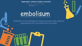
- 1. embolisum (Embolism is the blockage of a blood vessel by solid, liquid or gas(embolus) at a site distant from its origin. Bogomolets national medical university Discipline :- pathophysiology Semester :- 1
- 2. Embolus (Greek word :-"wedge", "plug") is an unattached mass that travels through the blood stream, and is capable of clogging arterial capillary beds (create an arterial occlusion) at a site distant from its point of origin There are a number of different types of emboli :- Fat embolism – embolism of bone fracture or fat droplets. Air embolism (also known as a gas embolism) – embolism of air bubbles. Cholesterol embolism – embolism of cholesterol, often from atherosclerotic plaque inside a vessel. Thromboembolism – embolism of thrombus (blood clot)
- 3. Classification :- Arterial embolism (Arterial embolism can cause occlusion in any part of the body. It is a major cause of infarction (tissue death from blockage of the blood supply). Venous embolisum embolus formed in a systemic vein will always impact in the lungs, This will form a pulmonary embolism that will result in a blockage of the main artery of the lungs and can be a complication of deep-vein thrombosis. Paradoxical (venous to arterial) In paradoxical embolism, also known as crossed embolism, an embolus from the veins crosses to the arterial blood system. This is generally found only with heart problems such as septal defects (holes in the cardiac septum)
- 4. Thrombosis Thrombosis is the formation of a solid mass of blood within the circulatory system Why does thrombosis occur? • (Abnormalities of the vessel wall) • atheroma • direct injury • inflammation
- 5. Arterial embolism Risk factors :- • advanced age • cigarette smoking • hypertension (high blood pressure) • obesity • hyperlipidaemia e.g. hypercholesterolemi a, hypertriglyceridemia, elevated lipoprotein or decreased levels of HDL cholesterol) • diabetes mellitus • Sedentary lifestyle • stress
- 6. Pathophysiological reason of arterial embolisum (Detection of Right Coronary Artery Air Embolism) An arterial embolism is caused by one or more emboli getting stuck in an artery and blocking blood flow, causing ischemia, possibly resulting in infarction with tissue death (necrosis). Individuals with arterial thrombosis or embolism often develop collateral circulation to compensate for the loss of arterial flow. However, it takes time for sufficient collateral circulation to develop, making affected areas more vulnerable for sudden occlusion by embolisation than for e.g. gradual occlusion as in atherosclerosis. Diagnosis :- Doppler ultrasound, duplex ultrasonography Echocardiography
- 7. Pulmonary embolism :- Risk factor :- • Alterations in blood flow • immobilization (after surgery, long-haul flight) • injury, pregnancy (also procoagulant), obesity • Factors in the vessel wall: endothelial injury" • surgery, catheterizations causing direct injury • Venous stasis • Estrogen,OCP,HRT
- 8. Complication :- • Passing out • abnormally low blood pressure • sudden death • Pulmonary infraction • Pulmonary hemorrhage • Increasing of intra arterial pressure • Pulmonary atherosclerosis
- 9. saddle embolism :- • Saddle pulmonary embolism (PE) is a form of large pulmonary thrombo-embolism that straddles the main pulmonary arterial trunk at its bifurcation. Its incidence among patients diagnosed with PE was found to be approximately 2.6%.
- 10. Diagonosis :- (CT pulmonary angiography) CT pulmonary angiography Ventilation/perfusion scan Fluoroscopic pulmonary angiography Echocardiography
- 11. Paradoxical embolism • A paradoxical embolism refers to an embolus which is carried from the venous side of circulation to the arterial side, or vice versa. • exm:- foramen ovale for any pathologically reasons couldn’t form ductus arteriosum, in this we can see this embolism.
- 12. Embolectomy :- (is the emergency surgical removal of emboli which are blocking blood circulation) Balloon embolectomy Aspiration embolectomy Surgical embolectomy
- 14. Thank you for your attention (have a really nice day) Created by ahamed sakil