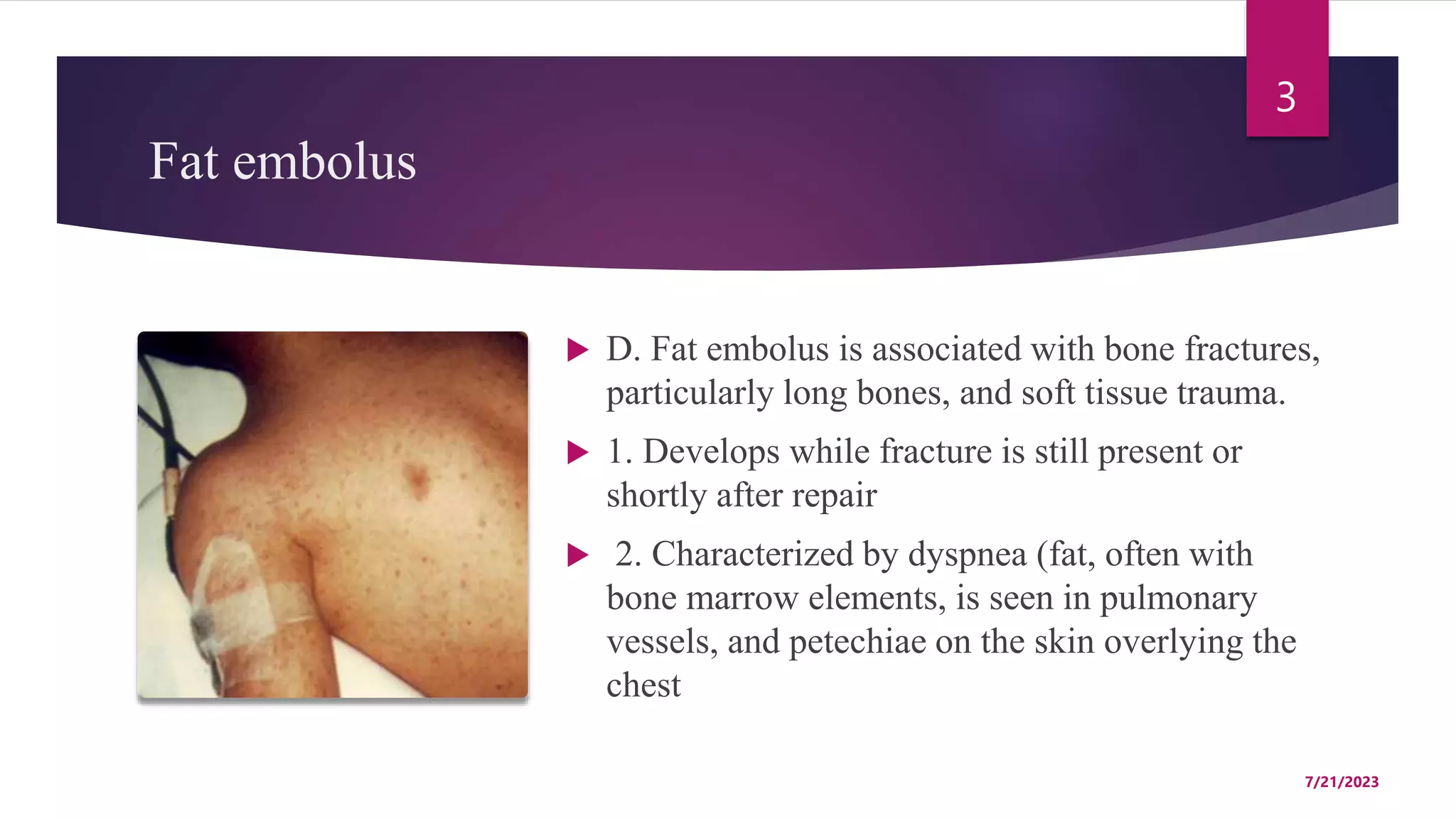
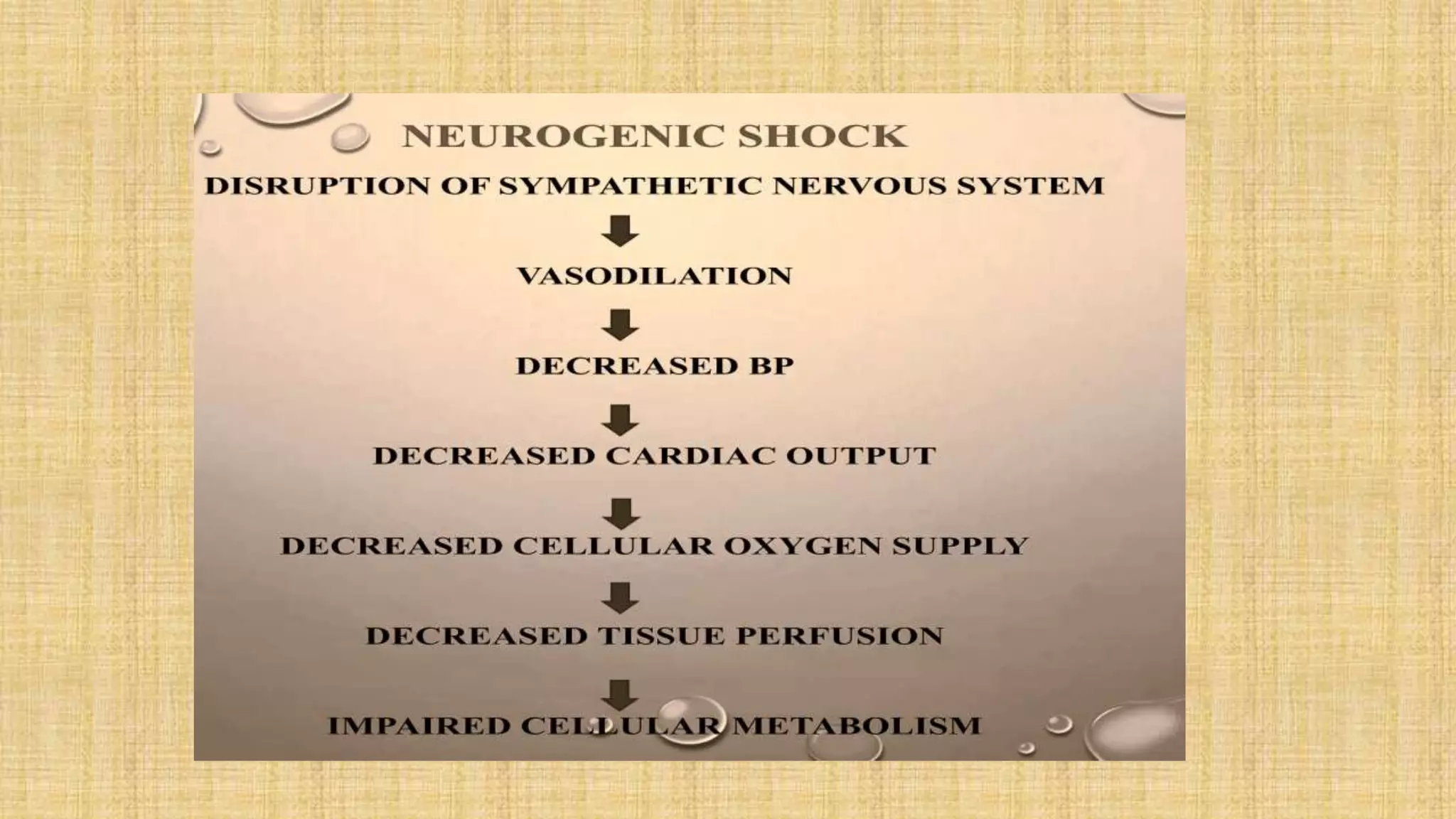
Hemostasis&
Hemodynamic disorders
EMBOLISM, INFARCTION AND SHOCK
Embolism
2
 A. Definition; Intravascular mass that travels
and occludes downstream vessels; symptoms
depend on the vessel involved.
 B. Thromboembolus is due to a thrombus that
dislodges; most common type of embolus
(>95%).
 C. Atherosclerotic embolus is due to an
atherosclerotic plaque that dislodges.
 1. Characterized by the presence of
cholesterol clefts in the embolus.
7/21/2023
Fat embolus
3
 D. Fat embolus is associated with bone fractures,
particularly long bones, and soft tissue trauma.
 1. Develops while fracture is still present or
shortly after repair
 2. Characterized by dyspnea (fat, often with
bone marrow elements, is seen in pulmonary
vessels, and petechiae on the skin overlying the
chest
7/21/2023
Gas embolus
4
 E. Gas embolus is classically seen in
decompression sickness.
 1. Nitrogen gas precipitates out of blood due
to rapid ascent by a diver.
 2. Presents with joint and muscle pain
('bends') and respiratory symptoms
('chokes’).
 3. Chronic form (Caisson disease) is
characterized by multifocal ischemic
necrosis of bone.
 4. Gas embolus may also occur during
laparoscopic surgery (air is pumped into the
abdomen).
7/21/2023
 F. Amniotic fluid embolus enters maternal circulation during labor
or delivery
 1. Presents with shortness of breath, neurologic symptoms, and DIC
(due to the tissue thromboplastin of amniotic fluid).
 2. Characterized by squamous cells and keratin debris, from fetal
skin, in embolus
PULMONARY EMBOLISM
6
 A. Usually due to thromboembolus; the most
common source is deep venous thrombus
(DVT) of the lower extremity, usually
involving the femoral, iliac, or popliteal veins.
 B. Most often clinically silent because (1) the
lung has a dual blood supply (via pulmonary
and bronchial arteries) and (2) the embolus is
usually small (self-resolves)
7/21/2023
 C. Pulmonary infarction occurs if a large- or medium-sized
artery is obstructed in patients with pre-existing
cardiopulmonary compromise; only 10% of PEs cause
infarction.
 1. Presents with shortness of breath, hemoptysis, pleuritic chest
pain, and pleural effusion
 2. V/Q lung scan shows mismatch; perfusion is abnormal.
 3. Spiral CT shows avascular filling defect in the lung.
 4. Lower extremity Doppler ultrasound is useful to detect DVT.
 5. D-dimer is elevated.
 6. Gross examination reveals hemorrhagic, wedge-shaped infarct.
 D. Sudden death occurs with a large saddle embolus that blocks both
left and right pulmonary arteries or with significant occlusion of a
large pulmonary artery
 Sudden death is due to electromechanical dissociation (the heart has
electrical activity but no mechanical contraction).
 Pulmonary hypertension may arise with chronic emboli that are
reorganized over time.
SYSTEMIC EMBOLISM
 A. Usually due to
thromboembolus.
 B. Most commonly arise in the
left heart.
 C. Travel down systemic
circulation to occlude flow to
organs, most commonly the
lower extremities.
7/21/2023 9
INFARCTION
 A. An infarct is an area of ischemic necrosis caused by
occlusion of the vascular supply to the affected tissue;
 1. classical examples are pulmonary infarction, myocardial
infarction, cerebral infarction, bowel infarction, and ischemic
necrosis of distal extremities (gangrene) that causes
substantial morbidity in the diabetic population.
Most common causes
 B. Most common causes are;
 1. Arterial thrombosis or arterial embolism underlies the vast majority of
infarctions.
 Local vasospasms,
 bleeding into an atheroma plaque and
 extrinsic vessel compression such as by a tumor,
 a dissecting aortic aneurysm or
 edema within confined space (anterior compartment syndrome).
 2.Other rare causes of tissue
infarction include vessel
twisting (e.g., in testicular
torsion or bowel volvulus).
 Hint; a volvulus is when a loop
of intestine twists around itself
and the mesentery that supports
it, resulting in
a bowel obstruction.
7/21/2023 12
13
 traumatic vascular rupture, and entrapment
in a hernia sac.
 A hernia is the abnormal exit of tissue or an
organ, such as the bowel, through the wall
of the cavity in which it normally resides
7/21/2023
venous thrombosis
 3. Although venous thrombosis can cause infarction,
 I. The more common outcome is simply congestion;
typically, bypass channels rapidly open to provide sufficient
outflow to restore the arterial inflow.
 II. Infarcts caused by venous thrombosis thus usually occur
only in organs with a single efferent vein (e.g., testis or
ovary).
Capillary obstruction
 Capillary obstruction can cause infarction.
 Vasculitis of the capillaries may narrow the lumen causing
ischemia and infarction of the affected area.
 DIC
 Frost bites
 Fat embolism
Factors That Influence Infarct Development
 A. The effects of vascular occlusion range from inconsequential to
tissue necrosis leading to organ dysfunction and sometimes death.
 B. The range of outcomes is influenced by
 (1) the anatomy of the vascular supply
 (2) the time over which the occlusion develops
 (3) Tissue vulnerability to ischemia
 (4) the blood oxygen content.
 Anatomy of the vascular supply; the presence or
absence of an alternative blood supplies is the most
important factor in determining whether the occlusion
of a vessel causes damage.
 II. Organs that have end-arterial circulation are more
vulnerable to tissue necrosis as the thrombus occludes
the arteries.
 Rate of occlusion; slowly developing occlusions are
less likely to cause infarction because they allow time
for the development of collateral blood supplies.
 The organs that have dual circulations are more persistent to the
infarctions (e.g., lungs, liver, hand and forearm.)
 Tissue vulnerability to ischemia;
 Neurons are highly susceptible to ischemic injury; whereas, skeletal
muscle is relatively more persistent.
 Myocardial cells, although hardier than neurons, still die after only 20 to
30 minutes of ischemia.
 Hypoxemia. Understandably, abnormally low blood O2 content
(regardless of cause) increases both the likelihood and extent of
infarction.
MORPHOLOGY
 Infarcts are classified on the basis of their color (reflecting the amount
of hemorrhage) and the presence or absence of microbial infection.
 Thus, infarcts may be either red (hemorrhagic) or white (anemic) and
may be either septic or bland.
 Red Infarcts occur (1) with venous occlusions (such as in ovarian
torsion); (2) in loose tissues (e.g., lung) where blood can collect in
infarcted zones; (3) in tissues with dual circulations such as lung and
small intestine, where partial, inadequate perfusion by collateral
arterial supplies is typical;
 (4) in previously congested tissues (as a
consequence of sluggish venous outflow); and
(5) when flow is reestablished after infarction
has occurred (e.g., after angioplasty of an
arterial obstruction).
 White infarcts occur with arterial occlusions in
solid organs with end-arterial circulations (e.g.,
heart, spleen, and kidney), and as the occlusion
occurs blood seeps into the affected area.
 Secondary infection leads to an abscess
formation (Abscess - Proteolytic enzymes from
neutrophils liquefy tissue).
21
 Infarcts tend to be wedge-
shaped
 Occluded vessel at the apex
 Periphery of the organ
forming the base.
 If the base is a serosal
surface, there is often
overlying fibrinous
exudate.
7/21/2023
 Acute infarcts are poorly defined and
slightly hemorrhagic
 With the time the margins are better
defined by a narrow rim of congestion
due to inflammation.
 In most tissues, the main histologic
finding associated with infarcts is
ischemic coagulative necrosis.
 In the brain;
 Infarcted are undergoes rapid
liquefactive necrosis.
Septic Infarcts
 Septic infarcts occur when infected cardiac valve
vegetations embolize, or when microbes seed
necrotic tissue.
 In these cases the infarct is converted into an
abscess, with a correspondingly greater
inflammatory response and healing by organization
and fibrosis.
Shock
 shock occurs when the body’s organs does not
get adequate oxygen and blood supply leading
to hypoxia.
 Life threatening condition
 Reversible if treated early
 Can quickly become irreversible and cause
multi-organ failure and death.
Clinical presentations
 Hypotension; low Bp (<90/60 mm Hg)
 Tachycardia; rapid HR( > 100 beats/min)
 Tachypnea; rapid breathing rate ( >25 breaths/min)
 Oliguria; low urine output ( < 400 ml/24 hrs)
 Changes in mental state; agitation, confusion, unresponsiveness,
etc.
 Skin changes; cool, clammy, pale with reduced peripheral
perfusion.
 Mean arterial pressure (MAP) is a good indicator of tissue
perfusion rather than systolic blood pressure.
 MAP is the average arterial pressure throughout one cardiac
cycle.
 MAP is influenced by cardiac output and systemic vascular
resistance
 Hence MAP; cardiac output X systemic vascular resistance
 Actual MAP; 2x diastolic + systolic/3.
 Normal MAP; 65 mm Hg----100 mm Hg.
 Stages of shock:
 1. pre-shock or initial nonprogressive stage
 A. early compensated shock.
 B. symptoms are absent or mild.
 2. Shock or progressive stage
 A. compensatory mechanisms are overwhelmed.
 Symptoms of organ dysfunction begin to appear
 worsening circulatory and metabolic derangement, including
acidosis.
 3. End-organ dysfunction
 A. An irreversible stage in which cellular and tissue injury is so
severe
 B. ultimately can lead to death.
Management
 Depends on the severity and cause of shock.
 Goal of treatment is to decrease the mortality and treat the
underlying causes.
 Fluid replacement and blood transfusions if it is needed.
 Medications to increase blood pressure, such as, metaraminol.
 Emergency surgery
 Iv antibiotics for septic shock
 IV steroids and allergy medications for anaphylactic shock
Types of shock
I. Hypovolemic shock
II. Cardiogenic shock
III. Obstructive shock
IV. Distributive shock
Hypovolemic shock
 Results from low cardiac output due to loss of blood or plasma
volume.
 It can be hemorrhagic or non-hemorrhagic.
 Hemorrhagic is decrease of blood volume due to blood loss of
blood.
 Examples are trauma, interstitial bleeding, intra-operative and post-
operative bleeding.
 non-hemorrhagic is decrease of blood volume due to loss of fluids.
 Burns, sodium and water loss from GIT, skin and kidney are
classical examples.
Cardiogenic shock
 Results from low cardiac output as a result of myocardial pump failure.
 Causes of cardiogenic shock are
 1. cardiomyopathies; problems with heart muscle,
 A. myocardial infarction and ischemia.
 B. myocarditis
 2. Arrythmia; problems with heart rate or rhythm,
 A. atrial and ventricular tachyarrhythmia.
 B. atrial or ventricular bradyarrhythmia.
mechanical causes are;
34
1. valve insufficient or
defects like mitral valve
stenosis.
 Aortic dissection of
aneurysm
 Ventricular septal
defect
7/21/2023
Aortic dissection
35
 Aortic dissection is an acute process in
which a tear in the internal face of the aorta
leads to dissection through the laminas and
formation of a new lumen (false lumen) and
acute hypotension.
 Most common cause is uncontrolled
hypertension.
 the most common complication of aortic
dissection is death due to severe internal
bleeding.
7/21/2023
Obstructive Shock
 Results from obstruction to cardiac outflow or filling
 This can result from pulmonary embolism, severe pulmonary
hypertension, and
 Cardiac tamponade which is when the fluid sac around your heart
fills with too much blood or other fluid and puts pressure on your
heart.
 Tension pneumothorax; a severe condition that results when air is
trapped in the pleural space under positive pressure, displacing
mediastinal structures and compromising cardiopulmonary function.
Distributive Shock
 Distributive shock, also known as vasodilatory shock.
 Systemic vasodilation leads to decreased blood flow to the brain,
heart, and kidneys causing damage to vital organs.
 They are three main types of distributive shock;
 1. septic shock.
 2. anaphylactic shock.
 Neurogenic shock.
Septic Shock
39
 Septic shock is triggered by microbial
infections and is associated with severe
systemic inflammatory response syndrome
(SIRS).
 The common pathogenic mechanism is a
massive production of inflammatory
mediators from innate and adaptive immune
cells that produce arterial vasodilation,
vascular leakage, and venous blood pooling.
7/21/2023
 These cardiovascular abnormalities result in tissue
hypoperfusion, cellular hypoxia, and metabolic
derangements that lead to organ dysfunction.
 if severe and persistent, organ failure and death
 Septic shock is most frequently triggered by gram -
positive bacterial infections, followed by gram-negative
bacteria and fungi.
 More common in ICU and immunosuppressed patients.
Pathogenesis of septic shock
Anaphylactic shock
 Results from severe allergic reactions.
 Allergen enters the blood stream and results in an
exaggerated inflammatory response.
 This includes massive release of histamine that
triggers systemic vasodilation, bronchiolar
constriction and tongue swelling.
Neurogenic shock
 Results from severe brain or spinal cord injury that
compromise the sympathetic nervous system.
 Occurs with spinal cord injury above the level of T6.
 Results in unopposed parasympathetic response and lead to
decrease in vascular resistance and vasodilation.
 Presentations
 Hypotension
 Bradyarrhythmia
 Temperature dysregulation
Embolism, Infarction and Shock ppt-1 slides.pptx

Embolism, Infarction and Shock ppt-1 slides.pptx

  • 1.
  • 2.
    Embolism 2  A. Definition;Intravascular mass that travels and occludes downstream vessels; symptoms depend on the vessel involved.  B. Thromboembolus is due to a thrombus that dislodges; most common type of embolus (>95%).  C. Atherosclerotic embolus is due to an atherosclerotic plaque that dislodges.  1. Characterized by the presence of cholesterol clefts in the embolus. 7/21/2023
  • 3.
    Fat embolus 3  D.Fat embolus is associated with bone fractures, particularly long bones, and soft tissue trauma.  1. Develops while fracture is still present or shortly after repair  2. Characterized by dyspnea (fat, often with bone marrow elements, is seen in pulmonary vessels, and petechiae on the skin overlying the chest 7/21/2023
  • 4.
    Gas embolus 4  E.Gas embolus is classically seen in decompression sickness.  1. Nitrogen gas precipitates out of blood due to rapid ascent by a diver.  2. Presents with joint and muscle pain ('bends') and respiratory symptoms ('chokes’).  3. Chronic form (Caisson disease) is characterized by multifocal ischemic necrosis of bone.  4. Gas embolus may also occur during laparoscopic surgery (air is pumped into the abdomen). 7/21/2023
  • 5.
     F. Amnioticfluid embolus enters maternal circulation during labor or delivery  1. Presents with shortness of breath, neurologic symptoms, and DIC (due to the tissue thromboplastin of amniotic fluid).  2. Characterized by squamous cells and keratin debris, from fetal skin, in embolus
  • 6.
    PULMONARY EMBOLISM 6  A.Usually due to thromboembolus; the most common source is deep venous thrombus (DVT) of the lower extremity, usually involving the femoral, iliac, or popliteal veins.  B. Most often clinically silent because (1) the lung has a dual blood supply (via pulmonary and bronchial arteries) and (2) the embolus is usually small (self-resolves) 7/21/2023
  • 7.
     C. Pulmonaryinfarction occurs if a large- or medium-sized artery is obstructed in patients with pre-existing cardiopulmonary compromise; only 10% of PEs cause infarction.  1. Presents with shortness of breath, hemoptysis, pleuritic chest pain, and pleural effusion  2. V/Q lung scan shows mismatch; perfusion is abnormal.  3. Spiral CT shows avascular filling defect in the lung.
  • 8.
     4. Lowerextremity Doppler ultrasound is useful to detect DVT.  5. D-dimer is elevated.  6. Gross examination reveals hemorrhagic, wedge-shaped infarct.  D. Sudden death occurs with a large saddle embolus that blocks both left and right pulmonary arteries or with significant occlusion of a large pulmonary artery  Sudden death is due to electromechanical dissociation (the heart has electrical activity but no mechanical contraction).  Pulmonary hypertension may arise with chronic emboli that are reorganized over time.
  • 9.
    SYSTEMIC EMBOLISM  A.Usually due to thromboembolus.  B. Most commonly arise in the left heart.  C. Travel down systemic circulation to occlude flow to organs, most commonly the lower extremities. 7/21/2023 9
  • 10.
    INFARCTION  A. Aninfarct is an area of ischemic necrosis caused by occlusion of the vascular supply to the affected tissue;  1. classical examples are pulmonary infarction, myocardial infarction, cerebral infarction, bowel infarction, and ischemic necrosis of distal extremities (gangrene) that causes substantial morbidity in the diabetic population.
  • 11.
    Most common causes B. Most common causes are;  1. Arterial thrombosis or arterial embolism underlies the vast majority of infarctions.  Local vasospasms,  bleeding into an atheroma plaque and  extrinsic vessel compression such as by a tumor,  a dissecting aortic aneurysm or  edema within confined space (anterior compartment syndrome).
  • 12.
     2.Other rarecauses of tissue infarction include vessel twisting (e.g., in testicular torsion or bowel volvulus).  Hint; a volvulus is when a loop of intestine twists around itself and the mesentery that supports it, resulting in a bowel obstruction. 7/21/2023 12
  • 13.
    13  traumatic vascularrupture, and entrapment in a hernia sac.  A hernia is the abnormal exit of tissue or an organ, such as the bowel, through the wall of the cavity in which it normally resides 7/21/2023
  • 14.
    venous thrombosis  3.Although venous thrombosis can cause infarction,  I. The more common outcome is simply congestion; typically, bypass channels rapidly open to provide sufficient outflow to restore the arterial inflow.  II. Infarcts caused by venous thrombosis thus usually occur only in organs with a single efferent vein (e.g., testis or ovary).
  • 15.
    Capillary obstruction  Capillaryobstruction can cause infarction.  Vasculitis of the capillaries may narrow the lumen causing ischemia and infarction of the affected area.  DIC  Frost bites  Fat embolism
  • 16.
    Factors That InfluenceInfarct Development  A. The effects of vascular occlusion range from inconsequential to tissue necrosis leading to organ dysfunction and sometimes death.  B. The range of outcomes is influenced by  (1) the anatomy of the vascular supply  (2) the time over which the occlusion develops  (3) Tissue vulnerability to ischemia  (4) the blood oxygen content.
  • 17.
     Anatomy ofthe vascular supply; the presence or absence of an alternative blood supplies is the most important factor in determining whether the occlusion of a vessel causes damage.  II. Organs that have end-arterial circulation are more vulnerable to tissue necrosis as the thrombus occludes the arteries.  Rate of occlusion; slowly developing occlusions are less likely to cause infarction because they allow time for the development of collateral blood supplies.
  • 18.
     The organsthat have dual circulations are more persistent to the infarctions (e.g., lungs, liver, hand and forearm.)  Tissue vulnerability to ischemia;  Neurons are highly susceptible to ischemic injury; whereas, skeletal muscle is relatively more persistent.  Myocardial cells, although hardier than neurons, still die after only 20 to 30 minutes of ischemia.  Hypoxemia. Understandably, abnormally low blood O2 content (regardless of cause) increases both the likelihood and extent of infarction.
  • 19.
    MORPHOLOGY  Infarcts areclassified on the basis of their color (reflecting the amount of hemorrhage) and the presence or absence of microbial infection.  Thus, infarcts may be either red (hemorrhagic) or white (anemic) and may be either septic or bland.  Red Infarcts occur (1) with venous occlusions (such as in ovarian torsion); (2) in loose tissues (e.g., lung) where blood can collect in infarcted zones; (3) in tissues with dual circulations such as lung and small intestine, where partial, inadequate perfusion by collateral arterial supplies is typical;
  • 20.
     (4) inpreviously congested tissues (as a consequence of sluggish venous outflow); and (5) when flow is reestablished after infarction has occurred (e.g., after angioplasty of an arterial obstruction).  White infarcts occur with arterial occlusions in solid organs with end-arterial circulations (e.g., heart, spleen, and kidney), and as the occlusion occurs blood seeps into the affected area.  Secondary infection leads to an abscess formation (Abscess - Proteolytic enzymes from neutrophils liquefy tissue).
  • 21.
    21  Infarcts tendto be wedge- shaped  Occluded vessel at the apex  Periphery of the organ forming the base.  If the base is a serosal surface, there is often overlying fibrinous exudate. 7/21/2023
  • 22.
     Acute infarctsare poorly defined and slightly hemorrhagic  With the time the margins are better defined by a narrow rim of congestion due to inflammation.  In most tissues, the main histologic finding associated with infarcts is ischemic coagulative necrosis.  In the brain;  Infarcted are undergoes rapid liquefactive necrosis.
  • 23.
    Septic Infarcts  Septicinfarcts occur when infected cardiac valve vegetations embolize, or when microbes seed necrotic tissue.  In these cases the infarct is converted into an abscess, with a correspondingly greater inflammatory response and healing by organization and fibrosis.
  • 24.
    Shock  shock occurswhen the body’s organs does not get adequate oxygen and blood supply leading to hypoxia.  Life threatening condition  Reversible if treated early  Can quickly become irreversible and cause multi-organ failure and death.
  • 25.
    Clinical presentations  Hypotension;low Bp (<90/60 mm Hg)  Tachycardia; rapid HR( > 100 beats/min)  Tachypnea; rapid breathing rate ( >25 breaths/min)  Oliguria; low urine output ( < 400 ml/24 hrs)  Changes in mental state; agitation, confusion, unresponsiveness, etc.  Skin changes; cool, clammy, pale with reduced peripheral perfusion.
  • 26.
     Mean arterialpressure (MAP) is a good indicator of tissue perfusion rather than systolic blood pressure.  MAP is the average arterial pressure throughout one cardiac cycle.  MAP is influenced by cardiac output and systemic vascular resistance  Hence MAP; cardiac output X systemic vascular resistance
  • 27.
     Actual MAP;2x diastolic + systolic/3.  Normal MAP; 65 mm Hg----100 mm Hg.  Stages of shock:  1. pre-shock or initial nonprogressive stage  A. early compensated shock.  B. symptoms are absent or mild.
  • 28.
     2. Shockor progressive stage  A. compensatory mechanisms are overwhelmed.  Symptoms of organ dysfunction begin to appear  worsening circulatory and metabolic derangement, including acidosis.  3. End-organ dysfunction  A. An irreversible stage in which cellular and tissue injury is so severe  B. ultimately can lead to death.
  • 29.
    Management  Depends onthe severity and cause of shock.  Goal of treatment is to decrease the mortality and treat the underlying causes.  Fluid replacement and blood transfusions if it is needed.  Medications to increase blood pressure, such as, metaraminol.  Emergency surgery  Iv antibiotics for septic shock  IV steroids and allergy medications for anaphylactic shock
  • 30.
    Types of shock I.Hypovolemic shock II. Cardiogenic shock III. Obstructive shock IV. Distributive shock
  • 31.
    Hypovolemic shock  Resultsfrom low cardiac output due to loss of blood or plasma volume.  It can be hemorrhagic or non-hemorrhagic.  Hemorrhagic is decrease of blood volume due to blood loss of blood.  Examples are trauma, interstitial bleeding, intra-operative and post- operative bleeding.  non-hemorrhagic is decrease of blood volume due to loss of fluids.  Burns, sodium and water loss from GIT, skin and kidney are classical examples.
  • 33.
    Cardiogenic shock  Resultsfrom low cardiac output as a result of myocardial pump failure.  Causes of cardiogenic shock are  1. cardiomyopathies; problems with heart muscle,  A. myocardial infarction and ischemia.  B. myocarditis  2. Arrythmia; problems with heart rate or rhythm,  A. atrial and ventricular tachyarrhythmia.  B. atrial or ventricular bradyarrhythmia.
  • 34.
    mechanical causes are; 34 1.valve insufficient or defects like mitral valve stenosis.  Aortic dissection of aneurysm  Ventricular septal defect 7/21/2023
  • 35.
    Aortic dissection 35  Aorticdissection is an acute process in which a tear in the internal face of the aorta leads to dissection through the laminas and formation of a new lumen (false lumen) and acute hypotension.  Most common cause is uncontrolled hypertension.  the most common complication of aortic dissection is death due to severe internal bleeding. 7/21/2023
  • 36.
    Obstructive Shock  Resultsfrom obstruction to cardiac outflow or filling  This can result from pulmonary embolism, severe pulmonary hypertension, and  Cardiac tamponade which is when the fluid sac around your heart fills with too much blood or other fluid and puts pressure on your heart.  Tension pneumothorax; a severe condition that results when air is trapped in the pleural space under positive pressure, displacing mediastinal structures and compromising cardiopulmonary function.
  • 38.
    Distributive Shock  Distributiveshock, also known as vasodilatory shock.  Systemic vasodilation leads to decreased blood flow to the brain, heart, and kidneys causing damage to vital organs.  They are three main types of distributive shock;  1. septic shock.  2. anaphylactic shock.  Neurogenic shock.
  • 39.
    Septic Shock 39  Septicshock is triggered by microbial infections and is associated with severe systemic inflammatory response syndrome (SIRS).  The common pathogenic mechanism is a massive production of inflammatory mediators from innate and adaptive immune cells that produce arterial vasodilation, vascular leakage, and venous blood pooling. 7/21/2023
  • 40.
     These cardiovascularabnormalities result in tissue hypoperfusion, cellular hypoxia, and metabolic derangements that lead to organ dysfunction.  if severe and persistent, organ failure and death  Septic shock is most frequently triggered by gram - positive bacterial infections, followed by gram-negative bacteria and fungi.  More common in ICU and immunosuppressed patients.
  • 41.
  • 42.
    Anaphylactic shock  Resultsfrom severe allergic reactions.  Allergen enters the blood stream and results in an exaggerated inflammatory response.  This includes massive release of histamine that triggers systemic vasodilation, bronchiolar constriction and tongue swelling.
  • 44.
    Neurogenic shock  Resultsfrom severe brain or spinal cord injury that compromise the sympathetic nervous system.  Occurs with spinal cord injury above the level of T6.  Results in unopposed parasympathetic response and lead to decrease in vascular resistance and vasodilation.  Presentations  Hypotension  Bradyarrhythmia  Temperature dysregulation