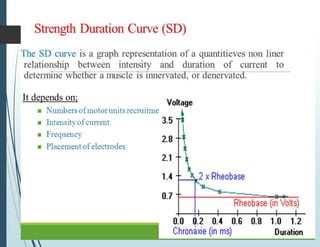
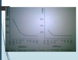
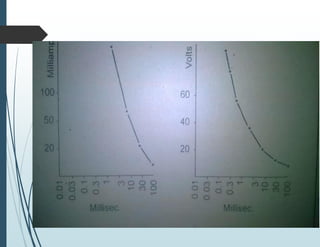
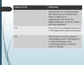
- The strength-duration curve is a graph that plots the electrical stimulus intensity against the time needed to elicit a muscle contraction. It can determine if a muscle is innervated, denervated, or partially denervated.
- The curve is generated by applying electrical stimuli of varying durations (0.01-300ms) to a muscle and recording the intensity needed to produce a minimal contraction. The shape of the curve indicates the muscle's innervation status.
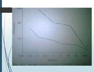
- A normal curve will have all longer duration stimuli producing a response at the same intensity, while shorter durations require more intensity. Complete denervation results in a steeply rising curve requiring more intensity for all shorter durations. Partial denervation produces