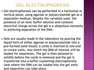
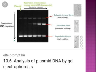
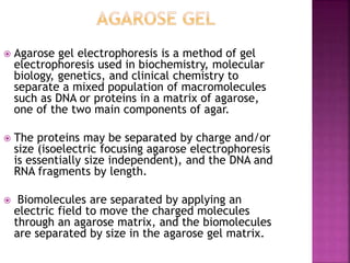
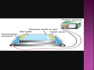
-BY DEEPIKA HAMAV
 Electrophoresis is the motion of dispersed particles
relative to a fluid under the influence of a spatially
uniform electric field.
 This electrokinetic phenomenon was first observed
in 1807 by Russian professors Peter Ivanovich
Strakhov and Ferdinand Frederic Reuss (Moscow
State University), who noticed that the application
of a constant electric field caused clay particles
dispersed in water to migrate.
 Electrophoresis of positively charged particles
(cations) is called cataphoresis while
electrophoresis of negatively charged particles
(anions) is called anaphoresis.
 It is a technique used in laboratories in order
to separate macromolecules based on size.
 This separation is facilitated by the negative
charge present on the DNA/RNA fragments
due to the release of positive hydrogen ions
from the phosphate groups that constitute
the ‘backbone’ of the molecule in the
presence of ionic buffer solutions such as Tris
Borate Ethylenediamine tetra-acetic acid
(EDTA) (TBE) or Tris Acetate EDTA (TAE).
 This is used for both DNA & RNA analysis.
 Electrophoresis is performed in buffer
solutions to reduce pH changes due to the
electric field, which is important because
the charge of DNA and RNA depends on pH,
but running for too long can exhaust the
buffering capacity of the solution. Further,
different preparations of genetic material
may not migrate consistently with each
other, for morphological or other reasons.
 Affinity electrophoresis
 Capillary electrophoresis
 Dielectrophoresis
 Electroblotting
 Gel electrophoresis
 Gel electrophoresis of nucleic acids
 Immunoelectrophoresis
 Iso electric focusing
 Isotachophoresis
 Pulsed-field gel electrophoresis
 Capillary electrophoresis (CE) is more accurately
described as a variation on the more established gel
electrophoresis methods rather than a new technique in
its own right. The main difference in the two
electrophoretic techniques is the use of a capillary
containing a polymer solution such as
hydroxyethylcellulose in place of the traditional
physical gel.
 Capillaries are made of fused silica and have an internal
diameter of only 50-100μm and can be 25-100cm in
length. Similarly to gel electrophoresis, size separation
is achieved via use of buffer solutions and application of
positive and negative charges at either end of the
capillary.
 Another major difference is in the method of
loading the DNA sample of interest in to the
capillary. With gel systems the samples must be
carefully loaded by an operator into the wells
created during casting of the gel. For CE, the
DNA sample is loaded into the separation
medium by electrokinetic injection whereby a
positive charge is applied to draw the negatively
charged DNA into the capillary. This method of
loading requires less operator time and is very
amenable to automated and high-throughput
processing.
 Capillary electrophoresis may be used for the simultaneous determination of the ions NH4+,, Na+, K+, Mg2+ and
Ca2+ in saliva.
 Application of capillary electrophoresis in forensic science
 One of the main application of CE in forensic science is the development of methods for amplification and
detection of DNA fragments using polymerase chain reaction (PCR) which has to lead to rapid and dramatic
advances in DNA typing in forensic. DNA separations are carried out using thin CE, 50-mm, fused silica capillaries
filled with a sieving buffer. These capillaries have excellent capabilities to dissipate heat, permitting much
higher electric field strengths to be used than slab gel electrophoresis. Therefore separations in capillaries are
rapid and efficient. Additionally, the capillaries can be easily refilled and changed for efficient and automated
injections. Detection occurs via fluorescence through a window etched in the capillary. Both single-capillary and
capillary-array instruments are available with array systems capable of running 16 or more samples
simultaneously for increased throughput.
 The major use of CE by a forensic biologist is typing of STR from biological samples to generate a profile from
highly polymorphic genetic markers which differ between individuals. Other emerging uses for CE include the
detection of specific mRNA fragments to help identify the biological fluid or tissue origin of a forensic sample.
 Another application of CE in forensic is ink analysis where the discrimination of inkjet printing inks is becoming
more necessary due to increasingly frequent counterfeiting of documents printed by inkjet printers. The
chemical composition of inks provides very important information in cases of fraudulent documents and
counterfeit banknotes. Micellar electrophoretic capillary chromatography (MECC) has been developed and
applied to the analysis of inks which extracted from paper, Due to its high resolving power relative to inks
containing several chemically very similar substances, differences between inks from the same manufacturer can
also be distinguished. This makes it suitable for evaluating the origin of documents based on the chemical
composition of inks. It is worth noting that because of the possible compatibility of the same cartridge with
different printer models, the differentiation of inks on the basis of their MECC electrophoretic profiles is a more
reliable method for the determination of the ink cartridge of origin (its producer and cartridge number) rather
than the printer model of origin.
 Gel electrophoresis can be performed in a horizontal or
vertical plane, using agarose or polyacrylamide gel as a
separation medium. Despite the variation used, the
presence of an ionic buffer solution and constant
electrical charge across the gel is a ubiquitous necessity
in achieving separation of the DNA.
 Gels are usually made in the laboratory by pouring the
liquid form of either agarose or polyacrylamide into a
pre-formed solid mould; a comb is inserted at one end
to create wells, into which the DNA of interest will be
loaded for separation. The gel is then allowed to
solidify before the comb is removed and the gel is
transferred into a buffer-containing electrophoresis
tank where the DNA can be loaded into the gel wells
and separation can take place.
 Agarose gel electrophoresis is a method of gel
electrophoresis used in biochemistry, molecular
biology, genetics, and clinical chemistry to
separate a mixed population of macromolecules
such as DNA or proteins in a matrix of agarose,
one of the two main components of agar.
 The proteins may be separated by charge and/or
size (isoelectric focusing agarose electrophoresis
is essentially size independent), and the DNA and
RNA fragments by length.
 Biomolecules are separated by applying an
electric field to move the charged molecules
through an agarose matrix, and the biomolecules
are separated by size in the agarose gel matrix.
 Agarose gel is easy to cast, has relatively fewer charged
groups, and is particularly suitable for separating DNA of
size range most often encountered in laboratories,
which accounts for the popularity of its use. The
separated DNA may be viewed with stain, most
commonly under UV light, and the DNA fragments can
be extracted from the gel with relative ease. Most
agarose gels used are between 0.7 - 2% dissolved in a
suitable electrophoresis buffer.
 The choice of gel used is largely dependent upon the
size and spacing of the DNA fragments under analysis.
Agarose is a polysaccharide which, together with
agaropectin, forms the seaweed-derived, gelatinous
substance agar. When set, the polysaccharide strands
form a matrix structure through which DNA molecules
can travel when a charge is present at either end of the
gel.
 The pore sizes within this matrix are considered
relatively large at approximately 100-300nm
depending upon the concentration of agarose,
and as such it does not allow for accurate
resolution of closely sized DNA fragments.
Agarose gels are generally used when larger
fragments, in the region of 500-20,000bp, are
required to be visualized.
 They can also be used to assess the quality of
extracted DNA, with degraded template
producing a smear when run on an agarose gel as
opposed to a tight band of high molecular weight
for high quality samples. Agarose gel
electrophoresis is used for fragment separation
during the DNA fingerprinting method described
above.
 Estimation of the size of DNA molecules following
restriction enzyme digestion, e.g. in restriction mapping
of cloned DNA.
 Analysis of PCR products, e.g. in molecular genetic
diagnosis or genetic fingerprinting
 Separation of DNA fragments for extraction and
purification.
 Separation of restricted genomic DNA prior to Southern
transfer, or of RNA prior to Northern transfer.
 Agarose gels are easily cast and handled compared to
other matrices and nucleic acids are not chemically
altered during electrophoresis. Samples are also easily
recovered. After the experiment is finished, the
resulting gel can be stored in a plastic bag in a
refrigerator.
 Polyacrylamide gels are made by inducing
polymerization of acrylamide and bisacrylamide
monomers in a process initialized by the presence
of Ammonium Persulphate and TEMED (N, N, N', N'-
tetramethylethylenediamine).
 The use of an artificial gel matrix in place of the
naturally extracted agarose produces smaller pore
sizes in the gel matrix at approximately 10-20nm in
a typical gel. This pore size reduction, along with
optimized running conditions, can allow for highly
accurate resolution of similarly sized DNA fragments
and under denaturing conditions achieves resolution
of single base-pair size differences.
 This level of accuracy led to polyacrylamide gels being
employed for separation of amplified STR markers
during DNA profiling development but has now been
largely replaced by more sensitive capillary
electrophoresis technologies.
 Polyacrylamide gel electrophoresis (PAGE) is conducted
in a manner very similar to agarose gel analysis. The un-
polymerized solution is poured between two closely
spaced glass plates, a gel comb is inserted to create the
wells into which the DNA will be loaded and the solution
is allowed to polymerize or set over 1-2 hours.
 Once set, the gel is moved to the running apparatus
where a buffer is placed at the top and bottom of the
gel, the DNA of interest is loaded into the wells created
during polymerization, and separation occurs in the
same way as for agarose gels when a fixed current is
applied across the gel apparatus.
 It has a clearer resolution than agarose and is
more suitable for quantitative analysis.
 In this technique, DNA foot-printing can
identify how proteins bind to DNA.
 It can be used to separate proteins by size,
density and purity.
 It can also be used for plasmid analysis,
which develops our understanding of bacteria
becoming resistant to antibiotics.
Electrophoresis

Electrophoresis

  • 1.
  • 2.
     Electrophoresis isthe motion of dispersed particles relative to a fluid under the influence of a spatially uniform electric field.  This electrokinetic phenomenon was first observed in 1807 by Russian professors Peter Ivanovich Strakhov and Ferdinand Frederic Reuss (Moscow State University), who noticed that the application of a constant electric field caused clay particles dispersed in water to migrate.  Electrophoresis of positively charged particles (cations) is called cataphoresis while electrophoresis of negatively charged particles (anions) is called anaphoresis.
  • 3.
     It isa technique used in laboratories in order to separate macromolecules based on size.  This separation is facilitated by the negative charge present on the DNA/RNA fragments due to the release of positive hydrogen ions from the phosphate groups that constitute the ‘backbone’ of the molecule in the presence of ionic buffer solutions such as Tris Borate Ethylenediamine tetra-acetic acid (EDTA) (TBE) or Tris Acetate EDTA (TAE).  This is used for both DNA & RNA analysis.
  • 4.
     Electrophoresis isperformed in buffer solutions to reduce pH changes due to the electric field, which is important because the charge of DNA and RNA depends on pH, but running for too long can exhaust the buffering capacity of the solution. Further, different preparations of genetic material may not migrate consistently with each other, for morphological or other reasons.
  • 5.
     Affinity electrophoresis Capillary electrophoresis  Dielectrophoresis  Electroblotting  Gel electrophoresis  Gel electrophoresis of nucleic acids  Immunoelectrophoresis  Iso electric focusing  Isotachophoresis  Pulsed-field gel electrophoresis
  • 6.
     Capillary electrophoresis(CE) is more accurately described as a variation on the more established gel electrophoresis methods rather than a new technique in its own right. The main difference in the two electrophoretic techniques is the use of a capillary containing a polymer solution such as hydroxyethylcellulose in place of the traditional physical gel.  Capillaries are made of fused silica and have an internal diameter of only 50-100μm and can be 25-100cm in length. Similarly to gel electrophoresis, size separation is achieved via use of buffer solutions and application of positive and negative charges at either end of the capillary.
  • 7.
     Another majordifference is in the method of loading the DNA sample of interest in to the capillary. With gel systems the samples must be carefully loaded by an operator into the wells created during casting of the gel. For CE, the DNA sample is loaded into the separation medium by electrokinetic injection whereby a positive charge is applied to draw the negatively charged DNA into the capillary. This method of loading requires less operator time and is very amenable to automated and high-throughput processing.
  • 9.
     Capillary electrophoresismay be used for the simultaneous determination of the ions NH4+,, Na+, K+, Mg2+ and Ca2+ in saliva.  Application of capillary electrophoresis in forensic science  One of the main application of CE in forensic science is the development of methods for amplification and detection of DNA fragments using polymerase chain reaction (PCR) which has to lead to rapid and dramatic advances in DNA typing in forensic. DNA separations are carried out using thin CE, 50-mm, fused silica capillaries filled with a sieving buffer. These capillaries have excellent capabilities to dissipate heat, permitting much higher electric field strengths to be used than slab gel electrophoresis. Therefore separations in capillaries are rapid and efficient. Additionally, the capillaries can be easily refilled and changed for efficient and automated injections. Detection occurs via fluorescence through a window etched in the capillary. Both single-capillary and capillary-array instruments are available with array systems capable of running 16 or more samples simultaneously for increased throughput.  The major use of CE by a forensic biologist is typing of STR from biological samples to generate a profile from highly polymorphic genetic markers which differ between individuals. Other emerging uses for CE include the detection of specific mRNA fragments to help identify the biological fluid or tissue origin of a forensic sample.  Another application of CE in forensic is ink analysis where the discrimination of inkjet printing inks is becoming more necessary due to increasingly frequent counterfeiting of documents printed by inkjet printers. The chemical composition of inks provides very important information in cases of fraudulent documents and counterfeit banknotes. Micellar electrophoretic capillary chromatography (MECC) has been developed and applied to the analysis of inks which extracted from paper, Due to its high resolving power relative to inks containing several chemically very similar substances, differences between inks from the same manufacturer can also be distinguished. This makes it suitable for evaluating the origin of documents based on the chemical composition of inks. It is worth noting that because of the possible compatibility of the same cartridge with different printer models, the differentiation of inks on the basis of their MECC electrophoretic profiles is a more reliable method for the determination of the ink cartridge of origin (its producer and cartridge number) rather than the printer model of origin.
  • 10.
     Gel electrophoresiscan be performed in a horizontal or vertical plane, using agarose or polyacrylamide gel as a separation medium. Despite the variation used, the presence of an ionic buffer solution and constant electrical charge across the gel is a ubiquitous necessity in achieving separation of the DNA.  Gels are usually made in the laboratory by pouring the liquid form of either agarose or polyacrylamide into a pre-formed solid mould; a comb is inserted at one end to create wells, into which the DNA of interest will be loaded for separation. The gel is then allowed to solidify before the comb is removed and the gel is transferred into a buffer-containing electrophoresis tank where the DNA can be loaded into the gel wells and separation can take place.
  • 12.
     Agarose gelelectrophoresis is a method of gel electrophoresis used in biochemistry, molecular biology, genetics, and clinical chemistry to separate a mixed population of macromolecules such as DNA or proteins in a matrix of agarose, one of the two main components of agar.  The proteins may be separated by charge and/or size (isoelectric focusing agarose electrophoresis is essentially size independent), and the DNA and RNA fragments by length.  Biomolecules are separated by applying an electric field to move the charged molecules through an agarose matrix, and the biomolecules are separated by size in the agarose gel matrix.
  • 14.
     Agarose gelis easy to cast, has relatively fewer charged groups, and is particularly suitable for separating DNA of size range most often encountered in laboratories, which accounts for the popularity of its use. The separated DNA may be viewed with stain, most commonly under UV light, and the DNA fragments can be extracted from the gel with relative ease. Most agarose gels used are between 0.7 - 2% dissolved in a suitable electrophoresis buffer.  The choice of gel used is largely dependent upon the size and spacing of the DNA fragments under analysis. Agarose is a polysaccharide which, together with agaropectin, forms the seaweed-derived, gelatinous substance agar. When set, the polysaccharide strands form a matrix structure through which DNA molecules can travel when a charge is present at either end of the gel.
  • 15.
     The poresizes within this matrix are considered relatively large at approximately 100-300nm depending upon the concentration of agarose, and as such it does not allow for accurate resolution of closely sized DNA fragments. Agarose gels are generally used when larger fragments, in the region of 500-20,000bp, are required to be visualized.  They can also be used to assess the quality of extracted DNA, with degraded template producing a smear when run on an agarose gel as opposed to a tight band of high molecular weight for high quality samples. Agarose gel electrophoresis is used for fragment separation during the DNA fingerprinting method described above.
  • 16.
     Estimation ofthe size of DNA molecules following restriction enzyme digestion, e.g. in restriction mapping of cloned DNA.  Analysis of PCR products, e.g. in molecular genetic diagnosis or genetic fingerprinting  Separation of DNA fragments for extraction and purification.  Separation of restricted genomic DNA prior to Southern transfer, or of RNA prior to Northern transfer.  Agarose gels are easily cast and handled compared to other matrices and nucleic acids are not chemically altered during electrophoresis. Samples are also easily recovered. After the experiment is finished, the resulting gel can be stored in a plastic bag in a refrigerator.
  • 17.
     Polyacrylamide gelsare made by inducing polymerization of acrylamide and bisacrylamide monomers in a process initialized by the presence of Ammonium Persulphate and TEMED (N, N, N', N'- tetramethylethylenediamine).  The use of an artificial gel matrix in place of the naturally extracted agarose produces smaller pore sizes in the gel matrix at approximately 10-20nm in a typical gel. This pore size reduction, along with optimized running conditions, can allow for highly accurate resolution of similarly sized DNA fragments and under denaturing conditions achieves resolution of single base-pair size differences.
  • 19.
     This levelof accuracy led to polyacrylamide gels being employed for separation of amplified STR markers during DNA profiling development but has now been largely replaced by more sensitive capillary electrophoresis technologies.  Polyacrylamide gel electrophoresis (PAGE) is conducted in a manner very similar to agarose gel analysis. The un- polymerized solution is poured between two closely spaced glass plates, a gel comb is inserted to create the wells into which the DNA will be loaded and the solution is allowed to polymerize or set over 1-2 hours.  Once set, the gel is moved to the running apparatus where a buffer is placed at the top and bottom of the gel, the DNA of interest is loaded into the wells created during polymerization, and separation occurs in the same way as for agarose gels when a fixed current is applied across the gel apparatus.
  • 20.
     It hasa clearer resolution than agarose and is more suitable for quantitative analysis.  In this technique, DNA foot-printing can identify how proteins bind to DNA.  It can be used to separate proteins by size, density and purity.  It can also be used for plasmid analysis, which develops our understanding of bacteria becoming resistant to antibiotics.