Download as PPSX, PPTX


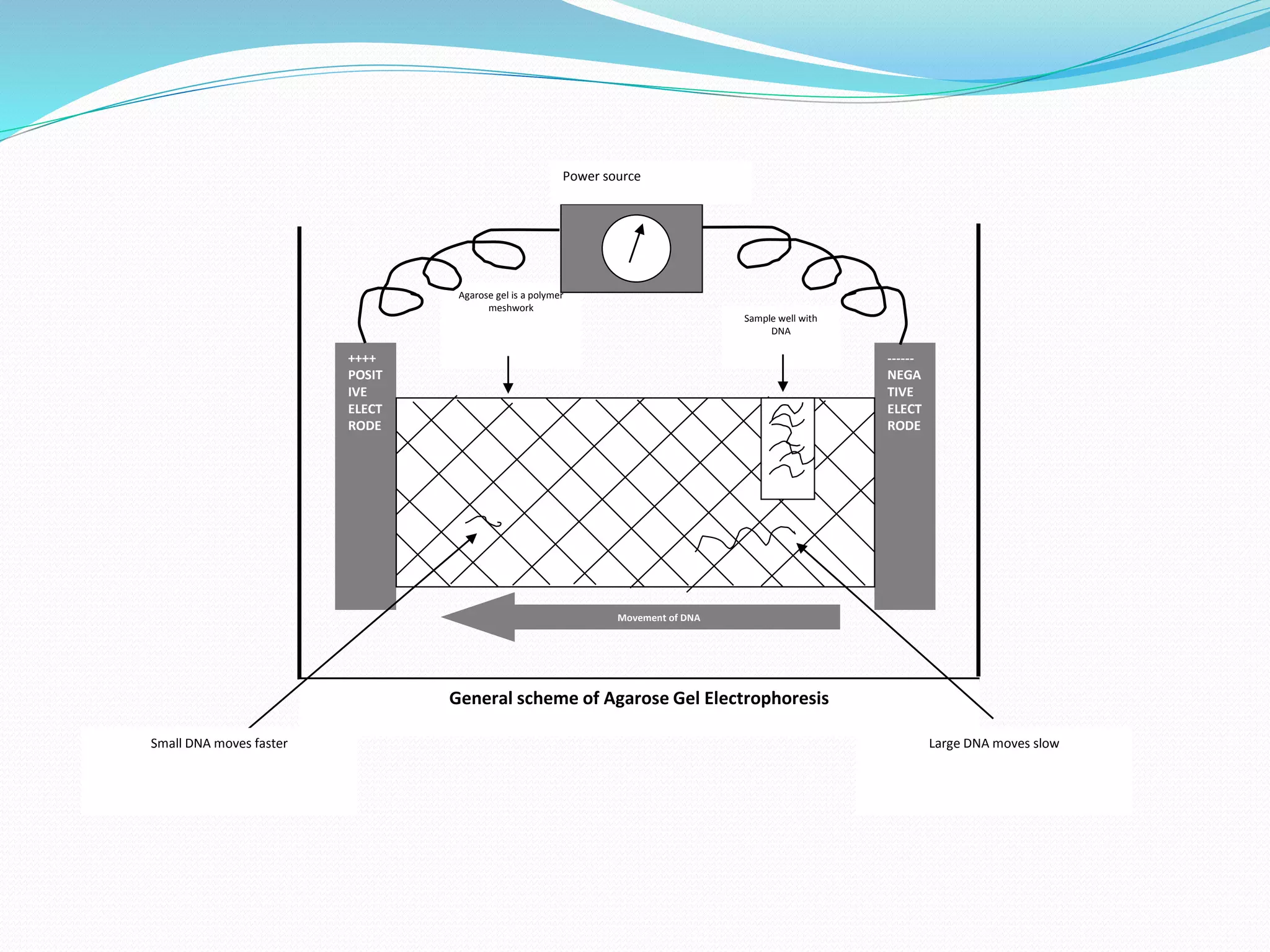


This document discusses agarose gel electrophoresis, which is used to separate DNA fragments by size. It begins by explaining the basic principle, which is that charged DNA fragments will move through an agarose gel under the influence of an electric field, with smaller fragments moving faster. It then describes how agarose gel is prepared by mixing agarose with buffer and casting it. Different types of gels, like agarose and polyacrylamide, can be used depending on the size of DNA fragments. The document outlines the steps for running samples on a gel, detecting DNA bands with ethidium bromide staining, and some applications like capillary and pulsed-field gel electrophoresis.