
Recommended
Recommended
More Related Content
Similar to Effects of Lactococcus lactis (L. lactis) subsp. lactis Supernatant on the Shelf Life of Vacuum-packaged Oncorhynchus mykiss Fillets
Similar to Effects of Lactococcus lactis (L. lactis) subsp. lactis Supernatant on the Shelf Life of Vacuum-packaged Oncorhynchus mykiss Fillets (20)
More from Iranian Food Science and Technology Research Journal
More from Iranian Food Science and Technology Research Journal (20)
Recently uploaded
Recently uploaded (20)
Effects of Lactococcus lactis (L. lactis) subsp. lactis Supernatant on the Shelf Life of Vacuum-packaged Oncorhynchus mykiss Fillets
- 1. Research Article Vol. 19, No. 6, Feb.-Mar., 2024, p. 111-124 Effects of Lactococcus lactis (L. lactis) subsp. lactis Supernatant on the Shelf Life of Vacuum-packaged Oncorhynchus mykiss Fillets A. Abbaspour Anbi 1 *, M. Seidgar 1 , M. Neyriz Naghadehi2 1- National Artemia Research Center, Iranian Fisheries Science Research Institute, Agricultural Research, Education and Extension Organization (AREEO), Urmia, Iran (*- Corresponding author: asadanbi1208@gmail.com) 2- Department of Food Hygiene and Quality Control, Urmia Branch, Islamic Azad University, Urmia, Iran Received: 08.08.2023 Revised: 05.11.2023 Accepted: 08.11.2023 Available Online: 08.11.2023 How to cite this article: Abbaspour Anbi, A., Seidgar, M., & Neyriz Naghadehi, M. (2024). Effects of Lactococcus lactis (L. lactis) subsp. lactis Supernatant on the shelf life of vacuum-packaged Oncorhynchus mykiss fillets. Iranian Food Science and Technology Research Journal, 19(6), 111-124. https://doi.org/10.22067/ifstrj.2023.83834.1274 Abstract The present investigation was done to study the effects of Lactococcus lactis (L. lactis) subsp. lactis on the shelf life of the vacuum-packaged Oncorhynchus mykiss. Fish fillets were prepared and divided into 5 different treatment groups including control (distilled water), 2% and 4% supernatant, and 106 CFU/g L. lactis subspecies lactis. The pH, Thiobarbituric Acid Reactive Substances (TBARS), Total volatile Nitrogen (TVN), and Peroxide Value (PV) of the fillets were determined on days 0, 5, 10, and 15 while maintained at 4˚C. Protein expression and destruction were analyzed using the SDS-PAGE. The organoleptic assessment was done using five expert sensory panelists. Contents of TBARS, TVN, pH, and PV were increased throughout the storage period (P <0.05). An increase in the concentration of supernatant caused a significant decrease in the content of TBARS, TVN, pH, and PV (P <0.05). The highest and lowest contents of TBARS, TVN, pH and PV on 15th day were belonged to the control (3.367±0.04 mg MDA/kg) and pure bacteria (0.70±0.02 mg MDA/kg), control (87.20±6.40 mg/100g) and 4% supernatant (40.79±0.61 mg/100g), pure bacteria (6.23±0.04) and 4% supernatant (5.44±0.07) and control (12.22±0.01 meq/kg) and 4% supernatant (3.08±0.06 meq/kg) groups, respectively. Protein destruction was lower in the fillet samples treated with pure bacteria and 4% supernatant. The highest scores of the odor, flavor, texture, and color were obtained for fillets treated with 4% supernatant, pure bacteria, pure bacteria, and 4% supernatant and pure bacteria, respectively. The results revealed that treating O. mykiss fillets with 4% supernatant and 106 CFU/g of pure L. lactis subsp. lactis can extend the shelf life of O. mykiss fillets. Keywords: Fish, L. lactis subsp. lactis, Oncorhynchus mykiss, Shelf life, Vacuum package 1 1 ©2023 The author(s). This is an open access article distributed under Creative Commons Attribution 4.0 International License (CC BY 4.0), which permits use, sharing, adaptation, distribution and reproduction in any medium or format, as long as you give appropriate credit to the original author(s) and the source. https://doi.org/10.22067/ifstrj.2023.83834.1274 Iranian Food Science and Technology Research Journal Homepage: https://ifstrj.um.ac.ir
- 2. 112 Iranian Food Science and Technology Research Journal Vol. 19, No. 6, 2024 Introduction It is well-known that fish is an important source of high-quality animal proteins for human nutrition. Furthermore, fish is a rich source of protein, fatty acids, vitamins, and minerals (Larsen et al., 2011). O. mykiss or Rainbow trout is one of the most extensively cultured and traded fish in Iran and many other countries of the world. Besides its nutritional value, the large and stable production quantity of rainbow trout makes it an important fish species for the seafood market. It is one of the major sources of protein, minerals, vitamins, and ω-3 long-chain polyunsaturated fatty acids (LC-PUFAs) including eicosapentaenoic acid (EPA, C20:5 n-3) and docosahexaenoic acid (DHA, C22:6 n-3) which have been demonstrated to have valuable and special health effects to inhibit cardiovascular disease, lower cholesterol levels, and blood viscosity, and reinforce memory and thinking ability for humans (Larsen et al., 2011). However, O. mykiss fillets are very prone to oxidation and rancidity. The oxidation reactions can cause alterations in the texture, color, and nutritional value of the final product (Chytiri et al., 2004; Fuentes-Amaya et al., 2015). Degradation of polyunsaturated fatty acids (PUFAs) caused by self-acting or enzymatic oxidation throughout different storage circumstances and processing operations can simply result in the development of undesirable oxidation products such as hydroperoxides, peroxides, aldehydes, conjugated dienes/trienes, ketones, and others (Chytiri et al., 2004; Fuentes-Amaya et al., 2015). Additionally, fish are very susceptible to microbial spoilage. The high water activity (aW) and the presence of corrosive tissues and proteins are the main factors that make it susceptible to chemical and microbial spoilage (Chytiri et al., 2004; Fuentes-Amaya et al., 2015). Therefore, microbiological, enzymatic, and chemical decomposition analyses are used to determine the shelf life of fish and marine products during processing and storage (Chytiri et al., 2004; Fuentes-Amaya et al., 2015). The competence of numerous techniques of seafood preservation such as low temperature, icing, and suitable packaging such as vacuum packaging, before the addition of synthetic or natural antioxidants, have been applied to monitor the progress of undesirable chemical, oxidative, enzymatic, and microbial changes in the product to increase its shelf-life (Gelman et al., 2001; Giuffrida et al., 2017). Vacuum packing is a technique to interrupt the microbial and chemical spoilage of fish. Vacuum packaging can be defined as the packaging of a product in a high-barrier package from which air is removed to avoid the growth of aerobic spoilage microorganisms, shrinkage, oxidation, and color deterioration (DeWitt & Oliveira, 2016; Özpolat et al., 2014). Novel research showed that the application of suitable packaging and using appropriate probiotic bacteria especially lactic acid bacteria (LAB) are efficient ways to prevent microbial and chemical spoilage of seafood products. LAB have long been used for changing the aromatic and textural properties of foods and for extending the shelf-life of various products such as milk, meat, poultry, fish, fruits, vegetables, and cereals. In most cases, the production of lactic and acetic acids and the resulting pH decrease are considered responsible for the inhibition of microbial and chemical spoilage (Chowdhury et al., 2016; Nath et al., 2014). LAB are gram-positive microorganisms safely used in the food industry. Certain LAB strains possess probiotic properties for human and animal health (Sałański et al. 2022; Fijan, 2014) Krishnamoorthi et al. (2022) isolated bacteriocins producing Lactococcus lactis strain CH3 and reported that bacteriocin has exhibited high antimicrobial, antibiofilm, and DPPH radical scavenging effects. LAB can produce various antimicrobials including lactic acid, acetic acid, carbon dioxide, hydrogen peroxide, and bacteriocins that can inhibit spoilage and pathogenic organisms leading to extending shelf-life and enhancing the safety of food (Amor et al., 2006) Despite the high importance of prevention from the microbial and chemical spoilage of fish
- 3. Abbaspour Anbi et al., Effects of Lactococcus lactis (L. lactis) subsp. lactis Supernatant … 113 fillets, there were no previously published data about the application of LAB as an improving factor on the shelf life of fish fillets. Therefore, the present research was done to study the effects of Lactobacillus lactis (L. lactis) subsp. lactis on the shelf life of vacuum-packaged O. mykiss fillets. Material and Methods Fish Fillet samples Forty kilograms of O. mykiss with an average weight of 600±5 g was randomly obtained from a Rainbow trout farm in Oshnavieh (Northwest of Iran) and immediately transferred to the National Artemia Research Center, Urmia, Iran in the ice box. Sample preparation was done according to the standard protocol of the Ministry of Health and Medical Education, Iran (Standard No. 1803929). All fishes were decapitated and eviscerated. Fish samples were then washed using sterile water. Washed samples were then filleted (100 g fillets) in a sterile hygienic condition. Bacterial culture and supernatant extraction L. lactis subsp. lactis (PTCC 11454) was prepared from the Persian Type Culture Collection, Research Organization for Science and Technology (IROST), Iran. Lactobacillus lactis (L. lactis) subsp. lactis was cultivated 24 h in Nutrient Broth (NB, Merck, Germany) and stored at -80°C in 50% glycerol until further experiment. The bacterium was then cultured in De Man Rogosa and Sharpe broth (MRS, Merck, Germany) medium according to Abbaspour et al. 2019. The bacterium was cultured again on MRS broth medium to obtain a concentration of 106 CFU/ml. The supernatant was obtained by refrigerator centrifuge (6000 rpm for 15 min) according to the method described by Scillinger et al. (1989) (Schillinger & Lücke, 1989). The supernatant was then filtered by a cellulose acetate filter (0.2 µl mesh size). The pH of the filtered supernatant was then adjusted to 6.5 using sodium hydroxide to hide the pH antibacterial effects (1N, Merck, Germany). The obtained supernatant concentrate was considered 100%. Distilled water was used to prepare 2% and 4% concentrations of bacterial supernatant (Erkan et al., 2007; Sarika et al., 2012; Shamloofar et al., 2015). Fish fillets inoculation Fillets of the O. mykiss were immersed in 2% and 4% supernatant and live cells (106 CFU/ml) of L. lactis subsp. lactis. Initially, 100 g of fish fillets were immersed in 2% and 4% supernatant and live cells and maintained for 15 min. Therefore, 4 different treatment groups were studied including fish fillet samples treated with 2% and 4% supernatant, pure bacteria, and also those of the control group. Plastic bags were then used for vacuum packaging according to the method described by Tufail et al., 2011. All samples were packed in nylon bags and vacuumed using Multivac- Germany apparatus at room temperature and stored at 4°C. Samplings were carried out on days 1, 5, 10, and 15 after packaging. All samples were analyzed for chemical characteristics (Thiobarbituric Acid Reactive Substances (TBARS), pH, Peroxide Value (PV), and Total Volatile Nitrogen (TVN)) and sensory properties. TBARS analysis Lipid oxidation was monitored by the evaluation of thiobarbituric acid reactive substances (TBARS) according to the procedure described by (Salih et al., 1987). Five g of fish fillet was minced and then mixed with 25 mL of trichloroacetic acid (20%) (Sigma Aldrich, USA) and 20 mL of distilled water and then centrifuging for 10 min with the revolving speed of 8000 rpm, and the filtrate was diluted with ultrapure water to 50 mL. The mixture of 10 mL of diluent and 10 mL of TBA solution was heated in a boiling water bath (95–100 °C) for 15 min to develop a pink color and then cooled with running tap water for 5 min. The absorbance of the cooled supernatant was measured at 532 nm by a spectrophotometer (UV-1800, Instruments of Mfg. Co. Ltd.,
- 4. 114 Iranian Food Science and Technology Research Journal Vol. 19, No. 6, 2024 Suzhou, China). The amount of TBARS was expressed as mg of MDA/kg sample. pH analysis The pH value was determined for the homogeneous mixtures of fish fillets with distilled water (1:10, w/v), using a digital pH meter (713 pH meter, Metrohm Herisau, Switzerland) as described by Benjakul et al., 2006. PV analysis The peroxide value of fish fillets was determined by the method described by Shon and Chin, 2008. Briefly, 5 g of fish fillet was heated in a water bath for 3 min at 60 °C followed by thorough mixing through agitation to dissolve the fat and homogenize the sample after the addition of 30 mL of acetic acid- chloroform solution (3:2 v/v). After filtration 0.5 mL saturated potassium iodide solution was added to the filtrate before transferring it into a burette. The sample was titrated against a standard solution of sodium thiosulfate (25 g/L) using starch solution as an indicator. The peroxide value was calculated and expressed in milliequivalent peroxides per kg (meq/kg) of the fish sample using the following formula: PV = (S×N/W) ×100 Whereas S= Volume of titration in mL, N= Normality of the sodium thiosulfate solution, W= Sample weight in kg. TVN analysis The total volatile basic nitrogen (TVN) of fish fillet was measured according to the method described by (Malle and Poumeyrol, 1989). Briefly, 10 g fish fillet, 1 g magnesium oxide (Merck, Darmstadt, Germany), and 60 mL distilled water were placed in a distilling flask. Samples were boiled and distilled into 40 mL of boric acid (Merck) containing methyl red as an indicator. After the distillation, the contents of the conical flask were titrated with H2So4 (Merck) and TVN was expressed as mg of N per 100 g muscle. Sensory evaluation Organoleptic analysis was performed according to the method described by Ndaw et al. (2008). The cooking procedure was done using liquid vegetable oil (Oila, Iran) and a constant amount of edible salt (Sepidan, Iran). The fresh samples of fish fillets were cooked at 270 °C for 20 min. The odor, texture, flavor, and color of fish fillet samples were evaluated by five expert panelists famed for the organoleptic properties of fish fillet samples. Three to ten-point scales were achieved by panelists according to Codex guidelines for the sensory evaluation of fish and shellfish in laboratories (Codex, 1999). Sodium Dodecyl Sulfate–Polyacrylamide Gel Electrophoresis (SDS-PAGE) analysis SDS-PAGE test was performed according to the method of Lammli (1970) Tissue denaturation of fish fillet samples was tracked using the SDS-PAGE. SDS-PAGE analysis was done using 5% polyacrylamide stacking gel (Thermofisher Scientific, Germany) and 15% resolving gel according to the method described by (Boulares et al., 2013). Statistical analysis Results of chemical and sensory analysis were reported as Mean ± Standard deviation (SD). Data were analyzed with SPSS 21.0 statistical software (SPSS Inc., Chicago, IL, USA) using the analysis of variance test (ANOVA). The Least Significant Differences (LSD) procedure was used to test for differences between means at the 0.05 significance level. Results Table 1 represents the results of the TBARS of the fish fillet samples treated with 2% and 4% supernatant, pure bacteria, and control group during storage conditions. The contents of TBARS of all samples were increased after 15 days of cold storage (P <0.05). The highest and lowest contents of the TBARS in fillet samples on the 15th day were related to the control group (3.367±0.04 mg/kg) and pure bacteria group (0.70±0.02 mg/kg). An increase
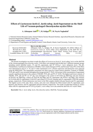
- 5. Abbaspour Anbi et al., Effects of Lactococcus lactis (L. lactis) subsp. lactis Supernatant … 115 in the concentration of supernatant caused a significant decrease in the content of TBARS (P <0.05). Table 2 represents the content of TVN of the fish fillet samples treated with 2% and 4% supernatant, pure bacteria, and control group during the maintenance period. The contents of TVN were increased during cold storage (P <0.05). The highest and lowest contents of the TVN on day 15 were related to fillet samples of the control group (87.20±6.40 mgN/100g) and 4% supernatant group (40.79±0.61 mgN/100g). An increase in the concentration of supernatant caused a significant decrease in the content of TVN (P <0.05). Table 3 represents the pH of the fish fillet samples treated with 2% and 4% supernatant, pure bacteria, and control group during the maintenance period. The pH values were increased during cold storage (P <0.05). The highest and lowest pH values at day 15 were related to fillet samples of pure bacteria (6.23±0.04) and 4% supernatant (5.44±0.07). An increase in the concentration of supernatant caused a significant decrease in the pH value (P <0.05). Table 4 represents the PV of the fish fillet samples treated with 2% and 4% supernatant, pure bacteria, and control group during the maintenance period. We found that the content of PV was increased in the maintenance period (P <0.05). The highest and lowest contents of the PV on day 15 were related to fillet samples of the control group (12.22±0.01 meq/kg) and 4% supernatant group (3.08±0.06 meq/kg). An increase in the concentration of supernatant caused a significant decrease in the content of PV (P <0.05). Table 5 represents the results of the organoleptic tests for different groups of fish fillet samples at the end of the maintenance period. Sensory evaluators gave the highest scores of the odor, flavor, texture, and color to fish fillet samples treated with 4% supernatant (8.00±0.00), pure bacteria (7.00±0.40), pure bacteria, and 4% supernatant (7.75±0.28 and 7.75±0.28, respectively) and pure bacteria (8.50±0.28), respectively. Fig. 1 represents the SDS-PAGE electrophoretic pattern of soluble proteins of different treatments of fish fillet samples on the first day of the maintenance period. There were no significant differences in the diversity of soluble proteins and their expression. A slight difference of about 80 KD was seen between the expression of proteins of fish fillet samples of the control and other groups. Furthermore, a slight difference was seen about 30 KD between the expression of proteins of fish fillet samples of pure bacteria and other groups. Fig. 2 represents the SDS-PAGE electrophoretic pattern of soluble proteins of different treatments of fish fillet samples at day 5 of the maintenance period. Severe differences were seen in the diversity of soluble proteins and their expression in 57 KD and higher than 93 KD bands. The severity of expression soluble proteins or their survival amongst other proteins was higher between studied samples. Fig. 3 represents the SDS-PAGE electrophoretic pattern of soluble proteins of different treatments of fish fillet samples on day 10 of the maintenance period. Severe differences were also seen in the diversity of soluble proteins and their expression in 57 KD and higher than 93 KD bands. The severity of expression soluble proteins or their survival amongst other proteins was higher between control and supernatant treatments (2% and 4%). Figure 4 represents the SDS-PAGE electrophoretic pattern of soluble proteins from different treatments of fish fillet samples on day 15 of the maintenance period. Severe differences were also seen in the diversity of soluble proteins and their expression especially in the expression of soluble proteins in the ranges between 57 to 93 KD which represented higher proteolysis.
- 6. 116 Iranian Food Science and Technology Research Journal Vol. 19, No. 6, 2024 Table 1- The TBARS of the treated fish fillet samples Storage period (day) TBARS (mg/kg) Pure bacteria (106 CFU/g) Supernatant Control 2% 4% 1 0.03±0/008A*d 0.015±0.00Bc 0.013±0.00Bc 0.014±0.00Bd 5 0.052±0.00Bc** 0.330±0.04Cc 0.188±0.05Db 0.914±0.03Ac 10 0.650±0.03Cb 1.520±0.11Ab 1.100±0.09Ba 1.668±0.04Ab 15 0.70±0.02Da 2.770±0.08Ba 0.960±0.08Ba 3.367±0.04Aa *Dissimilar capital leathers in each row show a significant statistical difference (P <0.05). **Dissimilar small leathers in each column show significant statistical differences (P <0.05). Table 2- The TVN of the treated fish fillet samples Storage period (day) TVN (mgN/100g) CFU/g) 6 Pure bacteria (10 Supernatant Control 2% 4% 1 10.30±0.11A*d 10.00.±0.35Ad 8.88±0.31Cd 9.50±0.50Bd 5 23.13±0.43Bc** 24.05±0.50Bc 18.86±1.39Cc 42.70±1.05Ac 10 33.50±0.87Db 43.80±0.91Cb 47.25±2.21Ba 55.9±2.10Ab 15 53.25±0.56Ca 78.68±0.60Ba 40.79±0.61Bb 87.20±6.40Aa *Dissimilar capital leathers in each row show significant statistical differences (P <0.05). **Dissimilar small leathers in each column show significant statistical differences (P <0.05). Table 3- The pH of the treated fish fillet samples Storage period (day) pH Pure bacteria (106 CFU/g) Supernatant Control 2% 4% 1 5.36±0.13A*b 4.96±0.07Bc 5.10±0.03Bb 5.10±0.04Ba 5 6.03±0.04Aa** 5.13±0.03Bc 5.18±0.06Bb 5.26±0.06Ba 10 6.42±0.03Ba 6.72±0.03Aa 5.83±0.03 Ca 5.49±0.24Ca 15 6.23±0.04Aa 5.65±0.12Bb 5.44±0.07Cb 5.61±0.16Ca *Dissimilar capital leathers in each row show significant statistical differences (P <0.05). **Dissimilar small leathers in each column show significant statistical differences (P <0.05). Table 4- The PV of the treated fish fillet samples Storage period (day) PV (meq/kg) Pure bacteria (106 CFU/g) Supernatant Control 2% 4% 1 1.09±0.03A*c 1.17±0.05Ac 0.00 0.00 5 1.13±0.02Bc** 1.37±0.21Bc 1.08±0.06Bc 3.82±0.13Ac 10 1.53±0.17Cb 3.56±0.13Bb 3.66±0.52Ba 4.20±0.00Ab 15 4.20±0.07Da 7.40±0.11Ba 3.08±0.06Cb 12.22±0.01Aa *Dissimilar capital leathers in each row show significant statistical differences (P <0.05). **Dissimilar small leathers in each column show significant statistical differences (P <0.05). Table 5- The sensory evaluation of the treated fish fillet samples period Treatments Organoleptic characters Odor Flavor Texture Color Pure bacteria (106 CFU/g) 4.25±0.25B* 7.00±0.40A 7.75±0.28A 8.50±0.28A Supernatant 2% 3.70±0.47C 3.70±0.25C 4.20±0.47B 4.00±0.40B 4% 8.00±0.00A 6.70±0.25B 7.75±0.25A 8.25±0.25A Control 3.00±0.00C 3.00±0.00C 3.00±0.00C 3.00±0.00C *Dissimilar capital leathers in each column show significant statistical differences (P <0.05).
- 7. Abbaspour Anbi et al., Effects of Lactococcus lactis (L. lactis) subsp. lactis Supernatant … 117 Fig. 1. SDS-PAGE electrophoretic pattern of soluble proteins of different treatments of fish fillet samples in the first day of storage period Fig. 2. SDS-PAGE electrophoretic pattern of soluble proteins of different treatments of fish fillet samples at day 5 of the storage period Fig. 3. SDS-PAGE electrophoretic pattern of soluble proteins of different treatments of fish fillet samples at day 10 of storage period
- 8. 118 Iranian Food Science and Technology Research Journal Vol. 19, No. 6, 2024 Fig. 4. SDS-PAGE electrophoretic pattern of soluble proteins of different treatments of fish fillet samples at day 15 of the storage period Discussion Application of LAB in combination with modified atmosphere packaging and even slight physicochemical treatments and low concentrations of natural traditional preservers may be considered as effective method to increase the shelf life and food safety of fish and marine products through the inhibition of chemical and microbial spoilage without changing the nutritional quality of food products (Pavlićević et al., 2013). The procedure of extension of shelf life by application of natural or controlled microorganisms and their antimicrobial components is recommended by novel scientific reports (Pavlićević et al., 2013). Abbaspour et al. (2018) reported that the number of spoilage bacteria in 4% acid live cells in the storage time of O. mykiss fillets was less than human consumption limits (7 logcfu/ml) compared with control and 2% supernatant of L. lactis subspecies lactis which improved the sensory characteristics of the fillets. The results revealed that the application of supernatant and pure culture of L. lactis subsp. lactis caused a significant decrease in the contents of TBARS, TVN, pH, and PV values compared with the control group. Furthermore, the reduction effects were dose-dependent so an increase in the concentration of bacterial supernatant caused a significant decrease in the contents of TBARS, TVN, pH, and PV. Additionally, fillet samples treated with bacterial supernatants and also pure bacteria had lower protein destruction in the SDS- PAGE analysis. Moreover, fillet samples treated with bacterial supernatants and pure bacteria caused an increase in their sensory scores. The increase in TVN value is attributed to autolytic enzymes and deamination, which lead to the formation of numerous volatile compounds such as dimethylamine, trimethylamine, ammonia, Trimethylamine Oxide (TMAO), hypoxanthine, and non- volatiles histamine, which are shaped by both bacterial and endogenous enzymes. In the treated samples of our study, the values of TVN were lower than in other groups, which may be attributed to the extended lag phase of the spoilage microorganisms as a result of competitive inhibition by LAB and at the same time effect of LAB acidification (Ndaw et al., 2008). Ibrahim & Salha (2009) (Ibrahim & Desouky, 2009) reported that the combined
- 9. Abbaspour Anbi et al., Effects of Lactococcus lactis (L. lactis) subsp. lactis Supernatant … 119 coating of LAB in tilapia fillets had reduced TVN values. Sudalayandi & Manja (2011) described that out of 7 different species of LAB tested for quality indices reduction, L. helveticus, L. lactis, and Pediococcus acidilactici (P. acidilactici) effectively monitored TVN content (Sudalayandi, 2011). Vacuum packaging together with low- temperature preservation is also reported to have the ability to inhibit the growth of spoilage bacteria (Goussault & Leveau, 2006). Similar findings have been reported from Turkey on Bonito fish (Sardasarda) fillets packaged with chitosan film (Alak, 2010). Oxidative rancidity is one of the most significant factors that effect on the acceptability of the fish throughout storage and processing. As general rule of Standard is that the level of PV in seafood should not exceed 10–20 meq/kg of fat (Lakshmanan, 2000). The control fillet samples of our study harbored the limit of acceptance of PV on day 10 of storage (4.20±0.00 meq/kg). In the presence of L. lactis subsp. lactis under vacuum, the sample treated with 2% and 4% supernatant and also those treated with pure bacteria harbored the limit of acceptability for PV on day 15 (7.40±0.11, 3.66±0.52 and 4.20±0.07 meq/kg, respectively). Nath et al. (2014) reported that the control fillets crossed the limit of acceptance of PV on day 6 of storage (14.11±0.37 meq/kg), while fillet groups treated with L. sakei under vacuum packaging crossed the limit of acceptability for PV on day 9 (18.78±0.10 meq/kg). As far as we know, the present research reported the lowest content of PV value in O. mykiss fillet samples treated with 4% supernatant of L. lactis subsp. lactis under vacuum packaging. A probable reason for this finding is the effects of low pH (which occurred by the activity of L. lactis subsp. lactis) on prevention from the production of PV in fish fillet samples. We found that the levels of pH in all studied groups have increased during the maintenance period, while the amounts of increase in the fish fillet samples treated with 4% supernatant of the L. lactis subsp. lactis bacteria were lower than other studied groups. This finding is mainly due to the activity of L. lactis subsp. lactis and production of acidic products. The pH value should not be above than 4.00-4.50 for soaked fish for products' safety. Acidic conditions make the tissue cathepsins much more active resulting in the degradation of some muscle proteins into peptides and amino acids. These components give the marinade its characteristic flavor and texture. Nath et al. (2014) reported that in the presence of L. sakei in aerobic or anaerobic (vacuum packaged) conditions all the samples exhibited a lowering of pH values from the initial level of 7.8 which was similar to our findings. Likewise, reductions in pH values were seen in several investigations on marinated anchovies (Sen & Temelli, 2003), sardines (Kilinc & Cakli, 2004), and Pacific saury (Sallam et al., 2007). TBA test measures malonaldehyde (MDA) produced due to the oxidation of fatty acids with three or more double bonds, and it measures other TBARS such as 2-alkenes and 2, 4-alkadienals. TBA is also usually applied as a quality marker in the fish industry and it has a close association with the sensory properties of fish fillets (Bogdanović et al., 2012). So far, no data have been reported to investigate the effects of L. lactis subsp. lactis on TBARS content of the vacuum-packaged O. mykiss fillets. We found that the fish fillet samples treated with pure L. lactis subsp. lactis and also those treated with 4% supernatant of L. lactis subsp. lactis had the lower contents of TBARS. A probable reason for this finding is the effects of low pH (which occurred by the activity of L. lactis subsp. lactis) on preventing TBARS production in fish fillet samples. Low pH is probably induced by the production of some kinds of acids especially lactic acid, acetic acid, and citric acid due to the activity of L. lactis subsp. lactis guarantee the higher antioxidant effects which decrease the content of TBARS in fish fillet samples. Furthermore, the lower TBARS values of some treatments might result from the direct microbial utilization of MDA and other TBARS or result from reactions
- 10. 120 Iranian Food Science and Technology Research Journal Vol. 19, No. 6, 2024 between TBARS and the amine compounds produced by bacterial metabolism.(Payap & Ommee, 2007). The increase in TBARS values in all treatments is probably due to the induced denaturation of muscle protein, leading to the release of from heme iron, a potential pro- oxidant in the muscle system (Greene & Cumuze, 1981; Payap & Ommee, 2007). Previous research showed that a TBARS value of at least 2.0 mg malonaldehyde/kg is essential for the perception of rancid taste and odors (Greene & Cumuze, 1981). We found that the contents of TBARS in fish fillet samples treated with pure L. lactis subsp. lactis and also 4% supernatant of L. lactis subsp. lactis on day 15 of the maintenance period were lower than 1.0 mg malonaldehyde/kg which could indirectly guarantee high scores given to the taste and odor of these treatments. Sensory evaluation showed that fish fillet samples treated with pure and also 4% supernatant of L. lactis subsp. lactis harbored the highest scores of odor, flavor, texture, and color. This finding is mainly due to the effects of L. lactis subsp. lactis on contents of TVN, TBARS, pH, and PV. Similar findings have been reported by (Soltanian et al., 2011; Boulares et al., 2013; Chanarat et al., 2014; Adilla et al., 2017 and Rahmatipoor et al., 2017). Conclusion The present research is the first report of the effects of L. lactis subsp. lactis on the shelf life of O. mykiss fillets. Results showed that using 4% supernatant and also pure L. lactis subsp. lactis bacteria caused a significant decrease in the contents of TBARS, TVN, pH, and PV values of O. mykiss fillets. Contents of the majority of studied chemical parameters were lower than the allowed limit reported by the standard organizations (Tables 1-4). Additionally, levels of protein destruction were studied by the SDS-PAGE in O. mykiss fillet samples treated with 4% supernatant and also pure L. lactis subsp. lactis bacteria were significantly lower than other studied groups. Furthermore, the mean scores given to the odor, flavor, texture, and color parameters were higher in the O. mykiss fillet samples treated with 4% supernatant and also pure L. lactis subsp. lactis bacteria. Therefore, soaking of O. mykiss fillet samples on 4% supernatant of L. lactis subsp. lactis and 106 CFU/g of pure bacteria is recommended as efficient method to extend the shelf life of O. mykiss fillet samples. However, further studies are required to analyze other chemical, sensory, and microbiological aspects of O. mykiss fillet samples treated with other LAB and their metabolites. Acknowledgments The authors would like to thank Dr. Amir Zeinali for his assistance. Also, we appreciate the Iranian Fisheries Science Research Institute's cooperation. References 1. Abbaspour, A.A., Rasavilar, V., Neyriz Naghadehi, M., Asadpour Osalou, Y.A. (2019). Study of the effect of incorporation of Lactococcus lactis on the shelf life of rainbow trout fillet in glacial condition. Journal of Food Microbiology, 6(4), 45-58. (In Persian) 2. Abbaspour Anbi, A., Razavilar, V., Neyriz Naghadehi, M., Seidgar, M., Nekuiefard, A., & Asadpour Osalou, Y.A. (2018). The effects of lactococcus lactis subsp. lactis and its supernatant on some bacteriological and sensory values in rainbow trout (Oncorhynchus mykiss) fillets. Microbiology Research, 9, 7431, 19-25. https://doi.org/10.4081/mr.2018.7431 3. Ammor, S., Tauveron, G., Dufour, E., & Chevallier, I. (2006). Antibacterial activity of lactic acid bacteria against spoilage and pathogenic bacteria isolated from the same meat small-scale facility 1-Screening and characterization of the antibacterial compounds. Food Control, 454–461. https://doi.org/10.1016/j.foodcont.2005.02.006
- 11. Abbaspour Anbi et al., Effects of Lactococcus lactis (L. lactis) subsp. lactis Supernatant … 121 4. Adilla, S.N, Utami, R., Nursiwi, A., & Nurhartadi, E.( 2016). The effect of nisin from Lactococcus lactis subsp. lactis on refrigerated patin fillet quality. In: International Conference On Food Science and Engineering. vol 193. IOP Publishing, https://doi.org/10.1088/1757- 899X/193/1/012014 5. Alak, G. (2010). Microbiological and chemical properties of modified atmosphere and vacuum. Kafk Üniver Vet Fak Dergisi, 16, S73-S80. https://doi.org/10.9775/kvfd.2009.1475 6. Benjakul, S., Seymour, T.A., Morrissey, M.T., & An, H.(2006). Physicochemical changes in Pacific whiting muscle proteins during iced storage. Journal Food Science, 62, 729-33. https://doi.org/10.1111/j.1365-2621.1997.tb15445.x 7. Bogdanović, T., Šimat, V., Frka Roić, A., & Marković, K. (2012). Development and application of quality index method scheme in a shelf life study of wild and fish farm affected bogue (Boops boops, L.). Journal Food Science, 77, 99-106. https://doi.org/10.1111/j.1750-3841.2011.02545.x 8. Boulares, M., Mankai, M., Belaam, Z., & Hassouna, M. (2013). Effect of inoculation of lactic acid bacteria on the proteolytic activity of psychrotrophic Gram-negative bacteria in fresh farmed sea bass (Dicentrarchus labrax) fillets during storage at 4° C under vacuum-packed conditions. Annal Microbiology, 63, 1493-500. https://doi.org/10.1007/s13213-013-0613-1 9. Chanarat, S., Benjakul, S., & Xiong, Y.I. (2014). Physicochemical changes of myosin and gelling properties of washed tilapia mince as influenced by oxidative stress and microbial transglutaminase. Journal Food Science Technology, 52, 3824–36. 10. Chanarat, S., Benjakul, S., & Xiong, Y.I. (2014). Physicochemical changes of myosin and gelling properties of washed tilapia mince as influenced by oxidative stress and microbial transglutaminase. Journal Food Science Technology, 52, 3824–36. 11. Chowdhury, S., Raychaudhuri, U., Nath, S., & Dora, K. (2016). Shelf life of lactic acid bacteria inoculated vacuum packed Tenualosa Elisha (Hamilton, 1822) at low temperature. Indian Journal of Fisheries, 63, 156-61. https://doi.org/10.21077/ijf.2016.63.1.27811-25 12. Chytiri, S., Chouliara, I., Savvaidis, I., Kontominas, M.(2004). Microbiological, chemical, and sensory assessment of iced whole and filleted aquacultured rainbow trout. Food Microbiol, 21: 157-65. https://doi.org/10.1016/S0740-0020(03)00059-5 13. Codex. (1999). Guidelines for the sensory evaluation of fish and shellfish in laboratories. CAC/GL 311999: 1-32. 14. DeWitt, C.A.M., & Oliveira, A. (2016). Modified atmosphere systems and shelf life extension of fish and fishery products. Foods, 5, 48. https://doi.org/10.3390/foods5030048 15. Erkan, N., Özden, Ö., & Inuğur, M. (2007). The effects of modified atmosphere and vacuum packaging on quality of chub mackerel. International Journal of Food Science & Technology, 42, 1297-304. https://doi.org/10.1111/j.1365-2621.2006.01325.x 16. Fijan, S. (2014). Microorganisms with claimed probiotic properties: an overview of recent literature. International Journal of Environmental Research and Public Health, 11, 4745–4767. https://doi.org/10.3390/ijerph110504745 17. Fuentes-Amaya, L.F., Munyard, S., Fernandez‐Piquer, J., & Howieson, J. (2015). Sensory, microbiological and chemical changes in vacuum‐packaged blue spotted Emperor (Lethrinus sp.), Saddletail Snapper (Lutjanus malabaricus), Crimson Snapper (Lutjanus erythropterus), Barramundi (Lates calcarifer) and Atlantic Salmon (Salmo salar) Fillets Stored at 4° C. Food Science Nutrient, 4, 479-89. https://doi.org/10.1002/fsn3.309 18. Gelman, A., Glatman, L., Drabkin, V., & Harpaz, S. (2001). Effects of storage temperature and preservative treatment on the shelf life of the pond-raised freshwater fish, silver perch (Bidyanus biryanis). Journal of Food Protection, 64, 1584-91. https://doi.org/10.4315/0362-028x- 64.10.1584
- 12. 122 Iranian Food Science and Technology Research Journal Vol. 19, No. 6, 2024 19. Giuffrida, A., Giarratana, F., Valenti, D., Muscolino, D., Parisi, R., Parco, A., Marotta, S., Ziino, G., & Panebianco, A. (2017). A new approach to predict the fish fillet shelf-life in the presence of natural preservative agents. Italian Journal Food Safety, 6, 6768. 20. Goussault, B., & Leveau, B. (2006). A guide to packaging technology for seafood value-addition. Eurofish, Copenhagen, 1-20. 21. Greene, B.E., & Cumuze, T.H. (1981). Relationship between TBA numbers and inexperienced panelists' assessments of oxidized flavor in cooked beef. Journal Food Science, 47, 52-8. https://doi.org/10.1111/j.1365-2621.1982.tb11025.x 22. Ibrahim, S., & Desouky, S.G. (2009). Effect of antimicrobial metabolites produced by lactic acid bacteria (Lab) on quality aspects of frozen tilapia (Oreochromis niloticus) fillets. World Journal of Fish and Marine Sciences, 1, 40-5. 23. Krishnamoorthi, R., Srinivas, M., Mahalingam, P.U., Malaikozhundan, B., Suganya, P., & Gurushankar, K. (2022) Antimicrobial, anti-biofilm, antioxidant and cytotoxic effects of bacteriocin by Lactococcus lactis strain CH3 isolated from fermented dairy products—An in vitro and silico approach. International Journal of Biological Macromolecules, 220, 291-306. https://doi.org/10.1016/j.ijbiomac.2022.08.087 24. Kilinc, B., & Cakli, S. (2004). Chemical, microbiological, and sensory changes in thawed fillets of sardine (Sardina pilchardus) during marination. Food Chemistry, 88, 275–80. https://doi.org/10.1016/j.foodchem.2004.01.044 25. Lakshmanan, P. (2000). Fish spoilage and quality assessment. Central Institute of Fisheries Technology and Society of Fisheries Technology, Cochin, India. 26. Larsen, R., Eilertsen, K.E, & Elvevoll, E.O. (2011). Health benefits of marine foods and ingredients. Biotechnology Advances, 29, 508-18. https://doi.org/10.1016/j.biotechadv.2011.05.017 27. Malle, P., & Poumeyrol, M. (1989). A new chemical criterion for the quality control of fish: trimethylamine/total volatile basic nitrogen (%). Journal of Food Protection, 52, 419-23. https://doi.org/10.4315/0362-028X-52.6.419 28. Nath, S., Chowdhury, S., & Dora, K. (2014). Effect of lactic acid bacteria application on shelf life and safety of fish fillet at 6±1 c. International Journal of Advanced Research, 2, 201-7. 29. Ndaw, A., Faid, M., Bouseta, A., & Zinedine, A. (2008). Effect of controlled lactic acid bacteria fermentation on the microbiological and chemical quality of Moroccan sardines (Sardina pilchardus). International Journal of Agriculture and Biology, 10, 21-7. https://doi.org/10.1556/AMicr.55.2008.3.2 30. Özpolat, E., Patır, B., Guran, H., & Gul, M. (2014). Effect of vacuum-packing method on the shelf–life of Capoeta umbrella sausages. Iranian Journal of Fisheries Sciences, 13, 178-84. https://doi.org/20.1001.1.15622916.2014.13.1.15.5 31. Pavlićević, N., Đorđević, V., Dimitrijević, M., Bošković, M., Marković, R., & Baltić, M.Ž. (2013). Lactic acid bacteria: Effect on the quality and safety of fishery products. In: 6th International Conference Water and Fish, Faculty of Agriculture, Belgrade-Zemun (Serbia), p 436-42 32. Julie, A.A.A. & Nacional, L.M. (2007). Effect of lactic, acetic, and citric acids on quality changes of refrigerated green mussel, Perna viridis (Linnaeus, 1758). Songklanakarin Journal of Science and Technology, 29, 171-179. https://doi.org/10.12944/crnfsj.6.3.29 · 33. Rahmatipoor, R., Roomiani, L., & Askary Sary, A. (2017). Effect of different packaging on the shelf life of silver carp (Hypophthalmichthys molitrix) fillets stored at 4 ºC. Iranian Journal of Aquatic Animal Health, 3, 22-35. https://doi.org/10.29252/ijaah.3.2.22 34. Sałański, P., Kowalczyk, M., & Bardowski, J.K. (2022). Health-promoting nature of Lactococcus lactis IBB109 and Lactococcus lactis IBB417 strains exhibiting proliferation inhibition and
- 13. Abbaspour Anbi et al., Effects of Lactococcus lactis (L. lactis) subsp. lactis Supernatant … 123 stimulation of interleukin-18 expression in Colorectal cancer cells, Frontiers in Microbiology, 13, 1-13. https://doi.org/10.3389/fmicb.2022.822912 35. Salih, A., Smith, D., Price, J., & Dawson, L. (1987). Modified extraction 2-thiobarbituric acid method for measuring lipid oxidation in poultry. Poultry Science, 66, 1483-8. https://doi.org/10.3382/ps.0661483 36. Sallam, K.I., Ahmed, A.M., Elgazzar, M.M., & Eldaly, E.A. (2007). Chemical quality and sensory attributes of marinated Pacific saury (Cololabis saira) during vacuum-packaged storage at 4o C. Food Chemistry, 102, 1061-70. https://doi.org/10.1016/j.foodchem.2006.06.044 37. Sarika, A., Lipton, A., Aishwarya, M., & Dhivya, R. (2012). Isolation of a bacteriocin-producing Lactococcus lactis and application of its bacteriocin to manage spoilage bacteria in high-value marine fish under different storage temperatures. Applied Biochemistry and Biotechnology, 167, 1280-9. https://doi.org/10.1128/aem.55.8.1901-1906.1989 38. Schillinger, U., & Lücke, F.K. (1989). Antibacterial activity of Lactobacillus sake isolated from meat. Applied and Environmental Microbiology, 55, 1901-6. https://doi.org/10.1128/aem.55.8.1901-1906.1989 39. Sen, M., & Temelli, S. (2003). Microbiological and chemical qualities of marinated anchovy prepared with different vegetable additives and sauce. Review Médicine Véterinary, 154, 703-8. 40. Shamloofar, M., Hoseini, E., Kamali, A., Motalebi Moghanjoghi, A., & Poorgholm, R. (2015). Antibacterial activities of nisin encapsulated in zein and modified atmosphere packaging on rainbow trout (Oncorhynchus mykiss) fillet during chilled storage 4 C. Iranian Journal of Fisheries Sciences, 14, 369-81. 41. Shon, J., & Chin, K. (2008). Effect of whey protein coating on quality attributes of low‐fat, aerobically packaged sausage during refrigerated storage. Journal Food Sciences, 73, 469-475. https://doi.org/10.1111/j.1750-3841.2008.00829.x 42. Soltanian, S., Behnam, S., Rezai, M., Safari, R., & Anvari, M. (2011). Nisin is a preservative for rainbow trout (Oncorhynchus mykis). Online Journal of Veterinary Research, 15, 354-65. 43. Sudalayandi, K. (2011). Efficacy of lactic acid bacteria in the reduction of trimethylamine- nitrogen and related spoilage derivatives of fresh Indian mackerel fish chunks. African Journal of Biotechnology, 10, 42-7. 44. Tufail, M., Hussain, S., Malik, F., Mirza, T., Parveen, G., Shafaat, S., Wajid, A., Mahmood, R., Channa, R.A., & Sadiq, A. (2011). Isolation and evaluation of antibacterial activity of bacteriocin produced by Lactobacillus bulgaricus from yogurt. African Journal of Microbiology Research, 5, 3842-7. https://doi.org/10.5897/AJMR11.846
- 14. 124 Iranian Food Science and Technology Research Journal Vol. 19, No. 6, 2024 پژوهشی مقاله جلد 19 شماره ، 6 ، بهمن - ،اسفند 1402 ، .ص 124 - 111 باکتری سوپرناتانت اثرات Lactococcus lactis (L. lactis) subsp. lactis فیله ماندگاری بر قزل رنگین آالی ( کمان Oncorhynchus mykiss بسته ) خالء در شده بندی انبی پور عباس اسد 1 * - صیدگر مسعود 1 - نقدهی نیریز مسلم 2 تا :دریافت ریخ 17 / 05 / 1402 :پذیرش تاریخ 17 / 08 / 1402 چکیده اثرات بررسی جهت حاضر تحقیق الکتیس الکتوباسیلوس زیرگونه الکتیس قزل فیله ماندگاری بر رنگین آالی بسته کمان فیله .شد انجام خالء در شده بندی به و تهیه ماهی های 5 باکتر ،)مقطر (آب کنترل شامل مختلف تیمار گروه و خالص ی 2 غوطه تیمار رویی مایع در ورشده 2 % و 4 % و 6 10 CFU/g actis l L. زیرگونه الکتیس طبقه مقادیر .شدند بندی pH اسید تیوباربیتوریک ، (TBARS) کل فرار نیتروژن ، (TVN) پراکسید مقدار و (PV) فیله روزهای در ها 0 ، 5 ، 10 و 15 دمای در نگهداری دوره 4 سانتی درجه گراد سولفات دودسیل سدیم ژل الکتروفورز از استفاده با پروتئین تخریب و بیان .شد بررسی - آکریل پلی آمید (SDS- PAGE) آن با ارگانولپتیک ارزیابی .شد الیز مقادیر .شد انجام خبره حسی ارزیاب پنج از استفاده TBARS ، TVN ، pH و PV یافته افزایش نگهداری دوره طول در است ( P <0.05 ) معنی کاهش باعث رویی مایع غلظت افزایش . مقادیر در داری TBARS ، TVN ، pH و PV شد ( P <0.05 مقادیر کمترین و بیشترین .) TBARS ، TVN ، pH و PV روز در 15 به گروه به مربوط ترتیب ( شاهد های 04 / 0 ± 367 / 3 میلی ( خالص باکتری و )کیلوگرم بر گرم 02 / 0 ± 70 / 0 میلی بر گرم کن ،)کیلوگرم ( ترل 40 / 6 ± 20 / 87 میلی )کیلوگرم بر گرم و .بود )بدن وزن 4 % ( رویی مایع 61 / 79 ± 0 / 40 mgN/100g) ( خالص باکتری ، 04 / 0 ± 23 / 6 و ) 4 % مایع ( رویی 07 / 0 ± 44 / 5 ( شاهد و ) 01 / 22 ± 0 / 12 ) meq/kg و 4 % رویی مایع ( 06 / 0 ± 08 / 3 ) meq/kg نمونه در پروتئین تخریب .بود باکت با شده تیمار فیله های ری و خالص 4 فیله به رنگ و بافت ،طعم ،بو امتیاز باالترین .بود کمتر رویی مایع درصد با شده تیمار های 4 ( رویی مایع درصد 00 / 0 ± 00 / 8 خالص باکتری ،) ( 40 / 0 ± 00 / 7 و خالص باکتری ،) 4 ( رویی مایع درصد 28 / 0 ± 75 / 7 و 28 / 0 ± 75 / 7 ( خالص باکتری و ) 28 / 0 ± 50 / 8 به ) .آمد دست فی خیساندن له های O. mykiss روی 4 % و رویی مایع 6 10 CFU/g از L. lactis . Subsp الکتیس می فیله مفید عمر تواند قزل های رنگین آالی .دهد افزایش را کمان واژه :کلیدی های باکتر ی اس ی د الکت ی ،ک بسته ،خالء در بندی ف ی له ماهی ، ماندگار ی ، Oncorhynchus mykiss 1 - مرکز م ،کشور آرتمیای تحقیقات ؤ تحقی سسه ایران ،ارومیه ،کشاورزی ترویج و آموزش ،تحقیقات سازمان ،کشور شیالتی علوم قات *( - :مسئول نویسنده Email: asadanbi1208@gmail.com ) 2 - ای ،ارومیه ،اسالمی آزاد دانشگاه ،ارومیه واحد ،غذایی مواد کیفی کنترل و بهداشت گروه ران https://doi.org/10.22067/ifstrj.2023.83834.1274 ایران غذایی صنایع و علوم پژوهشهای نشریه https://ifstrj.um.ac.ir