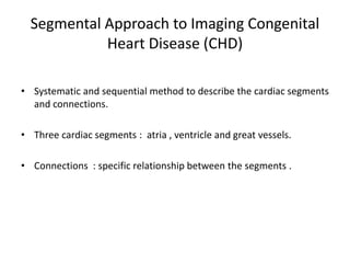
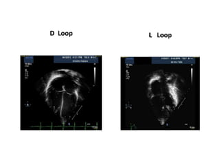
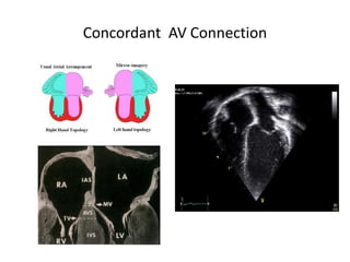
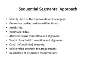
ECHO : SEGMENTAL APPROACH
Dr Binjo J Vazhappilly
SR Cardiology
MCH Calicut
Segmental Approach to Imaging Congenital
Heart Disease (CHD)
• Systematic and sequential method to describe the cardiac segments
and connections.
• Three cardiac segments : atria , ventricle and great vessels.
• Connections : specific relationship between the segments .
Determine visceroatrial situs
• Situs refers to spatial arrangement of the viscerae .
Three types of visceroatrial situs
• S – solitus (normal arrangement)
• I – inversus (or the mirror-image arrangement)
• A - ambiguus
• Situs ambiguus : defined as an abnormality in which thoracic and
abdominal organs are positioned with respect to each other, in such
a way that it cannot be clearly lateralized.
Situs Solitus
Situs Inversus
Right Isomerism(Asplenia)
Left Isomerism (polysplenia)
Cardiac Position & Orientation
• Position of the heart in the chest with regard to its location and
orientation of its apex.
Location of heart in the chest
• Levoposition : to the left
• Mesoposition : central
• Dextroposition : to the right
Cardiac orientation is the base to apex orientation of heart
• Levocardia : apex directed to left of midline.
• Mesocardia : apex oriented inferiorly in the midline
• Dextrocardia: apex directed to right of midline.
Atrial Morphology
Right Atrium
• Triangular, broad based,
anterior appendage .
• Receives IVC, SVC & coronary
sinus.
• Septum secundum (limbus of
fossa ovale) lies on RA side.
• Crista Terminalis is in RA.
Left Atrium
• Narrow, fingerlike posterior
appendage .
• LA receives all 4 pulmonary
veins
• Septum primum lies on LA
side .
• LA is smooth with fewer
trabeculations
The right & left atria are identified morphologically by their
respective atrial appendages and veins emptying into them.
Atrial Morphology
Pulmonary veins to LA
Atrial Situs
• Atrial Situs Solitus (S)
• Atrial Situs Inversus (I)
• Atrial Situs Ambiguous (A)
Right isomerism (bilateral right atria) or
Left isomerism (bilateral left atria).
Ventricular Looping
• Embryonic heart is a linear tube with bulbus cordis (future RV )
being cephalad to the developing left ventricle (LV) .
• In normal development the tube bends over on itself to right
forming a D-loop resulting in positioning of the bulbus cordis to the
anatomic right of the left ventricle
• Folding to left results in L-loop with the morphologic right ventricle
being to the left of the morphologic left ventricle
Ventricular Looping
D Loop L Loop
Identification of the ventricles
Tricuspid Valve ⇒ RV
• Septal chordal attachments
• Apical septal annular
attachment
• 3 leaflet
• Multiple small papillary
muscles
• Triangular orifice
Mitral valve ⇒ LV
• No septal chordal
attachments
• Basal septal annular
attachments
• 2 leaflet
• 2 large papillary muscles
• Elliptical orifice.
AV valve morphology directly correlates with ventricular type
RV Morphology
• Large apical trabaculations
• Coarse septal surface
• Moderator bands
• Cresentic in cross section
• Tricuspid – pulmonary
discontinuity
LV Morphology
• Small apical trabeculations
• Smooth upper surface
• No moderator band
• Mitral – Aortic continuity
Atrio-ventricular Connections
• Concordant ( Normal : RA to RV & LA to LV)
• Discordant ( RA to LV & LA to RV)
• Univentricular AV connections
Absent AV connection
Common AV valve
Double inlet connections
Concordant AV Connection
Univentricular AV Connections
• Absent AV connection (right / left)
Tricuspid / mitral atresia
• Common AV valve
With single ventricle
• Double inlet ventricle
Connection of both AV valve to the same ventricle
Anatomically left
Anatomically right
Undetermined
(rudimentary chamber always present)
Univentricular AV connections
Absent Rt AV Connection Absent Lt AV connection
Univentricular AV connections
Double inlet : RV Morphology Double inlet : LV Morphology
Overriding & Straddling
• Overriding :
Biventricular emptying of AV Valve or biventricular origin of a
semilunar valve.
• Straddling :
Chordae or papillary muscle of the valve attach to contralateral
ventricle.
Involve AV Valves and requires presence of VSD.
Arterial Morphology
• Aorta : artery that gives rise to the coronary arteries and the
brachiocephalic vessels.
• Pulmonary artery : branches into two but does not give rise to any
vessels.
Ventriculo-Arterial Connection
• Concordant :
Aorta connected to LV and pulmonary artery to RV .
• Discordant :
Aorta connected to RV and pulmonary artery to LV .
• Double Outlet : Both great arteries arise from a single ventricle.
• If more than 50% of an artery overrides a ventricle it is said to be
committed to it.
Commitment
• Commitment describes possible abnormalities of flow through
valves into ventricles and great vessels.
• In TOF, atria, AV valves & ventricles are positioned normally and
concordant.
Aorta overrides a VSD and is doubly committed to both ventricles.
• In univentricular heart both AV valves are usually doubly
committed to the single ventricle.
Ventriculo-arterial connections
• Concordant (normal)
Ventricular septal defect, tetralogy of Fallot
• Discordant
Transposition
• Double outlet ventricle
With subaortic VSD
subpulmonary VSD
doubly committed VSD
non-committed VSD
• Common arterial trunk
Conus
• Subpulmonary:
Absence of subaortic infundibular free wall, found in normal heart.
• Subaortic:
Absence of subpulmonary infundibular free wall, found in D-loop
TGA.
• Bilaterally present :
In double outlet RV , but rarely in TGA.
• Bilaterally Absent:
In double outlet LV.
Relation b/w semilunar Valves
• Solitus : Aorta posterior and to right.
• Inversus : mirror image : Aorta posterior and to left
• D-malposition : aortic valve anterior and to the right.
• L-malposition : aortic valve anterior and to the left.
• Anterior malposition : aortic valve anterior in the middle.
• Parasternal and high parasternal short axis and subcostal short axis
views are used to recognize the positions of AV and PV.
Possible Great vessel relations
Normal
PSX Suprasternal long and short axis
d TGA
cc TGA
DORV
TOF
Truncus Arteriosus
Assessment of Associated Anomalies
Abnormalities in each segment should be assessed
 Septal defects
 AV valve abnormalities like Ebstein’s
 Outflow tract stenosis
 Aortic arch branching and sidedness
 Pulmonary artery and branches
 PDA
 Coronary abnormalities
Ebstein anomaly
Pulmonary valve pathology
LV outflow
Subaortic membrane Supravalvular AS
COARCTATION OF AORTA
Sequential Segmental Approach
• Identify situs of the thoraco-abdominal organs.
• Determine cardiac position within thorax.
• Atrial Situs.
• Ventricular Situs.
• Atrioventricular connection and alignment.
• Ventriculo-arterial connection and alignment.
• Conal (infundibular) anatomy.
• Relationship between the great arteries.
• Description of associated malformations
Segmental Expression
1 . Visceroatrial situs : S , I or A.
2 . Ventricular situs : D or L
3 . Position of great vessels : S , I , D , L or A
 Normal : S , D , S
 Situs inversus Dextrocardia : I , L , I
 D TGA : S , D , D
 D TGA with situs inversus : I , L ,L
 L TGA with situs solitus : S , L , L
QUIZ
1. Interrupted IVC is seen in
A) Situs inversus
B) Situs solitus
C) Right isomerism
D) Left isomerism
2. Morphological feature of right atrium
A) Finger like posterior appendage
B) Smooth surface
C) Presence of cristae terminalis
D) Septum primum lies on RA side
3. True about right atrial appendage
A) Narrow based
B) Triangular
C) Posterior
D) None of the above
4.True about ventricular morphology
A) LV is cresentic in cross section
B) Moderator band is seen in RV
C) Trabaculations are less in RV
D) RV has smooth septal surface
5. Straddling means
A) Papillary muscle of the valve attached to contralateral
ventricle.
B) Papillary muscle attached to septum
C) Absence of papillary muscle
D) Fusion of papillary muscle
6. In d TGA true about connection is
A) AV discordant , VA concordant
B) AV concordant , VA discordant
C) Both AV and VA concordant
D) Both AV and VA discordant
7. In TOF true about connection is
A) AV discordant , VA concordant
B) AV concordant , VA discordant
C) Both AV and VA concordant
D) Both AV and VA discordant
8. Conus is bilaterally absent in
A)DORV
B) TGA
C) DOLV
D) Normal heart
9. In d malposition of great arteries
A) Aorta posterior and to right
B) Aorta posterior and to left
C) Aorta anterior and to right
D) Aorta anterior and to left
10.Segmental expression of L TGA is
A) S , D , S
B) I , L , I
C) S, D , D
D) S , L , L
1. Interrupted IVC is seen in
A) Situs inversus
B) Situs solitus
C) Right isomerism
D) Left isomerism
2. Morphological feature of right atrium
A) Finger like posterior appendage
B) Smooth surface
C) Presence of cristae terminalis
D) Septum primum lies on RA side
3. True about right atrial appendage
A) Narrow based
B) Triangular
C) Posterior
D) None of the above
4. True about ventricular morphology
A) LV is cresentic in cross section
B) Moderator band is seen in RV
C) Trabaculations are less in RV
D) RV has smooth septal surface
5. Straddling means
A) Papillary muscle of the valve attached to contralateral
ventricle.
B) Papillary muscle attached to septum
C) Absence of papillary muscle
D) Fusion of papillary muscle
6. In d TGA true about connection is
A) AV discordant , VA concordant
B) AV concordant , VA discordant
C) Both AV and VA concordant
D) Both AV and VA discordant
7. In TOF true about connection is
A) AV discordant , VA concordant
B) AV concordant , VA discordant
C) Both AV and VA concordant
D) Both AV and VA discordant
8. Conus is bilaterally absent in
A)DORV
B) TGA
C) DOLV
D) Normal heart
9. In d malposition of great arteries
A) Aorta posterior and to right
B) Aorta posterior and to left
C) Aorta anterior and to right
D) Aorta anterior and to left
10.Segmental expression of L TGA is
A) S , D , S
B) I , L , I
C) S, D , D
D) S , L , L
THANK YOU

ECHO SEGMENTAL APPROACH- DR BINJO.ppsx

  • 1.
    ECHO : SEGMENTALAPPROACH Dr Binjo J Vazhappilly SR Cardiology MCH Calicut
  • 2.
    Segmental Approach toImaging Congenital Heart Disease (CHD) • Systematic and sequential method to describe the cardiac segments and connections. • Three cardiac segments : atria , ventricle and great vessels. • Connections : specific relationship between the segments .
  • 5.
    Determine visceroatrial situs •Situs refers to spatial arrangement of the viscerae . Three types of visceroatrial situs • S – solitus (normal arrangement) • I – inversus (or the mirror-image arrangement) • A - ambiguus • Situs ambiguus : defined as an abnormality in which thoracic and abdominal organs are positioned with respect to each other, in such a way that it cannot be clearly lateralized.
  • 6.
  • 7.
  • 8.
  • 9.
  • 10.
    Cardiac Position &Orientation • Position of the heart in the chest with regard to its location and orientation of its apex. Location of heart in the chest • Levoposition : to the left • Mesoposition : central • Dextroposition : to the right Cardiac orientation is the base to apex orientation of heart • Levocardia : apex directed to left of midline. • Mesocardia : apex oriented inferiorly in the midline • Dextrocardia: apex directed to right of midline.
  • 11.
    Atrial Morphology Right Atrium •Triangular, broad based, anterior appendage . • Receives IVC, SVC & coronary sinus. • Septum secundum (limbus of fossa ovale) lies on RA side. • Crista Terminalis is in RA. Left Atrium • Narrow, fingerlike posterior appendage . • LA receives all 4 pulmonary veins • Septum primum lies on LA side . • LA is smooth with fewer trabeculations The right & left atria are identified morphologically by their respective atrial appendages and veins emptying into them.
  • 12.
  • 13.
  • 14.
    Atrial Situs • AtrialSitus Solitus (S) • Atrial Situs Inversus (I) • Atrial Situs Ambiguous (A) Right isomerism (bilateral right atria) or Left isomerism (bilateral left atria).
  • 15.
    Ventricular Looping • Embryonicheart is a linear tube with bulbus cordis (future RV ) being cephalad to the developing left ventricle (LV) . • In normal development the tube bends over on itself to right forming a D-loop resulting in positioning of the bulbus cordis to the anatomic right of the left ventricle • Folding to left results in L-loop with the morphologic right ventricle being to the left of the morphologic left ventricle
  • 16.
  • 17.
  • 18.
    Identification of theventricles Tricuspid Valve ⇒ RV • Septal chordal attachments • Apical septal annular attachment • 3 leaflet • Multiple small papillary muscles • Triangular orifice Mitral valve ⇒ LV • No septal chordal attachments • Basal septal annular attachments • 2 leaflet • 2 large papillary muscles • Elliptical orifice. AV valve morphology directly correlates with ventricular type
  • 19.
    RV Morphology • Largeapical trabaculations • Coarse septal surface • Moderator bands • Cresentic in cross section • Tricuspid – pulmonary discontinuity
  • 20.
    LV Morphology • Smallapical trabeculations • Smooth upper surface • No moderator band • Mitral – Aortic continuity
  • 21.
    Atrio-ventricular Connections • Concordant( Normal : RA to RV & LA to LV) • Discordant ( RA to LV & LA to RV) • Univentricular AV connections Absent AV connection Common AV valve Double inlet connections
  • 22.
  • 23.
    Univentricular AV Connections •Absent AV connection (right / left) Tricuspid / mitral atresia • Common AV valve With single ventricle • Double inlet ventricle Connection of both AV valve to the same ventricle Anatomically left Anatomically right Undetermined (rudimentary chamber always present)
  • 24.
  • 25.
    Univentricular AV connections AbsentRt AV Connection Absent Lt AV connection
  • 26.
    Univentricular AV connections Doubleinlet : RV Morphology Double inlet : LV Morphology
  • 27.
    Overriding & Straddling •Overriding : Biventricular emptying of AV Valve or biventricular origin of a semilunar valve. • Straddling : Chordae or papillary muscle of the valve attach to contralateral ventricle. Involve AV Valves and requires presence of VSD.
  • 28.
    Arterial Morphology • Aorta: artery that gives rise to the coronary arteries and the brachiocephalic vessels. • Pulmonary artery : branches into two but does not give rise to any vessels.
  • 29.
    Ventriculo-Arterial Connection • Concordant: Aorta connected to LV and pulmonary artery to RV . • Discordant : Aorta connected to RV and pulmonary artery to LV . • Double Outlet : Both great arteries arise from a single ventricle. • If more than 50% of an artery overrides a ventricle it is said to be committed to it.
  • 30.
    Commitment • Commitment describespossible abnormalities of flow through valves into ventricles and great vessels. • In TOF, atria, AV valves & ventricles are positioned normally and concordant. Aorta overrides a VSD and is doubly committed to both ventricles. • In univentricular heart both AV valves are usually doubly committed to the single ventricle.
  • 31.
    Ventriculo-arterial connections • Concordant(normal) Ventricular septal defect, tetralogy of Fallot • Discordant Transposition • Double outlet ventricle With subaortic VSD subpulmonary VSD doubly committed VSD non-committed VSD • Common arterial trunk
  • 32.
    Conus • Subpulmonary: Absence ofsubaortic infundibular free wall, found in normal heart. • Subaortic: Absence of subpulmonary infundibular free wall, found in D-loop TGA. • Bilaterally present : In double outlet RV , but rarely in TGA. • Bilaterally Absent: In double outlet LV.
  • 33.
    Relation b/w semilunarValves • Solitus : Aorta posterior and to right. • Inversus : mirror image : Aorta posterior and to left • D-malposition : aortic valve anterior and to the right. • L-malposition : aortic valve anterior and to the left. • Anterior malposition : aortic valve anterior in the middle. • Parasternal and high parasternal short axis and subcostal short axis views are used to recognize the positions of AV and PV.
  • 34.
  • 35.
  • 36.
  • 37.
  • 38.
  • 39.
  • 40.
  • 41.
    Assessment of AssociatedAnomalies Abnormalities in each segment should be assessed  Septal defects  AV valve abnormalities like Ebstein’s  Outflow tract stenosis  Aortic arch branching and sidedness  Pulmonary artery and branches  PDA  Coronary abnormalities
  • 42.
  • 43.
  • 44.
  • 45.
  • 46.
    Sequential Segmental Approach •Identify situs of the thoraco-abdominal organs. • Determine cardiac position within thorax. • Atrial Situs. • Ventricular Situs. • Atrioventricular connection and alignment. • Ventriculo-arterial connection and alignment. • Conal (infundibular) anatomy. • Relationship between the great arteries. • Description of associated malformations
  • 47.
    Segmental Expression 1 .Visceroatrial situs : S , I or A. 2 . Ventricular situs : D or L 3 . Position of great vessels : S , I , D , L or A  Normal : S , D , S  Situs inversus Dextrocardia : I , L , I  D TGA : S , D , D  D TGA with situs inversus : I , L ,L  L TGA with situs solitus : S , L , L
  • 48.
  • 49.
    1. Interrupted IVCis seen in A) Situs inversus B) Situs solitus C) Right isomerism D) Left isomerism
  • 50.
    2. Morphological featureof right atrium A) Finger like posterior appendage B) Smooth surface C) Presence of cristae terminalis D) Septum primum lies on RA side
  • 51.
    3. True aboutright atrial appendage A) Narrow based B) Triangular C) Posterior D) None of the above
  • 52.
    4.True about ventricularmorphology A) LV is cresentic in cross section B) Moderator band is seen in RV C) Trabaculations are less in RV D) RV has smooth septal surface
  • 53.
    5. Straddling means A)Papillary muscle of the valve attached to contralateral ventricle. B) Papillary muscle attached to septum C) Absence of papillary muscle D) Fusion of papillary muscle
  • 54.
    6. In dTGA true about connection is A) AV discordant , VA concordant B) AV concordant , VA discordant C) Both AV and VA concordant D) Both AV and VA discordant
  • 55.
    7. In TOFtrue about connection is A) AV discordant , VA concordant B) AV concordant , VA discordant C) Both AV and VA concordant D) Both AV and VA discordant
  • 56.
    8. Conus isbilaterally absent in A)DORV B) TGA C) DOLV D) Normal heart
  • 57.
    9. In dmalposition of great arteries A) Aorta posterior and to right B) Aorta posterior and to left C) Aorta anterior and to right D) Aorta anterior and to left
  • 58.
    10.Segmental expression ofL TGA is A) S , D , S B) I , L , I C) S, D , D D) S , L , L
  • 59.
    1. Interrupted IVCis seen in A) Situs inversus B) Situs solitus C) Right isomerism D) Left isomerism
  • 60.
    2. Morphological featureof right atrium A) Finger like posterior appendage B) Smooth surface C) Presence of cristae terminalis D) Septum primum lies on RA side
  • 61.
    3. True aboutright atrial appendage A) Narrow based B) Triangular C) Posterior D) None of the above
  • 62.
    4. True aboutventricular morphology A) LV is cresentic in cross section B) Moderator band is seen in RV C) Trabaculations are less in RV D) RV has smooth septal surface
  • 63.
    5. Straddling means A)Papillary muscle of the valve attached to contralateral ventricle. B) Papillary muscle attached to septum C) Absence of papillary muscle D) Fusion of papillary muscle
  • 64.
    6. In dTGA true about connection is A) AV discordant , VA concordant B) AV concordant , VA discordant C) Both AV and VA concordant D) Both AV and VA discordant
  • 65.
    7. In TOFtrue about connection is A) AV discordant , VA concordant B) AV concordant , VA discordant C) Both AV and VA concordant D) Both AV and VA discordant
  • 66.
    8. Conus isbilaterally absent in A)DORV B) TGA C) DOLV D) Normal heart
  • 67.
    9. In dmalposition of great arteries A) Aorta posterior and to right B) Aorta posterior and to left C) Aorta anterior and to right D) Aorta anterior and to left
  • 68.
    10.Segmental expression ofL TGA is A) S , D , S B) I , L , I C) S, D , D D) S , L , L
  • 69.